| J. Kieser corresponding author. e-mail: jules.kieser@stonebow.otago.ac.nz phone: +64-3-479-7083; fax: +64-3-479-7070 Published online 16 April 2004 in J-STAGE (www.jstage.jst.go.jp) DOI: 10.1537/ase.00087 |
Studies on Egyptian mummies have had a long and often controversial history (Brier, 1994; Cockburn and Cockburn, 1998; Pringle, 2001) and have enjoyed a resurgence of interest in recent years (e.g. Nerlich et al., 1997; Klys et al., 1999a, b; Colombini et al., 2000; Zink et al., 2000). Of particular interest is the use of non-invasive techniques such as radiography and computed tomographic (CT) scan imaging which have highlighted numerous embalming, paleopathological, osteometric and craniodental features. The earliest radiological studies of mummified Egyptian remains were those of König in 1896 and Petrie in 1898, (quoted by Notman et al., 1986), soon after Roentgen’s historic discovery. Vahey and Brown (1984) performed the first CT scans of an Egyptian mummy, after which Lewin (1988) produced the first three-dimensional CT images. Since then a number of CT studies on Egyptian remains have been undertaken (Notman et al., 1986; Pickering et al., 1990; Baldock et al., 1994; Melcher et al., 1997; Dennison, 1999; Macleod et al., 2000; Thekkaniyil et al., 2000; Ruhli et al., 2002).
Our present study was of an Egyptian mummy housed in the Otago Museum, Dunedin, New Zealand. Originally thought to be from the 19th Dynasty (1200–1340 BC), it was donated to the Museum in 1893, where it has been located ever since.
The mummy was transported to the Dunedin Hospital for CT scanning. Handling of the cartonnage was reduced to a minimum by construction of a custom-made wooden cradle which fitted into the scanner but had no influence on the quality of the images. Prior to scanning, the mask was carefully removed, and a number of radiographic exposures made. Scans were then performed in a Somatom Plus imager (Erentous, Erlangen, Germany) with the mummy in the supine position.
To date the mummy, we removed a small fragment of woven textile which became detached, from which the contaminants were subsequently removed by soxhlet extraction with chloroform/ethanol, 100% ethanol, acetone and distilled water in an elutrope sequence. The sample was washed in 10% HCl, rinsed and treated with 1% NaOH. The NaOH-insoluble fraction was then treated with hot HCl, filtered, rinsed and dried. Radiocarbon dating was then performed at the University of Waikato Radiocarbon Dating Laboratory.
Small samples of the woven textile dressing were mounted on aluminium stubs, sputter-coated with gold (Polaron 270, SEM coating unit E5100), examined under a scanning electron microscope (Cambridge Steroscan 360), in a back-scattered electron mode.
The mummy is supine, face directed forwards, feet together and toes pointing forwards. The arms are extended and slightly bent at the elbows, with the hands over the thighs; the right hand is in the midline, its thumb overlying the index finger of the left hand whose fingers are flexed, so that the wrapped distal phalanges are visible (Figure 1). The arms and legs are individually wrapped, with packing material placed between the legs. While there appears to be some remnant lung tissue at the thoracic inlet, the mediastinal plurae appear to be undisturbed (Figure 2). The thoracic cavity appears to be empty, save for the aorta (Figure 3) which shows evidence of heavy plaque formation (Figure 4).
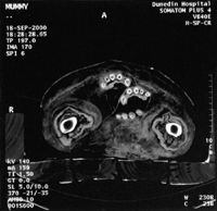 View Details | Figure 1. Axial spiral CT image at the level of the upper thigh, showing the hands. |
 View Details | Figure 2. Axial CT images showing residual lung tissue at the thoracic inlet (left) and undisturbed mediastinal plurae (right). |
 View Details | Figure 3. Axial CT images showing an empty thoracic cavity with dura (meningeal covering of the spinal chord) visible (left) and atheromatous aorta (right). |
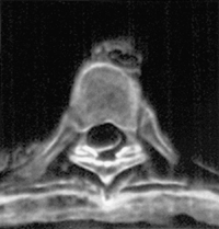 View Details | Figure 4. Enlarged view of atheromatous aorta, from Figure 3. |
The vertebral column deviates towards the right (Figure 5) with no angled gaps between adjacent vertebral bodies. There is a single transverse fracture of the right femur at the level of the lesser trochanter (Figure 6) and also subluxation of the sacroiliac joints (Figure 7). An antero-posterior view of the skull shows a degree of hypertelorism, with an interorbital distance of 25.7 mm calculated (Figure 8). There is no evidence of damage to the nasal septum or the cribriform plate (Figure 9) which suggests that the brain had not been extracted through the nose. Figure 10 shows a lateral radiograph and CT image of the dentition. The maxillary dentition consists, of a first molar, a first premolar and a canine on the left side, and a canine, some root remnants and a first molar on the right side. The mandibular dentition is represented by a number of retained root stubs. No teeth occlude in the mouth and some degree of alveolar resorption is present in the mandible. From the gynaecoid pelvis (Figure 6) and the sharp supra orbital margins, the skeleton is clearly that of a woman (Brothwell, 1963).
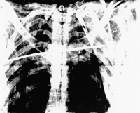 View Details | Figure 5. Coronal radiograph of the upper vertebral column, showing a deviation to the right. |
 View Details | Figure 6. Coronal radiograph showing transverse fracture of the right femur at the level of the lesser trochanter. |
 View Details | Figure 7. Axial spiral CT image showing subluxation of the sacroiliac joints. |
 View Details | Figure 8. Anterioposterior view of the skull, showing a degree of hypertelorism. |
 View Details | Figure 9. Axial spiral CT image of the skull showing undisturbed ethmoidal air cells (arrows), eyes, ear ossicles and residual brain tissue. |
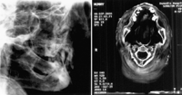 View Details | Figure 10. Oblique lateral radiograph and axial spiral CT scan of the dentition. |
All of her teeth had erupted and, at time of death, very few teeth remained. All of the secondary ossification centres had long fused. This indicates that she was well-matured and probably already elderly.
Radiocarbon age determination gave a result of 2,358±53 years before present.
The general design of the wrapping is an outer single-layer with double strips along each of the longer sides and three horizontal bands formed by folded strips of textile. The principal textile is woven; a loose, plain weave (approximately 10 warp yarns and 10–14 weft yarns/10 mm) but with a fancy weave in the lower third (Figure 11). This fancy weave is rib-like, with each of the three ribs spaced at approximately 100 mm intervals and the ribs created by groups (two, three, two) of slivers (i.e. assemblages of fibres in continuous form, without twist (Denton and Daniels, 2002).
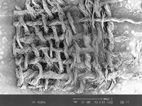 View Details | Figure 11. SEM of wrapping. The textile is woven in a loose, plain weave with a fancy weave in the lower third. |
Mummification has a long history in ancient Egypt, with preservation considered to be essential for the survival of the deceased in the afterlife (Peck, 1998). Initially, mummification was due to rarification in the desert sand and natural dehydration, but from around 4600 BP bodies were artificially prepared to ensure preservation (Macleod et al., 2000). The practice continued until 1600 BP, when the traditional mummification was forbidden by the Emperor Theodosius after the adoption of Christianity as the official religion of the Roman Empire (Macleod et al., 2000). The mummy examined here was dated at 2,358 BP, the period of the 30th Dynasty (356 BC) of Nakhthorhebe (Nectanebo II), and prior to the second Persian domination with the conquest by Alexander in 332 BC (Hallo and Simpson, 1971).
Computed tomography has proved a useful non-destructive method in the investigation of ancient Egyptian mummies (Pickering et al., 1990; Dennison, 1999). Our study shows that the mummy’s arms were extended, palms down over the thighs. From a review of 111 mummies, Gray (1972) found that those from the start of the 21st Dynasty tended to follow this pattern. The thoracic cavity appears empty, which is unusual, as the heart was considered to be the source of wisdom and emotions, and was normally left in situ (Millet et al., 1998).
That the body may have been forced into a narrow coffin is shown by the transverse fracture of the right femur, the fractured ankles and possibly by the subluxed sacroiliac joints.
Figure 5 shows that in each vertebral body the inferior surface maintains a parallel interface with the superior surface of the vertebral body below. Were these simply the result of the body having been crushed into a small coffin after death, there would have appeared angular gaps between the adjoining vertebral bodies. The fact that there are no gaps indicates that the curvature to the right occurred while the woman was alive because bone growth of the vertebral bodies had compensated for the spinal curvature. Close examination of the thoracolumbar region showed degenerative changes in the intervertebral discs. This was particularly marked at the lumbo-sacral level by osteoarthritic changes in the articular joints.
Usually during the embalming process most of the abdominal viscera were removed, dessicated, wrapped, and returned to the body cavity (Foster et al., 1997). The heart was usually left in situ, to be weighed in the Final Judgement, (Zabalgoita, 1996). Figure 3 shows an apparently empty thoracic cavity. The embalmer’s incision for removal of the viscera was made in the left flank (Figure 3).
Although brains were often removed through the nose (Peck, 1998), the present specimen had intact nasal septa and cribriform plates which suggests that the brain had been left in situ. The orbital breadth for this individual (46.2 mm) is at the upper limit of the values recorded from ancient Egyptian males (Oetteking, 1909; cited by Martin and Saller, 1959). While both the anterior interorbital breadth (25.7 mm) and the biorbital breadth (118.9 mm) are greater than those cited by Oetteking, the interorbital index (21.6) is identical to the value given for Egyptians. The appearance of hypertelorism therefore appears to be due to an increase in nasal breadth.
The mummy examined here suffered from extensive dental disease. Of the 32 teeth normally present in the mouth only six are identifiable; a small number of retained root apices are also present. That there was extensive periodontal disease is revealed by the appreciable loss of bony support of the molar tooth. The diet of ancient Egyptians, particularly bread, reportedly had an excessive sand content, whose silica is thought to be a major cause of dental attrition (Melcher et al., 1997; Thekkaniyil et al., 2000). Egyptian mummies are characterised by high-wear rates and low caries, and a high prevalence of periodontal disease (Harris et al., 1998), which accords well with our observation.
Close examination of a small fragment of textile from a non-defined position of the wrapping showed irregularity both in yarn twist (S-twist; Denton and Daniels, 2002) and in the number of warp and weft yarns per 10 mm (Figure 11). Note that one set of yarns curving around the other at one edge suggests that this textile fragment includes a selvage. Fibrillation, as described by Hearle et al., (1998) is also evident in Figure 12. The fibre is cellulose, probably linen, as shown by the horizontal nodes irregularly spaced along the fibre length (Figure 12). A large number of species of linen and flax are known to exist, and fibres from other plants may also have been used to manufacture mummy-wrappings. Textile was not recorded in the Mediterranean region before the time of Christ, with linen being the dominant textile at the time (Cockburn and Cockburn, 1998).
 View Details | Figure 12. SEM of individual textile fibres showing horizontal nodes and a hollow core, which suggest linen. |
The authors thank Mr. S. Paul, Director of the Otago Museum for allowing us access to the mummy.
|