| M. G. Hossain, corresponding author. e-mail: hossain95@yahoo.com phone: +880-721-750597; fax: +880-721-750064 Published online 13 July 2004 in J-STAGE (www.jstage.jst.go.jp) DOI: 10.1537/ase.00066 |
Brachycephalization is the name given for an increase in head breadth relative to head length. (Brachycephaly is defined as short-headed with a relatively broad skull.) In effect, head form, when viewed from above, becomes more rounded. The cephalic index is commonly used to quantify brachycephalization.
In an early work, Weidenreich (1945) recognized and described secular changes of human head form tending towards brachycephaly. According to Weidenreich, brachycephalization had occurred over time in various populations. Thereafter, many researchers reported the same phenomenon worldwide (e.g. Bear, 1956; Bielicki and Welon, 1964; Huizinga and Slob, 1965; Jorgensen et al., 1974; Skrobak-Kaczynski et al., 1977).
With respect to Japanese populations, according to Suzuki (1969), brachycephalization seems to have occurred in both adult sexes after the Kamakura era (1192–1333 AD). Over the last hundred years, in Japanese adults, the rate of increase in the cephalic index has been extremely high (Morita and Ohtsuki, 1973; Yanagisawa and Kondo, 1973; Ohtsuki and Ito, 1980; Ohtsuki and Iwamura, 1980; Kouchi, 1986; Ashizawa, 1988; Kondo et al., 1999). In contrast, debrachycephalization (the opposite of brachycephalization) has also been observed in numerous ethnic groups (Miller, 1970; Tobias and Netscher, 1977; Susanne et al., 1988; Cameron et al., 1990; Vercauteren, 1990; Gazi-Coklica and Muretic, 1991; Gyenis, 1994; Zellner et al., 1998; Constant, 1999).
Kouchi (2000) looked at the head dimensions of 9008 Japanese males and 3430 females, measured between 1942 and 1998. Head length was found to have remained relatively unchanged with time, while head breadth for both sexes started to increase in the cohort of those born in 1890–1900, increased rapidly in the 1910–1949 male cohorts, and increased similarly in the 1920–1959 female cohorts. The cephalic index showed changes similar to those observed for head breadth. The increase of cephalic index values then seems to have ceased, with head form having reached a steady condition in Japanese males born after 1950 and Japanese females born after 1960.
The purpose of the present study was to look at more recent measurements of head dimensions, and compare them with previous values reported for Japanese adult females.
The total female sample used in the current study consisted of 832 healthy Japanese adults. The average age at the time of measurement was 19.32 ± 0.95 years (range 18–25 years). The samples, drawn from a student population, were collected between 1998 and 2001 from several universities in the Tokyo and Kyoto areas. Four head dimensions were measured: head length, head breadth, head height, head circumference. The height and body weight of the subjects were also recorded. These were measured by one of us (F.O.). Measurements were taken following the technique of Martin and Saller (1957).
Two previous studies were available for comparative purposes. Study I comprises materials originally collected by Matsumura (1925) and Study II comprises a measurement series collected more recently (Ohtsuki and Ito, 1980)
Study I
Comparisons with the Matsumura (1925) series were done for head length, head breadth, cephalic index, and height. The Matsumura study was conducted at numerous institutions in various districts of Japan from 1910 to 1917, and comprises measurements taken from 2000 adult Japanese female students. A single observer made the measurements. This series, taken about 85 years before the current series, consisted of subjects aged 18 years and older.
Study II
Comparisons with the Ohtsuki and Ito (1980) series were done for head length, head breadth, head height, head circumference, cephalic index, and height and weight. The Ohtsuki and Ito study comprises measurements taken from 1547 Japanese female students, ranging in age from 18 to 25 years. Again, a single observer (F.O.) took the measurements over the period 1975–1979. The technique of Martin and Saller (1957) was used. This series, taken at various colleges and universities in Tokyo, was collected about 21 years prior to the current series.
For comparison with the previous studies, head form (cephalic index, x) in the current series was divided into six categories (Knussmann, 1988). The cephalic index categories were: hyper-dolichocephalic (x ≤ 71.9), dolichocephalic (72.0 ≤ x ≤ 76.9), mesocephalic (77.0 ≤ x ≤ 81.9), brachycephalic (82.0 ≤ x ≤ 86.4), hyper-brachycephalic (86.5 ≤ x ≤ 91.9), and ultra-brachycephalic (x ≥ 92.0). To facilitate comparison, the cephalic index values of the Matsumura series were also rearranged according to Knussmann (1988).
Descriptive statistics were calculated for the following seven variables: head length, head breadth, head height, cephalic index, head circumference, and height and weight. Comparisons were made between the current study, Study I of Matsumura (1925), and Study II of Ohtsuki and Ito (1980).
The data set of the current study was subdivided according to birth year cohorts from 1978 to 1983 (see Table 1) to discern the presence of any trends in head dimensions during the measurement period. The birth year of the subjects actually ranged from 1976 to 1983. However, as the sample sizes for 1976 and 1977 cohorts were very small, these were excluded from the present analysis.
The current series was then subjected to further statistical analysis. To examine the possibility of a birth year cohort effect on head dimensions, height, and weight, a one-way analysis of variance (ANOVA) was conducted. The underlying linear model corresponding to each variable is
Yij = μ + αi + εij (i = 1, 2, …, p and j = 1, 2, …, q)
where Yij is the jth observation (response variable) for the ith birth year cohort, μ is the general mean effect, αi (=μi−μ) is the additional effect of the ith birth year cohort, μi is the average effect of the ith birth year cohort, εij is the error term, p is the number of cohorts, and q is the number of observations in each cohort.
The ANOVA procedure tests the hypothesis that:
H0: α1 = α2 = α3 = … = αp = 0 and equivalently μ1 = μ2 = … = μp = μ by means of a single F test.
If the hypothesis of equality of cohort means is rejected, one may presume that there are cohort mean differences. The randomness, normality, and homogeneity of cohort variances were tested prior to the ANOVA by using the Kolmogorov-Smirnov nonparametric test, use of a normal probability plot, and the Levene test, respectively.
Finally, linear regression analysis was applied to the current study data set to try to detect the presence of trends in the variables among the birth year cohorts from 1978 to 1983. All statistical analyses were carried out using Statistica 5.0 software.
Although the primary emphasis of this investigation was on head dimensions, the other two variables, height and body weight, are discussed first. Table 2 displays the means and standard deviations of height and weight. The other five variables—head length, head breadth, cephalic index, head height, and head circumference—also shown in Table 2, are discussed later.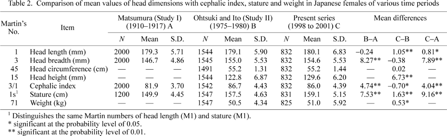
Comparison of Study I with the current study
Comparison of the current study with Study I (the 1925 series) showed that the subjects of the current study were taller by 9.16 cm than their predecessors of 85 years ago (Table 2). This height difference was statistically significant (P < 0.01). Study I did not include body weight, so no comparisons of this were possible.
Comparison of Study II with the current study
Comparison of the current study with Study II (the 1980 series of Ohtsuki and Ito) showed that height had increased significantly (P < 0.01) by 1.63 cm. Differences in mean weight recorded by the two studies were also significant, with the current study showing a significant (P < 0.05) increase of 0.53 kg (Table 2).
Comparison of Study I with Study II
When Study I was compared with Study II, a significant difference in height was found, with an increase in stature of 7.53 cm from the 1925 to the 1980 studies (Table 2).
Comparison of Study I with the current study
Comparisons showed a significant increase in three variables: head length (P < 0.05), head breadth (P < 0.01), and cephalic index (P < 0.01). Head length increased by 0.81 mm, head breadth by 7.89 mm, and the cephalic index by 4.04 units (Table 2). Study I did not contain a measure of head height and head circumference, so comparisons of these data were not possible.
Comparison of Study II with the current study
Comparisons showed increases in head length, head circumference, and head height. Head breadth and cephalic index had decreased. Head length increased by 1.05 mm, head circumference by 0.02 cm, and head height by 6.73 mm. Of these measures, only the differences seen in head length and height were significant (P < 0.01). Head breadth decreased by 0.38 mm and the cephalic index by 0.70 units. Of the latter two variables, only the decrease in cephalic index was significant (P < 0.05) (Table 2).
Comparison of Study I with Study II
Comparison of Study I with Study II displayed increases in head breadth and cephalic index. Head length showed a slight decrease, which was not statistically significant. Head breadth increased by 8.27 mm and cephalic index by 4.74 units, while head length decreased by only 0.24 mm. Changes in head breadth and cephalic index were statistically significant at P < 0.01 (Table 2).
Table 3 shows the frequency distribution of the cephalic index by type in the various studies of Japanese females. The frequencies of hyper-dolichocephalic, dolichocephalic, mesocephalic, and brachycephalic head forms in Study I were higher than in Study II. In contrast, the hyper-brachycephalic and ultra-brachycephalic head forms in Study I were less frequent than in Study II. The frequencies of hyper-dolichocephalic, dolichocephalic, and mesocephalic head forms in Study I were higher than in the current study. In addition, the brachycephalic, hyper-brachycephalic, and ultra-brachycephalic frequencies in Study I were lower than in the current study. Finally, the differences in the six categories of the cephalic index between Study II and the current study were very small (Table 3).
Subdivision into cohorts by birth year facilitated further study of possible trends over time in the current study sample. Table 4 gives the results of the analysis of variance in head and body dimensions as well as in the cephalic index of Japanese adult females. Of the seven variables, only head length (P < 0.01), head breadth (P < 0.01), and head circumference (P < 0.01) were found to vary statistically significantly over the birth year cohorts from 1978 to 1983.
Regression analysis was used to examine for the presence of trends in the current study sample. The means of all dimensions and the cephalic index are displayed in Figure 1. It can be seen that there were yearly fluctuations in the dimensions, including cephalic index. This is often a characteristic of cohort studies. These fluctuations in the head and body dimensions were further examined with linear regression analysis. Table 5 shows the regression coefficients of the head and body dimensions in the Japanese adult females. All the dimensions, as well as the cephalic index, displayed negative regression coefficients. These negative coefficients indicate a tendency for decreasing values within the 1978–1983 birth year cohorts.
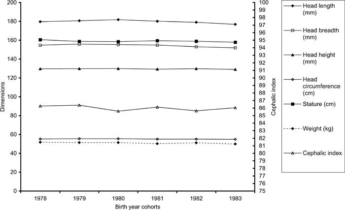 View Details | Figure 1. Mean values of head dimensions, height, weight, and cephalic index of adult Japanese females. |

The present study demonstrates that secular changes in the head form of Japanese adult females have occurred during the last eight decades. With respect to head dimensions between Study I and Study II, head breadth increased substantially, while head length decreased only slightly (Table 2). As expected, these changes in head dimensions were also reflected in head form (cephalic index). That this change in the cephalic index has been extensive during the first six decades can be seen in the difference between Study I and Study II. The difference in the cephalic index was 4.74 units, which corresponds to an incremental change of 0.7 units per decade. From Study II to the current study, head breadth decreased only slightly, while head length increased by 1.05 mm and cephalic index decreased 0.3 units per decade. These findings suggest that the tendency toward brachycephalization in Japanese adult females may have ended. Hossain et al. (2004) found the same phenomenon in a Japanese male sample. These results are in accord with those of Kouchi (2000), who reported that the increase in the mean cephalic index had ceased for Japanese females born after 1960. The present results are also in agreement with Miller (1970), Tobias and Netscher (1977), Susanne et al. (1988), Cameron et al. (1990), Constant (1999), and Zellner et al. (1999). There is now agreement that recent Japanese adult females are brachycephalic in head form (Yanagisawa and Kondo, 1973; Ohtsuki and Ito, 1980; Ohtsuki and Iwamura, 1980; Kouchi, 1986, 2000; Kondo et al., 1999). The current study also showed that the present-day Japanese adult females have greater mean head height, as well as height and weight, than their predecessors of two and eight decades ago.
Analysis of variance of the head and body dimensions and the cephalic index across cohorts of birth years from 1978 to 1983 showed that head length, head breadth, and head circumference displayed statistically significant differences (Table 4). A regression analysis was used to identify the presence of any trends in the cohort data. The slope of the regression lines for the head and body dimensions and the cephalic index showed slightly decreasing tendencies during the investigated period, especially with respect to head length and head breadth (Figure 1, Table 5). These results are in agreement with those of Gyenis (1994), who reported that the head form of university students in Hungary also displayed a slight decrease in values, and with studies of head form in German children (Zellner et al., 1998, 1999). Susanne et al. (1988) also found significant decreases in head form in three populations: Belgium (1960–1980), East Germany (1954–1980), and Czechoslovakia (1957–1987). Finally, the present results in females parallel those found in a study of Japanese males (Hossain et al., 2004).
While Kouchi (2000) suggested that the tendency for the cephalic index to increase over time seemed to be absent in Japanese females born after 1960, it is important to note that the current study was limited to females born between 1976 and 1983. Nevertheless, there is some agreement with the findings of Kouchi in that the cephalic index exhibits a very slight tendency to decrease in the 1978–1983 birth year cohorts (Figure 1, Table 5).
The present study showed that the increase in mean cephalic index in Japanese adult females was primarily influenced by the increase of head breadth. This pattern was the same as that seen in our previous study of Japanese males (Hossain et al., 2004) and in other previous studies (Ohtsuki and Ito, 1980; Kouchi, 2000). It is not known exactly which factors are responsible for the changes in human head form over time (Tanner, 1962). Many hypotheses have been proposed for the secular changes in head form, such as environmental factors (Abbie, 1947), increased protein in the diet (Miller, 1970), decreased psychological and physiological stress (Miller, 1970), and increased medical facilities and care (Miller, 1970). Some investigators (Beals, 1972; Crognier, 1981; Beals et al., 1983; Bharati et al., 2001) have suggested that the occupation of cold climates was one of the circumstances that may have led to the increasing frequency of brachycephalic head form over time. Other researchers (Bielicki and Welon, 1964; Henneberg, 1976) believe that the brachycephalic head form have been selected as a consequence of evolutionary forces. Nevertheless at the moment the causes of secular changes in head form remain incompletely known.
|