| Kazuhiro Sakaue, corresponding author. e-mail: k-sakaue@mail.tains.tohoku.ac.jp phone: +81-22-717-8027; fax: +81-22-717-8030 Published online 30 November 2005 in J-STAGE (www.jstage.jst.go.jp) DOI: 10.1537/ase.00098 |
In forensic and physical anthropology, estimation of age at death of individuals from osteological features is an important research theme. In particular, the pubic symphyseal face has been widely used in estimating age (Todd, 1920; Hanihara, 1952; McKern and Stewart, 1957; Nemeskeri et al., 1960; Gilbert and McKern, 1973; Suchey, 1979; Meindl et al., 1985; Katz and Suchey, 1986; Zhang, 1986; Brooks and Suchey, 1990; Klepinger et al., 1992; Sinha and Gupta, 1995; Hoppa, 2000). Classification methods based on age-related changes of the pubic symphyseal region have been established using skeletal series comprised of individuals with known ages at death (Todd, 1920; McKern and Stewart, 1957; Nemeskeri et al., 1960; Gilbert and McKern, 1973; Katz and Suchey, 1986). These methods can be used to estimate age in individuals of unknown age at death.
The Suchey–Brooks system was developed with a large reference sample of known age (n = 1255), at least four times as large as those of other studies, comprising modern individuals autopsied at the Department of Coroner, County of Los Angeles between 1977 and 1979 (Brooks and Suchey, 1990). These individuals had birth and/or death certificates, their sex was known, and the population/ethnic group to which they belonged were estimated (the majority was considered American White, American Black, or Mexican). The methodology of the Suchey–Brooks system is comparatively unambiguous. The pubic symphyseal face is classified into six phases according to age-related osteological features common to both males and females, and the mean age and standard deviation of each phase are given by sex (Brooks and Suchey, 1990). For the larger male sample, means and standard deviations of each population/ethnic group (American White, American Black, and Mexican) were also given (Katz and Suchey, 1989). In addition, casts of model cases of each phase have been made available, enhancing the clarity of the methodology (Suchey et al., 1988).
Previous studies have discussed possible influences of population variation on age estimation based on the pubic symphyseal region (Todd, 1920, 1921; Meindl et al., 1985; Katz and Suchey, 1989; Sinha and Gupta, 1995; Hoppa, 2000; Schmitt, 2004). Katz and Suchey (1989) observed increased differences among populations with advanced age, reporting significant differences after phase III. Klepinger et al. (1992) recommended a “racially specific” refinement of the Suchey–Brooks system. However, Katz and Suchey (1989) and Suchey and Katz (1998) cautioned that it is not possible to determine whether the observed differences relate to genetic or environmental factors such as nutrition or drug/alcohol use. Schmitt (2004) attempted a test of the Suchey–Brooks system in a Thai population with known ages and reported a remarkable retention of earlier phase morphologies in advanced age (their Figure 2), resulting in drastically lower estimated ages.
With Japanese skeletal material, Hanihara (1952) and Hanihara and Suzuki (1978) have studied the pubic symphyseal face in relation to age in males. However, their sample sizes were small (135 and 70 individuals, respectively), and since they used a revised Todd’s system as the standard of evaluation, their results cannot be compared with those of other studies based on the original Todd or the Suchey–Brooks systems.
The purpose of this study was to apply the Suchey–Brooks system of evaluating the pubic symphyseal region in a relatively large Japanese sample, and to determine mean ages and degree of variation for each of the six phases. The present study is based on a male sample over twice the size of previous Japanese samples, and is the first to examine Japanese females. Finally, bias and inaccuracy that accompany age estimations by the six-phase Suchey–Brooks system was investigated.
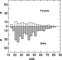
The materials used were pubic bones of 416 Japanese skeletons (326 males and 90 females) with known sex and age at death, collected between 1885 and 1944. The age range of the series was 14–83 years (Figure 1). The skeletal materials are stored in the University Museum, the University of Tokyo; the Department of Anatomy, Chiba University School of Medicine; the Graduate School of Social and Cultural Studies, Kyushu University; and the Kyoto University Museum. The right os pubis was observed, but when the right side was damaged, the left side was used. Figure 2 shows the representative morphology observed in the recent Japanese series for each of the six Suchey–Brooks phases.
 View Details | Figure 1. Age distribution of the recent Japanese sample of the present study. |
 View Details | Figure 2. Characteristic morphology of each phase of the Suchey–Brooks system, reproduced from Brooks and Suchey (1990) and observed in the recent Japanese sample. The key features are shown in italics. Phase I: Symphyseal face has a billowing surface (ridges and furrows) which usually extends to include the pubic tubercle. The horizontal ridges are well-marked, and ventral beveling may be commencing. Although ossific nodules may occur on the upper extremity, a key to the recognition of this phase is the lack of delimitation of either extremity (upper or lower). Phase II: The symphyseal face may still show ridge development. The face has commencing delimitation of lower and/or upper extremities occurring with or without ossific nodules. The ventral rampart may be in beginning phases as an extension of the bony activity at either or both extremities. Phase III: Symphyseal face shows lower extremity and ventral rampart in process of completion. There can be a continuation of fusing ossific nodules forming the upper extremity and along the ventral border. Symphyseal face is smooth or can continue to show distinct ridges. Dorsal plateau is complete. Absence of lipping of symphyseal doral margin; no bony ligamentous outgrowths. Phase IV: Symphyseal face is generally fine grained although remnants of the old ridge and furrow system may still remain. Usually the oval outline is complete at this stage, but a hiatus can occur in upper ventral rim. Pubic tubercle is fully separated from the symphyseal face by definition of upper extremity. The symphyseal face may have a distinct rim. Ventrally, bony ligamentous outgrowths may occur on inferior portion of pubic bone adjacent to symphyseal face. If any lipping occurs, it will be slight and located on the dorsal border. Phase V: Symphyseal face is completely rimmed with some slight depression of the face itself, relative to the rim. Moderate lipping is usually found on the dorsal border with more prominent ligamentous outgrowths on the ventral border. There is little or no rim erosion. Breakdown may occur on superior ventral border. Phase VI: Symphyseal face may show ongoing depression as rim erodes. Ventral ligamentous attachments are marked. In many individuals the pubic tubercle appears as a separate bony knob. The face may be pitted or porous, giving an appearance of disfigurement with the ongoing process of erratic ossification. Crenulations may occur. The shape of the face is often irregular at this stage. |
The mean age and standard deviation of each phase were calculated. Kolmogorov–Smirnov tests, however, resulted in rejection of a normal distribution in phases II, III, and V in males, and phases II and III in females, at the 1% significance level. Therefore, to determine whether adjacent phases were significantly different in age, the Mann–Whitney U test was performed. Differences in age between males and females were also analyzed by the Mann–Whitney U test.
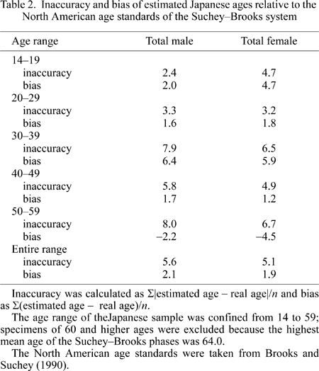
The mean ages of the Suchey–Brooks total male and female series were then given, as age estimations, to individual recent Japanese specimens attributed to each of the six phases. Mean errors between the estimated and real ages (inaccuracy and bias) were calculated at 10-year age intervals (Lovejoy et al., 1985; Schmitt, 2004). Inaccuracy was calculated as Σ|estimated age − real age|/n, showing the average magnitude of absolute error. Bias was calculated as Σ(estimated age − real age)/n, expressing the tendency for either over- or under-estimation of age.
All tests were performed using the statistical software package SYSTAT 10.2.
Table 1 shows the mean age and standard deviation of each of the six phases of the recent Japanese series of the present study. Age differences between each of the two adjacent phases were significant (P < 0.05), except between phases III and IV in the females. As suggested by previous studies (McKern and Stewart, 1957; Katz and Suchey, 1986; Klepinger et al., 1992), variation as expressed by the standard deviation tended to increase with age. Differences between males and females were not significant in any of the phases at the 5% level, but were significant in phases III and IV at the 10% level.
Table 2 shows inaccuracy and bias at 10-year intervals, when the mean sex-specific ages of the Suchey–Brooks series (Brooks and Suchey, 1990) were applied to the recent Japanese series. In the recent Japanese males, inaccuracy did not exceed 8.0 in any age group (range 2.4–8.0). Bias in males tended to be low, at around two years (although higher in the 30–39 year age group), and positive in all age groups except for the 50–59 year age group (bias of −2.2). In the recent Japanese females, inaccuracy was relatively high (4.7) in the 10–19 year age group, but did not exceed 7.0 in any age group (range 3.2–6.7). Bias in females ranged from one to six years, and was positive in all age groups except for the 50–59 year age group (bias of −4.5).
In the recent Japanese series of the present study, age distribution of each phase differed significantly between adjacent phases with the exception of the female Phase III/IV contrast based on small samples. This suggests that the Suchey–Brooks method of evaluating age-related changes of the pubic symphyseal region is effective in recent Japanese skeletal series. Katz and Suchey (1986) noted that age estimation based on the pubic symphyseal region is more useful in individuals below 40 years, which is also the conclusion of the present study judging from the large standard deviation values of over 10 years in phases V and VI (> 45 years).
The morphological features used in the Suchey–Brooks system supposedly do not require consideration of sex, because they focused their definition of phases on features that apparently did not vary between sexes. Thus, when this standard is applied, the difference in male and female mean ages in each of the phases is expected to be insignificant. In the present study, this expectancy was largely met, although a difference between sex, significant at the 10% level, was observed in phases III and IV. As Gilbert and McKern (1973) suggested, the development of the ventral rampart of the female pubis tends to be delayed compared with that of the male pubis. In the Suchey–Brooks system, development and form of the ventral rampart is a key morphological feature of phases III and IV, perhaps leading to the potential sexual differences observed in the present study.
Schmitt (2004) applied the mean ages of the Suchey–Brooks series (Brooks and Suchey, 1990) to a Thai population supposedly of known age. He reported that, in the Thai series that he examined, inaccuracy observed at 10-year age intervals between 30 and 60 years of age ranged up to 17 years. The accuracy values of the present Japanese series were just over 3 years in the 20–29 year age group, and 8.0 or less in the 10-year intervals between ages 30 and 60. Thus, compared to the Thai series, the errors involved in estimating age by means of the Suchey–Brooks age standards (Brooks and Suchey, 1990) were relatively slight in the Japanese series. Indeed, the difference between the recent Japanese and the Suchey–Brooks series in mean ages by phase was less than 3 years (Table 3). Therefore, the application of the Suchey–Brooks system to the recent Japanese may cause no major problems, contrary to the claims of Schmitt (2004) that application to Asian populations in general may be problematic.
As part of the Suchey–Brooks system, Katz and Suchey (1989) reported the mean ages and standard deviations by phase separately for their American White, American Black, and Mexican male subsamples. Since age estimation is potentially affected by population variation, and some researchers recommend “racially specific” methods (Klepinger et al., 1992), it is of interest to known how the mean ages of the recent Japanese compare with those of the above three populations. Figure 3 shows that the Japanese male means plot closest to those of the American Black males. The difference between the Japanese and American White males was significant (with a t-test) at the 1% level in phases IV–VI, although the validity of such tests are open to question because of the lack of conformity to a normal distribution. Despite such ambiguity in populational comparisons, the mean Japanese ages of the Suchey–Brooks phases presented above can be considered as a valid age scale for application to Japanese skeletal materials of unknown age.
 View Details | Figure 3. Mean ages and standard deviations of recent Japanese males, and means of the American White, American Black, and Mexican males (from Katz and Suchey, 1989). |
I wish to express my gratitude to Dr G. Suwa (The University Museum of the University of Tokyo), Dr T. Chiba (Chiba University School of Medicine), Dr Y. Tanaka and Dr T. Nakahashi (Graduate School of Social and Cultural Studies, Kyushu University), Dr H. Ishida and Dr M. Nakatsukasa (Kyoto University) and Dr Y. Dodo (Tohoku University School of Medicine), for permission to access skeletal materials and for valuable advice.