| Yousuke Kaifu, corresponding author. e-mail: kaifu@kahaku.go.jp phone: +81-3-3364-7140; fax: +81-3-3364-7104 Published online 28 February 2007 in J-STAGE (www.jstage.jst.go.jp) DOI: 10.1537/ase.061208 |
Minatogawa 1, which dates from 18000 BP (ca. 21500 years ago), was unearthed from the Minatogawa Fissure site on Okinawa Island in 1970. Minatogawa 1 is one of the rare complete skeletons of Late Paleolithic Homo sapiens from East Asia, and thus is central to the investigation on the earlier phase of peopling in this region (Suzuki and Hanihara, 1982; Baba et al., 1998; Kondo and Matsu’ura, 2005). Recently, Kodera (2006) examined the occlusion of Minatogawa 1, and questioned whether its cranium and mandible came from the same individual. Because of the potential importance of this claim on both past and future studies, I re-examined the original specimen to test it. A revised description on the occlusion and tooth wear of Minatogawa 1 is also presented.
According to Suzuki (1982a), the skeleton of Minatogawa 1 was discovered in “an almost anatomically normal state.” However, details of the spatial distribution of each bone within the sediments have not been reported. In addition, because three isolated mandibles were also unearthed from the vicinity of Minatogawa 1 (Minatogawa mandible A, B, and C: Suzuki, 1982b), Kodera’s claim cannot be rejected based on the published information of the excavation. Thus, a test of his claim should be based on direct observation of the specimen itself. If the two elements are from a single individual, the portions of their contact (the temporo-mandibular joint and occlusion) should show a good fit.
The major bases of Kodera’s assessment were (1) inconsistency of the occlusal wear pattern between the left maxillary and mandibular first molars, (2) absence of reasonable contact or occlusal relations between the maxillary and mandibular dentitions, and (3) disparity between the bi-mandibular fossa breadth on the cranial base and bi-condylar breadth of the mandible (Kodera, 2006). However, these arguments are not well-founded because sufficient examination was not made for the process of tooth wear and distortion of the specimen.
Kodera (2006) pointed out the inconsistency between the bi-mandibular fossa breadth of the cranial base and bi-condylar breadth of the mandible. However, this aspect does not serve as a good guide in examining the identity of the cranium and mandible, because both bones suffer from minor but significant damage as is usually seen in a fossil specimen. In particular, the basal symphyseal part of the mandible of Minatogawa 1 is crushed, and this probably affects the breadth of the mandible. When the shape of the condyle and mandibular fossa are compared on each side, they are a good match and there is no reason to suspect their identity (Figure 1).
 View Details | Figure 1. (a) Articular eminence and mandibular condyle of Minatogawa 1. The parts indicated by the arrows are restorations. (b) Occlusion on the right side of Minatogawa 1. |
There are some misalignments of the maxillary incisors in the current reconstruction of Minatogawa 1. Also, I could not be sure whether the left canine glued to the mandible belonged to this individual. However, these uncertainties do not affect the following examination of the occlusion.
Many of the teeth of the maxilla and mandible of Minatogawa 1 were missing; some had been lost before death. Dental occlusion can be examined only on the left molar rows, and partially at the area of the right maxillary canine (C1) and second premolar (P2). The latter teeth fit well onto the area between the mandibular first premolar (P1) and first molar (M1) when the right mandibular condyle is articulated with the right mandibular fossa. However, the left jaw bones are damaged and many of the left molars are more or less dislocated from their original positions (Figure 2).
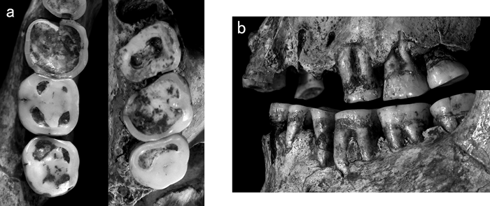 View Details | Figure 2. Left molar rows of Minatogawa 1. (a) Occlusal views of the mandibular (left) and maxillary (right) rows; (b) buccal view. |
Because it was difficult to correct the tooth alignments directly on the fragile fossil specimen, I decided to reconstruct the original dentition and occlusal relation of Minatogawa 1 using plaster casts. I prepared high-quality plaster casts of the left molar rows using dental silicone as the mold material. The casts were then cut and the three molars on each row were separated from each other, except for M1/M2 where the original contact relation is retained. The excess plaster on the mesial and distal interproximal facets, where present, was carefully ground off, and the cast of each molar was glued together to reconstruct the original dentitions (Figure 3). The interproximal facets between the maxillary and mandibular M2s and M3s were distinct enough to find their original positional relationships. Although only tiny facets remain at the buccal portion of the M1/M2 contact area, the positional relationships between these teeth can be easily reconstructed by smoothly connecting their worn occlusal surfaces.
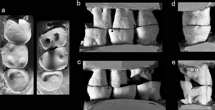 View Details | Figure 3. (a) The reconstructed casts of the left maxillary (right) and mandibular (left) molar rows of Minatogawa 1. The distortion in the original specimen has been corrected following the procedure described in the text. (b)–(e) The buccal, lingual, posterior, and anterior views of the reconstructed casts of the molar rows in centric occlusion. The arrows in (a)–(c) indicate slight enamel projections that fit to inter-spaces between their opposing molars. |
Next, I examined the occlusion between these reconstructed casts of the molar rows. Although much of the occlusal contact has been lost between the M1s due to extreme wear on the M1, the centric occlusion (the occlusal position that attains maximum contact between the maxillary and mandibular dentitions) can be easily located by the M3s and the buccal enamel of the M2s (Figure 3). In this position, the maxillary and mandibular molar rows show a good fit in buccal view. In addition, the buccolingually undulating occlusal surfaces of the M3s fit well in posterior view. When the occlusal surfaces are observed closely, the slightly projecting portions of the enamel at the distobuccal corner of M2, near the mesiobuccal corner of M2, and at the mesiolingual corner of M3 fit respectively to the inter-spaces between their opposing molars (Figure 3).
Thus, when a proper correction is made for the distortion, the maxillary and mandibular molar rows of Minatogawa 1 occlude perfectly. Other details of occulsal surface morphology of the left maxillary and mandibular molars are also consistent with the interpretation that they belong to a single individual (see below).
These reconstructed dentitions enable a valid evaluation of the occlusal relation in Minatogawa 1. Both the maxillary and mandibular molar rows show helicoidal occlusal surfaces along their long axes (Smith, 1986). Figure 3 indicates that Minatogawa 1 approximates a Class I anteroposterior molar relation, in which the mesiobuccal cusp of M1 is aligned over the buccal groove of M1. Although the latter structure seems to be shifted slightly anteriorly relative to the former, this degree of discrepancy is not unusual in people who suffer from extensive interproximal wear and subsequent mesial drift of the buccal teeth (Kaifu et al., 2003). However, the buccolingual molar relation of this individual is shifted somewhat from the normal state: the maxillary molars occlude almost directly onto the mandibular molars, so that their buccal and lingual enamel surfaces are almost in the same vertical planes. This probably resulted from the partial decay of M1 and M2 due to severe occlusal wear, which is described below.
The state of tooth wear provides anthropologists with important information on both the masticatory and non-masticatory activities of populations. In this regard, what we can learn from the tooth wear of Minatogawa 1 is an intriguing question. In this section, I describe occlusal wear on the left molars of Minatogawa 1 and discuss possible causes of the disparity in wear between the maxillary and mandibular M1s.
The M1 is extremely worn, exposing the pulp cavities of the buccal and lingual roots. The occlusal surface is steeply sloping buccolingually. Low enamel remains on the buccal portion of the crown, but the wear reaches deep into the root on the lingual side, particularly at the mesiolingual corner. The worn occlusal dentine surface is gently concave both buccolingually and mesiodistally. The M2 also suffers from severe occlusal wear, leaving good amounts of enamel only at the buccal, distal, and lingual rims. The occlusal enamel remains only at the buccal end of the buccal occlusal groove, and most of the mesial enamel wall is worn away. The buccolingually concave occlusal dentine surface slopes mesially to continue to the occlusal surface of M1. The lingual enamel forms a step and its distal half is higher than the mesial half. This step existed during the individual’s life, because its edge is slightly worn. The M3 is worn flat, and dentine is exposed along its mesial border, connecting the mesiolingual and mesiobuccal cusps.
The occlusal enamel of the M1 is completely worn away. The remaining enamel rim is disproportionately higher on the lingual than on the buccal side. The occlusal dentine surface is gently concave. The occlusal surface of the M2 is worn flat and large dentine patches are exposed on the four major cusps. The occlusal surface of M3 is also worn flat. Large to moderate patches of dentine are exposed on the mesiobuccal, mesiolingual, and mesiodistal cusps.
The disparity of worn occlusal surfaces between the jaws in archeological human remains is often ascribed to the practice of using teeth as a tool (Hillson, 1996; Larsen, 1997). What, then, can we say for the case of the left molars in Minatogawa 1? Although the possibility of a partial involvement of non-masticatory function cannot be ruled out, normal masticatory activity alone can explain the observed situation.
Dentine wears more easily and rapidly than enamel. This explains the steep buccolingual wear plane of the M1: its loss of lingual enamel probably increased the rate of wear on this side, resulting in the development of the steep slope. The activity of biting and pulling out various materials may produce similar patterns of wear, but the absence of substantial occlusal clearance between the buccal enamels of the maxillary and mandibuar M1s in this individual (Figure 3) contradicts such an explanation. Furthermore, the concavities of the occlusal dentine surfaces on M1, M2, and M1 were probably formed primarily through a vertical, masticatory crushing motion rather than by horizontal friction with non-dietary materials. The lingual enamel of M1 remains disproportionately high probably because of the disappearance of the lingual enamel of M1. This partial decay of the lingual two-thirds of M1 crown probably led to the diminution of occlusal support in this part, and accelerated wear on the mesial M2 to a degree that resulted in the breakdown of its enamel, as described above. Additionally, this early breakdown of the enamel of the M2 enabled its antagonist, M2, to retain much of its occlusal enamel.
Similarly, the remaining maxillary incisors, maxillary and mandibular premolars, and mandibular right molars show no obvious signs of non-masticatory function. Their occlusal surfaces are worn flat (with varying degree of labiolingual inclinations), and can be smoothly connected between the adjacent teeth.
The recent claim (Kodera, 2006) that the cranium and mandible of Minatogawa 1 belong to different individuals did not appropriately consider the distortion present in its jaws and dentitions. When a proper correction is made for such distortions, the maxillary and mandibular molar rows of Minatogawa 1 occlude perfectly in a normal Class I relation, with a harmonious fit of the minute details of their occlusal wear patterns. Thus, the cranium and mandible of Minatogawa 1 no doubt belong to the same individual.
An interesting suggestion that arose from the present examination is the considerably rapid rate of tooth wear in this individual. Suzuki (1982b), and Hanihara and Ueda (1982) first noted this view in their original descriptions of the Minatogawa remains.
Great differences in the wear scores between the M1 and M2 of Minatogawa 1 (Hanihara and Ueda, 1982) do not necessarily indicate a rapid wear rate in this individual. This is because the wear severity of a tooth could be highly dependent on other adjacent teeth, particularly in this advanced stage of wear as is exemplified above, and because a wear scoring system is subjective in nature so that difference between, for example, 8 and 7, and that between 7 and 6, are not necessarily equal to each other in terms of physical wear and tear. However, Minatogawa 1 probably experienced rapid tooth wear during his life, even compared to the Jomon people who are often characterized by extensive wear on their dentitions (Kaifu, 1999). The age at death of Minatogawa 1 as estimated from its state of cranial suture closure is 25–30 years (Suzuki, 1982b). Kaifu (2000) reported that the wear severity of the M1, M2, M1, and M2 of Minatogawa 1 are respectively 8, 7, 6.5, and 4, based on the Smith’s 8-grade scoring system (Smith 1984). These figures are far greater than the average scores for the dentally adult sample from the Holocene Jomon hunter-gathers of Japan (M1 = 4.6, M2 = 4.0, M1 = 4.5 and M2 = 3.8, n = 61–63 for each tooth: Kaifu, 2000). This observation is also consistent with the strong postorbital constriction in Minatogawa 1, which indicates a marked development of its temporal muscles (Baba et al., 1998).
In the above examination on the process of tooth wear, no affirmative evidence for non-masticatory oral activities was found in Minatogawa 1. This suggests that daily masticatory activities were mostly, if not exclusively, responsible for the severe tooth wear in this individual. In other words, the degree of tooth wear in Minatogawa 1 directly reflects the abrasiveness and toughness of the foods consumed (and food contaminants such as particles of sand when present) and/or the preparation technique of them.
I thank Dr. Gen Suwa for the access to the original specimen, and Ms. Elizabeth Hollar for her kind editorial advice.
|