| Correspondence to: Naomichi Ogihara, Laboratory of Physical Anthropology, Graduate School of Science, Kyoto University, Kitashirakawa-oiwakecho, Sakyo-ku, Kyoto 606-8502, Japan. E-mail: ogihara@anthro.zool.kyoto-u.ac.jp Published online 1 August 2008 in J-STAGE (www.jstage.jst.go.jp) DOI: 10.1537/ase.080130 |
The ancient Mesopotamian civilization developed in what is now Iraq. Obviously, due to its significance as the ‘cradle of civilization’ a large number of archeological investigations have been conducted in this area and many precious artifacts have been discovered. However, anthropological studies on human remains excavated from Mesopotamia remain extremely scarce. Details of anthropological characteristics and temporal variations of those who established and developed the Mesopotamian civilization have yet to be elucidated.
Morphological studies on Mesopotamian human remains are confined to those excavated from al-Ubaid (c. 4000 BC) (Keith, 1927), Ur (1900–1700 BC) (Keith, 1927), Kish (c. 3000 BC) (Buxton and Rice, 1931; Penniman, 1934), and Nippur (c. 900–500 BC and 9th century AD) (Swindler, 1956) in southern Mesopotamia, and from Yorgan Tepa (c. 3rd century AD) (Ehrich, 1939) in northern Mesopotamia. Based on cephalic index (maximum head breadth/maximum head length), these studies have suggested that inhabitants in southern Mesopotamia have been basically dolichocranic and largely unaltered over a period of thousands of years to the present day (Keith, 1927; Ehrich, 1939; Swindler, 1956). However, temporal variations in craniofacial morphology in northern Mesopotamia remain unclear due to the limited availability of specimens.
In 1978–1980, the Kokushikan University archeological expedition excavated human remains in the Himrin basin and neighboring areas of northern Iraq (Figure 1) (Fujii, 1981; Ishida and Wada, 1981; Wada, 1986). This skeletal collection, now housed at Kyoto University, Japan, comprises approximately 600 individuals, with more than half from the Islamic period, and the remainder dating back to the Samarra, Ubaid, Jamdat Nasr, Isin Larsa/Old Babylonian, Neo-Assyrian, and Parthian periods, demonstrating the temporal diversity of the collection (Fujii, 1981; Ishida and Wada, 1981; Ikeda et al., 1984; Wada, 1986). Many of specimens before the Islamic period are unfortunately fragmentary, and hence the number of the pre-Islamic specimens that can be morphologically analyzed is quite limited. However, this skeletal collection is of great importance for understanding the craniofacial characteristics of inhabitants in northern Mesopotamia and its temporal variations since the Mesopotamian human remains from the northern areas are so far confined to those excavated from Yorgan Tepa (Ehrich, 1939). This study therefore aimed to document temporal variations in craniofacial form in northern Mesopotamia based on this skeletal collection. Specifically, we tried to test the hypothesis that no temporal differences in craniofacial shape exist based on a three-dimensional (3D) morphometric technique that has recently been used to investigate into regional variation of human craniofacial morphology (Hennessy and Stringer, 2002; Badawi-Fayad and Cabanis, 2007).
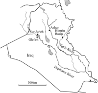 View Details | Figure 1. Map of present Iraq and locations of the Himrin basin and other sites where the crania analyzed in the present study were excavated. |
Previously, using these skeletal remains from the Himrin basin and neighboring areas of northern Iraq, Wada conducted intensive craniometric studies of the skulls from the Islamic period, and found that the Himrin population could be broadly divided into two groups, i.e. dolichocranic and brachycranic (Wada, 1986, 1989), by the time of the Islamic period. Wada et al. (1987a, b) also published several paleopathological studies. However, no studies have been conducted to investigate variations in craniofacial form based on this skeletal collection before the Islamic period. Ishida and Wada (1981) provided a preliminary report on temporal variations in cephalic index, but detailed changes in 3D craniofacial shape have not been investigated.
In order to test the hypothesis of temporal variation, a total of 45 crania in the skeletal collection from the Samarra (c. 5500–5000 BC), Jamdat Nasr (c. 3200–3000 BC), Isin Larsa/Old Babylonian (c. 2000–1600 BC), Neo-Assyrian (c. 1000–600 BC), Parthian (c. 240 BC–230 AD) and Islamic (after c. 650 AD) periods were investigated (Table 1). Initially, we tried to select only undeformed adult crania that preserve near-complete craniofacial morphology. However, such well-preserved specimens before the Islamic period are very rare in this collection. In this study, we therefore analyzed the morphological variations of the crania in two ways: (1) a craniofacial analysis using crania that preserve both the viscero- and neurocranium but with a smaller sample of 28; and (2) a vault analysis using crania that preserve just the neurocranium but with a larger sample of 45 (Table 1). The second vault analysis included almost all adult crania in this collection from the pre-Islamic period that can be used for analysis. It must be noted that not all crania in the present analysis were excavated from the Himrin basin: 6 crania of the Neo-Assyrian period came from Sur Jur’eh and Glei’eh (in Haditha), and 5 crania of the Islamic period came from Ashur (Figure 1). However, for the sake of simplicity we refer to all samples as Himrin crania. Sex determination of crania was based on Wada (1986, 1989, personal communication). However, because the number of specimens studied here is small, we do not consider the sexual dimorphism in the present study. In addition, for comparative purposes, 10 modern Japanese adult crania (5 females and 5 males) housed at Kyoto University were also included in the two analyses.
Each cranium was scanned using a helical computed tomography (CT) scanner (TSX-002A/4I; Toshiba Medical Systems, Japan) in the Laboratory of Physical Anthropology at Kyoto University. Tube voltage, current and slice thickness were set at 120 kV, 100 mA, and 2.0 mm, respectively. Cross-sectional images were reconstructed at 0.5 mm intervals, with a pixel size of 0.5 mm. Cross-sectional images were then transferred to commercial software (Analyze 6.0; Mayo Clinic, USA) and the 3D surface of the cranium was reconstructed using a triangular mesh model. A total of 35 landmarks (Figure 2, Table 2) were digitized on the surface of each cranium using commercial software (Rapidform 2004; INUS Technology, Korea) for the first craniofacial analysis. However, if some of the landmarks on the face and the cranial base were completely missing due to damage, we just used 9 landmarks on the cranial vault for the second vault analysis (Table 2). After digitizing landmarks, positions on the mesh model were double-checked with reference to the corresponding actual cranium. As shape variance due to asymmetry was not considered in this study, positions of all landmarks were symmetrized for further comparisons (Zollikofer and Ponce de León, 2002). For some crania, one of the bilateral landmarks was absent due to damage. For such cases, the median sagittal plane was calculated based on acquired midsagittal landmarks and the missing landmark was restored based on a mirror image, assuming the cranium to display essentially bilateral symmetry (Ogihara et al., 2006).
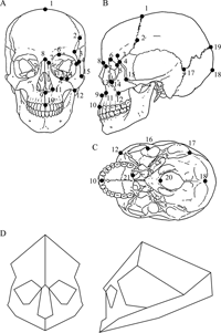 View Details | Figure 2. Landmarks used in the present study. (A) Frontal plane; (B) sagittal plane; (C) horizontal plane; and (D) wireframe connecting landmarks for visualizing variations in craniofacial shape. |

To describe variations in craniofacial shape, 3D coordinates of landmarks were analyzed using Morphologika geometric morphometric software, version 2.3.1 (O’Higgins and Jones, 2006). Landmark coordinates of each specimen were normalized by centroid size for size-independent shape analysis and registered using the Generalized Procrustes method. Principal components (PCs) of shape variations among specimens were then calculated using the variance-covariance matrix of the Procrustes residuals of all crania. More details of the calculation method can be found in O’Higgins and Jones (1998) and O’Higgins (2000).
Because the number of crania from the pre-Islamic period was small, we placed all the pre-Islamic crania into one ‘pre-Islamic’ group in the first craniofacial analysis for statistical testing. In the second vault analysis, the crania from the Samarra, Jamdat Nasr, and Isin Larsa/Old Babylonian periods were placed into ‘Chalcolithic and Bronze Age’, whereas those from the Neo-Assyrian and Parthian were placed into ‘Iron Age’. To test for significant differences in PC scores among the population groups, one-way analysis of variance (ANOVA) and post-hoc Tukey’s honestly significant difference (HSD) multiple comparisons tests were performed using Statistica 06J software (StatSoft, USA). Normality was tested using the Kolmogorov-Smirnov test. Homogeneity of variance was tested using the Bartlett test (Bartlett, 1937).
Results of PC analysis (PCA) of the first craniofacial analysis are shown in Figure 3 as plots of the first principal component (PC1) vs. PC2, and PC2 vs. PC3. PC1 accounted for 16.6% of total variance, PC2 for 11.5%, and PC3 for 10.6%. Approximately 70% of variance was incorporated into the first eight PCs.
 View Details | Figure 3. Result of principal component analysis in the craniofacial analysis. (A) PC1 (x-axis) vs. PC2 (y-axis); and (B) PC2 (x-axis) vs. PC3 (y-axis). f, female; m, male. |
The pattern of shape variations shown in Figure 3 indicates that scores on PC1 were apparently independent of temporal variations in cranial shape, while scores on PC2 seemed to be related to such variations. Crania from the pre-Islamic period (Isin Larsa/Old Babylonian, Neo-Assyrian, and Parthian periods) displayed relatively lower scores on PC2 and appeared to be separated from those of the Islamic period, although slight overlap was apparent. Along the PC3 axis, Japanese individuals were separated from both pre-Islamic and Islamic individuals, indicating that this axis was linked to geographical variations in cranial morphology. We also performed a PCA without Japanese crania and confirmed that the results were consistent with those of the combined analysis presented here.
Comparisons of mean values for the first three PCs are provided in Figure 4. It was confirmed that there is no significant difference between the distribution of the first three PC scores and a normal distribution. Variances were also confirmed to be homogeneous. ANOVA indicated that mean values of PC scores differed significantly among the three populations [F(6,66), 7.98; P < 0.001]. Post-hoc Tukey’s HSD test revealed that mean PC2 score was significantly lower in the pre-Islamic population than in the other two populations (P < 0.005), and that of PC3 was significantly smaller in the Japanese population than in the other two (P < 0.001). No significant differences were detected for PC1. Differences in craniofacial shape thus actually exist between pre-Islamic and Islamic periods, and between the Mesopotamian and Japanese populations.
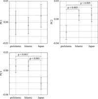 View Details | Figure 4. Comparison of mean scores for the first three PCs among pre-Islamic and Islamic Himrin populations and the modern Japanese population. Lines represent 95% confidence intervals. |
Figure 5A–C shows 3D shape variations along PC1, PC2, and PC3, respectively (other PCs = 0) by warping the cranial shape represented by the wireframe connecting landmarks. As visualized, with increasing PC1, relative decreases in cerebral (but not facial) breadth and cranial height, and increases in upper facial and nasal heights were observed. The shape of the orbit was largely unaltered along PC1, and hence relative elongation of upper facial height was the result of maxillary elongation. Furthermore, a large difference existed in position of the stephanion, which was located relatively more superiorly with increasing PC1, despite the fact that cranial height decreased.
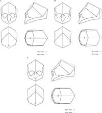 View Details | Figure 5. Variations in craniofacial shape represented by the first three PCs. Shape variations are visualized with 3D deformation of the wireframe connecting landmarks. Variations along (A) PC1, (B) PC2, and (C) PC3, respectively. |
PC2 clearly described the relative contraction of the cranial length and relative expansion of craniofacial breadth with an increase in PC2. In addition, upper facial, orbital and nasal heights became relatively larger.
With increasing PC3, relative sagittal inclination of the upper face (profile angle) increased, indicating that prognathism increased as PC3 decreased. Posterior border of the palate accordingly shifted anteriorly. Moreover, with an increase in PC3, cranial height became relatively shorter and downward displacement of lambda was observed. Relative inclination of the segment connecting the nasion and bregma was decreased as PC3 increased, suggesting that the forehead was relatively receding.
Results of PCA of the second vault analysis on a larger sample of 45 Mesopotamian crania (but with the smaller set of neurocranial landmarks) are shown in Figure 6, Figure 7, and Figure 8. PC1 accounted for 27.0% of total variance, and PC2 for 18.1%. Approximately 70% of variance was incorporated into the first four PCs.
 View Details | Figure 6. Result of principal component analysis in the vault analysis. PC1 (x-axis) vs. PC2 (y-axis). f, female; m, male. |
 View Details | Figure 7. Comparison of mean scores for PC1 and PC2 among the four population groups. Lines represent 95% confidence intervals. |
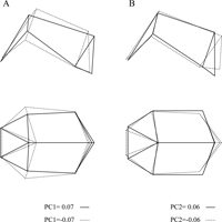 View Details | Figure 8. Variations in vault shape represented by PC1 and PC2. Shape variations are visualized with 3D deformation of the wireframe connecting landmarks. Variations along (A) PC1 and (B) PC2. |
The pattern of shape variations shown in Figure 6 indicates that the results of the vault analysis are generally consistent with those of the first craniofacial analysis. With increasing PC1, a relative decrease in the breadth of frontal bone and a relative elevation of the stephanion were noticed as in the first craniofacial analysis (Figure 8). The mean PC1 scores did not differ significantly among the four groups, but the pre-Islamic populations tended to display a larger score (Figure 7), inferring that the cranial vaults of the pre-Islamic populations were relatively narrow. The PC2 scores seemed to be related to temporal variations. Crania from the Chalcolithic and Bronze Age (Samarra, Jamdat Nasr, and Isin Larsa/Old Babylonian periods) displayed relatively lower scores on PC2, while those from the Islamic period showed relatively higher PC2 scores, although the difference was less prominent compared to the first craniofacial analysis. The PC2 axis described the relative contraction of the cranial length and the relative expansion of cerebral breadth. ANOVA and post-hoc Tukey’s HSD test indicated that the mean PC2 score was significantly smaller in the Chalcolithic and Bronze Age than in the Islamic populations (P < 0.01). No significant differences were detected for other pairs, but the mean PC2 scores of the crania from the Iron Age (Neo-Assyrian and Parthian) were closer to those from the Chalcolithic and Bronze Age, indicating that the pre-Islamic populations tended to display a dolichocranic cranium as also suggested by the first craniofacial analysis. However, in this vault analysis, no clear separation between the Mesopotamian and Japanese crania was observed in the PC3 score, unlike in the first craniofacial analysis.
The results of both analyses suggest that temporal changes in the craniofacial form exist among the Himrin population from the Chalcolithic and Bronze Age to the Islamic period. The change in PC2 score for both the craniofacial and vault analyses was generally related to time history (Figure 3A, Figure 4, Figure 6, Figure 7); pre-Islamic individuals showed lower PC2 scores, while those from the Islamic period generally had higher scores in the craniofacial analysis. If the PC2 score is negative, the cranium is relatively dolichocranic (long-headed) (Figure 5, Figure 8). Conversely, PC2 scores of most Islamic individuals were positive, representing a relatively brachycranic population (short-headed), although some individuals were long-headed as before (Figure 3). This tendency was observed to be less prominent in the vault analysis (Figure 6), probably due to the fact that the number of landmarks used was not sufficient to extract clear morphological variations. However, the pre-Islamic individuals tended to display higher PC1 and lower PC2 scores (i.e. relatively dolichocranic), and vice versa in the Islamic individuals, suggesting that the vault analysis is also generally consistent with the craniofacial analysis. Consequently, although the pre-Islamic sample size was small due to the limited availability of materials, this study suggests that the Himrin population was relatively dolichocranic and generally unaltered until the Parthian period as in southern Mesopotamia (Keith, 1927; Ehrich, 1939; Swindler, 1956), but sometime in or after the Parthian period a more brachycranic population came into this northern Mesopotamian area and craniofacial characteristics within the inhabitants in this area probably became more diverse, as preliminarily suggested by Ishida and Wada (1981) and Wada (1986). It has been suggested based on archeological data that the population of Mesopotamia began to be influenced by Persians after the Achaemenean domination, and more foreigners were settled and mixed with the native population in the Parthian period (Roux, 1992). The present results do not contradict this view. Furthermore, this study depicts the dolichocranic population as tending to have a relatively lower orbit and broader (lower) nose, and vice versa in the brachycranic population. These results are consistent with the findings of Wada (1986), indicating that the present morphometric analysis successfully extracted the morphological characteristics derived from conventional craniometry.
However, this PC axis classifying dolichocranic and brachycranic groups accounts for only 11.5% of total variance in the craniofacial analysis (18.1% in the vault analysis). In addition, this is not the PC1 of the present analysis, indicating that morphological variability of the Himrin crania is actually quite large, and simple classification into dolichocranic and brachycranic groups is not valid. Furthermore, a certain tendency of craniofacial variability was found to exist along a specific direction (PC1) independent of temporal change, and such directional variability was larger than variations through time.
In the present analysis, the modern Japanese population was included for comparison to provide a better picture of craniofacial characteristics of the Himrin population. Our geometric morphometric analysis successfully characterized craniofacial shape differences between the two populations along PC3 (Figure 5). Himrin crania were found to differ from modern Japanese in terms of relative orthognathism of the maxilla and declination of the forehead. The magnitude of prognathism in Mesopotamian inhabitants was known to be slight (Ehrich, 1939; Swindler, 1956), while that of the Japanese was suggested to be increasing over time (Kaifu, 1999). This difference in relative orthognathism between the two groups was clearly visualized here. However, the PC scores of the Himrin population from the Islamic period and the modern Japanese population are not separated in the second vault analysis, indicating the morphological differences between the two populations are mainly characterized by variability in their facial shape. By adding Japanese crania as an out-group, morphological characteristics of the Himrin crania were clearly contrasted in the present study.
This study is the first attempt to apply a geometric morphometric technique to analyze the temporal variations of Mesopotamian crania. Although direct comparison was difficult between the present results and those of the conventional craniometric studies that have previously been published, the present cranial shape analysis based on a geometric morphometric technique has some advantages. First, the present analysis enables comparisons of size-independent covariation in cranial shape because landmark coordinates of each specimen were normalized by the centroid size. Second, this geometric morphometric technique can extract subtle differences that are sometimes unexpected (such as the systematic craniofacial variability detected along the PC1), as overall spatial relationships of landmarks in each cranium were completely preserved and fully incorporated into the analysis. Third, the extracted shape difference could be visualized to facilitate interpretation, which is quite difficult using conventional craniometric methods. One limitation of this study is that the number of the Mesopotamian crania analyzed is relatively small (Table 1). However, it should be noted that the human remains in the collection are very fragmentary and we herein included almost all specimens from the pre-Islamic periods that can be used for the present analysis of temporal cranial variations. In addition, skeletal remains from the northern Mesopotamia are indeed very scarce as explained previously. The present accurate description of craniofacial variability therefore offers an important basis for future comparative research with neighboring populations to better clarify the origin and characteristics of Mesopotamian inhabitants. In the future, we aim to perform CT scans of crania from other neighboring areas such as Telul eth-Thalathat, northern Iraq (Ikeda, 1958, 1970) and Dailaman, northern Iran (Egami and Ikeda, 1963; Ikeda, 1968) that are housed at the University of Tokyo, and include these crania in the present analysis for comparison.
The authors wish to sincerely thank Yo Wada (Osaka Isen), Hiroo Kumakura (Osaka University), Eishi Hirasaki (Osaka University), Osamu Kondo (University of Tokyo), Hiroshi Tsujikawa (Tohoku University), Naoki Morimoto (University of Zurich) and Hiroko Hashimoto (Nara National Research Institute for Cultural Properties) for their invaluable assistance in the present study, and Tsunehiko Hanihara and anonymous reviewers for their comments and suggestions for improving this manuscript. This study was supported by a MEXT Grant-in-Aid for Scientific Research on Priority Area ‘Formation of tribal communities in the Bishri mountains, middle Euphrates’.
|