| Correspondence to: Marc Oxenham, College of Arts and Social Sciences, Australian National University, Canberra, ACT 0200, Australia. E-mail: marc.oxenham@anu.edu.au Published online 10 April 2009 in J-STAGE (www.jstage.jst.go.jp) DOI: 10.1537/ase.081114 |
While a number of studies in palaeopathology have inferred disabling complications as a consequence of disease and trauma in prehistory (Trinkaus, 1983; Dickel and Doran, 1989; Luna et al., 2008), until now only an individual from early historic period New Mexico (Hawkey, 1998) and one case from late Jomon period (~3000–4000 years BP) Hokkaido, Japan (Suzuki et al., 1984) have provided unequivocal evidence of extreme and long-term physical impairment in the past. Lack of archaeological evidence for severe disability, reducing our ability to study human behaviour in these circumstances, may reflect: (i) the limited number of diseases that manifest in bone; (ii) death of prehistoric individuals with severe pathologies prior to a skeletal manifestation; or (iii) the poorer preservational potential of pathologically altered skeletal tissues.
In this paper we document the case of MB07H1M09 (M9), an adult male excavated from a Neolithic cemetery (≥3500 years BP) in Northern Vietnam, who minimally suffered lower limb paralysis, and maximally quadriplegia, acquired as a young child. M9 represents one of the earliest known demonstrable instances of survival with a disability so severe as to be inconsistent with life without the long-term intervention of a dedicated caregiver(s).
The site of Man Bac, located in Bach Lien Village, Yen Thanh Commune, Yen Mo District (20°08′00″ N, 109°59′017″ E), between 1999 and 2007 furnished 95 excavated burials in a generally very good state of preservation. The burials are dated between 1524 and 1867 BC (Oxenham et al., 2008); temporally and material culturally belonging to a series of late Neolithic sites (Phung Nguyen Culture) bordering the current Red River delta. An agricultural subsistence base, supplemented with riverine, marine, and terrestrial fauna, is implied for Man Bac; while craft specialization extended to ceramic, jade, and other lithics, as well as shell artefact production (Oxenham et al., 2008). There is evidence for regional trade that may have extended as far as Shang Dynasty China (Higham, 1996). With the exception of 12 poorly preserved burials from Lung Hoa, Man Bac is the only north Vietnamese skeletal assemblage providing a window on the biology and health of people during the missing millennia between the early Neolithic and developed Bronze Age in the region.
M9 was one of only three flexed interments (the remainder being supine/extended and free of serious pathology), but otherwise received a similar mortuary treatment to other adults; his burial position may reflect postural constraints during life. Figure 1 and Figure 2 summarize the preservation of M9; post-mortem loss of the lower thoracic region, including most ribs, majority of lumbar vertebrae and lower appendicular joints makes a definitive diagnosis problematic. Although assessment of sex and age-at-death in skeletal remains displaying evidence of severe long-term pathology is always problematic, the expression of cranial secondary sexual characteristics (nuchal, mastoid and supraorbital size and morphology particularly; as well as frontal bone slope and lack of bossing) is consistent with a male. The sex assessment of male is further supported through the use of Walrath et al.’s method for cranial sex estimation (Walrath et al., 2004). Significant (>30%) ecto- and endocranial suture synostosis and intra-sample comparative molar wear, in addition to full fusion of all preserved epiphyses, suggests age-at-death in the third decade. Pathological cranial and dental conditions include bilateral remodelled cribra orbitalia, slight to moderate calculus deposits on all preserved teeth, and presence of enamel hypoplasia on LC LC1, RI1, RI2, LP3, and LC1. No evidence for antemortem tooth loss (including tooth ablation), caries, abscessing, or periodontal disease was seen. The mandibular condyles display osteoarthritis (OA) and are strongly asymmetrical, but there is no evidence for bilateral asymmetry in tooth wear.
 View Details | Figure 1. Schematic summarizing the skeletal preservation (black represents missing portions) of MB07H1M09. |
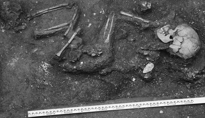 View Details | Figure 2. Photograph of MB07H1M09 in situ prior to removal. Note extreme gracility of limbs. |
Preserved vertebrae were ankylosed (includes centra, zygapophyses and laminae) from the first cervical (C1) to third thoracic (T3). While these presented as a continuous block of vertebrae in situ (Figure 3a), fragmentation occurred during lifting. Figure 3b shows constriction at the intervertebral junctions of C2/3 and C3/4 (a feature seen throughout the in situ vertebral column prior to removal). Figure 3c illustrates complete fusion at the interarticular processes and adjacent laminae of C5–C7 as well as extremely small, often completely obliterated, and somewhat oval in shape intervertebral foramina (again, this was seen throughout the in situ vertebral column). Additional complications included occipitalization and atlantoaxial rotatory (>30°) fixation (subluxation) with the anterior aspect of the dens separated from the corresponding C2 facet by 8 mm; resultant C1/C2 stenosis may have been mitigated by the apparent aplasia of C1 posterior neural arch (Figure 4). Stenosis is indicated by the relatively small anteroposterior neural canal diameters of C2 (13.1 mm) and C3 (11.7 mm). It was not possible to assess the presence or absence of lower vertebral or sacral spina bifida due to poor preservation of this region. A fragmentary, very gracile, right os coxae was ankylosed at the auricular area.
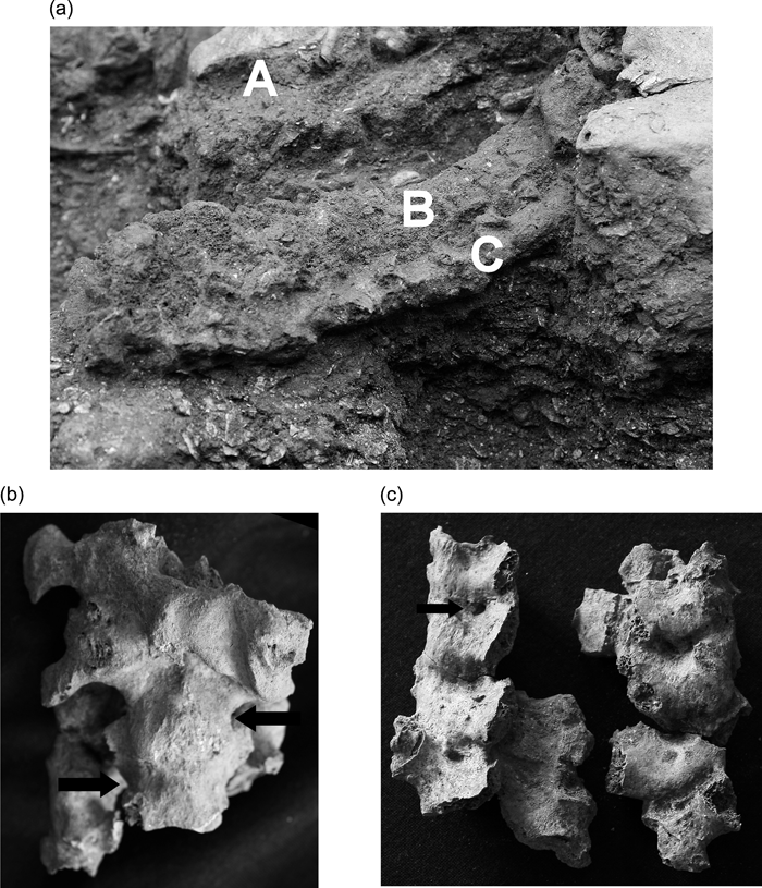 View Details | Figure 3. (a) Left oblique view of the fused vertebrae (C1–T1 in situ). The posterior body of the mandible (damaged post-mortem) has been removed (A) to facilitate examination of the superior cervical vertebrae. Note the flattened, fused vertebral bodies (B) and continuous fusion along the transverse processes (C). (b) Anterior aspect of ankylosed C1 to superior aspect of C4. Arrows indicate areas of intervertebral junction constriction (characteristic of congenital vertebral fusion). (c) Posterior aspect of C5–C7. The vertebral bodies (centra) disintegrated upon removal. Complete fusion at zygapophyseal joints and laminae, as well as small oval intervertebral foramina (see black arrow) is indicative of congenital fusion. |
 View Details | Figure 4. C1 and C2 illustrating occipitalization (the roughened bone at point A is the broken right occipital condyle fused to the superior C1 condylar facet) and rotatory fixation. Note ossified connective tissue (point B) between the anterior dens and C1 dens facet. C1 neural arch is missing either post-mortem or due to arch aplasia. The two lines help to identify the degree and direction of C1 rotation on C2. |
All preserved major long bones have significantly reduced diaphyseal diameters relative to other adult Man Bac individuals (Table 1). Only the right humerus, radius, and ulna have preserved articular portions. The humeral olecranon fossa and trochlea is free of OA, while the head displays marked anteromedial osteophytosis and porosity. There is a sharp, prominent deltoid crest (~40 mm long, raised ~5 mm) and a very deep intertubercular groove extending to the deltoid insertion. The radial proximal ulnar facet and capitular surface display OA, while distally OA free, and there is minimal development of the biceps tuberosity. The right ulna has a roughened triceps insertion, but no ossification of this tendon, and proximal, but not distal, OA.
Both femoral diaphyses are significantly compressed anteroposteriorly, with the left (better preserved) displaying marked flaring superiorly, initiating just inferior to the lesser trochanter. The lesser trochanter of the left femur (not preserved in the right) manifests unusually as a sharp medial ridge (21.6 mm superoinferior length, projecting ~9.0 mm). Linea aspera in both femora manifests as very slight and sharpish ridges. Both tibiae are also very gracile with minimal expression of muscle insertions. The fibulae appear exceptionally fragile (minimum diameter of the neck in the right is only 2.9 mm), with the better preserved right diaphysis showing marked proximal metaphyseal flaring.
Proximal and middle hand phalanges are superoinferiorly compressed, with the proximal phalanges manifesting flaring proximal articular regions and mediolaterally compressed distal regions with the addition of slight anterior palmar curvature. OA was not seen on assessable right or left foot elements, although the proximal phalanges of the second, third, and fourth rays of the right foot have relatively narrow diaphyses with flaring and slightly cupped metatarsophalangeal facets.
In developing a differential diagnosis, the exceptionally slender nature of the limbs, particularly M9’s legs, is consistent with clinical descriptions of adult patients with spinal cord injury (SCI) occurring as a juvenile (Giangregorio and McCartney, 2007). SCI occurring post-adolescence typically leads to a significant loss of bone endosteally, resulting in an enlarged medullary cavity and thinner cortical walls, but the maintenance of diaphyseal diameter (Modlesky et al., 2005). The post-mortem loss of the epiphyseal areas of the lower limbs may be explained by much greater bone loss in this region relative to the diaphysis in the unloaded limbs of SCI individuals (Eser et al., 2004). The gracility of the preserved upper limbs also suggests some level of unloading before adulthood (hand and foot bone diaphyseal atrophy is also relevant in this regard), given that bone loss is restricted to unloaded limbs and is not a systemic condition in SCI individuals (Zehnder et al., 2004). However, the greater robustness of these limbs relative to the lower limbs, as well as evidence for OA and deltoid muscle use, indicates functional quadriparesis with intermittent or limited upper limb function. M9 also suffered from a permanent and severe torticollis and right rotation of the head, which may have led to restricted right side temporomandibular function.
Given that M9 suffered from quadriplegia or quadriparesis from some time prior to adolescence, there is a question regarding the underlying cause(s) of this seriously debilitating condition and whether neurological impairment preceded or was a consequence of the ankylosed spine. While spinal ankylosis, as well as sacroiliac fusion, has been observed to develop in paralysed individuals (Park et al., 1993), the extent and nature of the fused vertebral column in this case is not consistent with such a scenario. If spinal ankylosis preceded or was coincidental with neurological impairment, several juvenile-onset conditions need to be considered: Klippel–Feil syndrome (KFS), fibrodysplasia ossificans progressiva (FOP) and juvenile idiopathic arthritis (JIA).
Two forms of juvenile-onset arthritis that can cause severe vertebral ankylosis, juvenile rheumatoid arthritis (JRA) and juvenile-onset ankylosing spondylitis (JOAS), are now classified under the ILAR system as subcategories of JIA (Tse and Laxer, 2003; Duffy et al., 2005). However, from a palaeopathological standpoint, there is value in differentiating between JOAS and JRA as these conditions have quite distinct skeletal signatures. In adult RA vertebral lesions can include occipitalization, erosion of cervical apophyseal joints, and, rarely, sacroiliac lesions, while in JRA vertebral ankylosis can occur at the destroyed apophyseal joints, leaving the centra unaffected (Hadley, 1976). With JOAS occipitalization is rare, while sacroiliac lesions and widespread enthesitis are characteristic with the potential for ankylosis of the lumbar then thoracic vertebrae (Hadley, 1976). The morphology of early vertebral ankylosis, in childhood, may mimic congenital fusion or non-segmentation (Hadley, 1976). In JRA, appendicular joint disease is much more common in the upper extremities (particularly wrist, interphalangeal and metacarpophalangeal), while tarsal involvement and enthesitis is very common with JOAS (Burgos-Vargas and Vazquez-Mellado, 1995). While poor preservation of appendicular epiphyseal areas in M9 may be masking erosive lesions and ankylosed joints, the lack of such lesions in the preserved joints tends to exclude JIA in general, and JOAS and JRA specifically, from further consideration.
FOP is a rare, 1 in 1.6 million, condition characterized by vertebral fusion as well as extensive and ultimately severely disabling heterotopic ossification and appendicular joint ankylosis (Kaplan et al., 2005). While both FOP and KFS are characterized by ankylosis of the cervical spine, and as few as two contiguous vertebrae can be involved in both conditions, vertebral centra in FOP are typically narrow with enlarged pedicles and spinous processes and occipitalization does not occur (Schaffer et al., 2005). Moreover, the lack of heterotopic ossification, appendicular joint ankylosis and congenital malformation of the great toes in this case (Kaplan et al., 2005) excludes FOP from further consideration.
KFS is a segmentation disorder characterized by the congenital fusion of two or more cervical vertebrae with an estimated prevalence of 1 in 40,000–42,000 (Samartzis et al., 2006). Two important features of KFS vertebrae are: (i) an apparent restriction at the interspace of the fused vertebral bodies (Hadley, 1976, Fig. III: 12, p. 81), also described as ‘wasp-waist sign’ (Nguyen and Tyrrel, 1993); and (ii) relatively small, round or oval intervertebral foramina (Hadley, 1976). M9 meets the criteria for KFS Type III (multiple continuous fused cervical vertebrae), which is associated with a relatively high risk of radiculopathic and myelopathic symptoms (Samartzis et al., 2006). The association of Type III and occipitalization is also correlated with a much greater risk of superior ondontoid migration (SOM) and concomitant neural compression (Samartzis et al., 2007). While post-mortem damage to the occipital region makes determination of SOM difficult, some level of stenosis is apparent at C2 and C3, although C1 neural arch aplasia would have relieved compression potentially resulting from C1/C2 subluxation. Serious neurological impairment in both children and adults has been recorded in association with KFS, e.g. intermittent quadriparesis (Ritterbusch et al., 1991). Further, there is an increased risk of serious SCI subsequent to trauma in KFS individuals (Strax and Baran, 1975).
KFS in its milder Type I and II forms has been documented in a number of archaeological sites globally from as early as 5000 BC through to medieval times: Japan (Fukushima, 1988); Portugal (Fernandes and Costa, 2007; Silva and Ferreira, 2008); Hungary (Pany and Teschler-Nicola, 2007); Greece (Papathanasiou, 2005); Central and South America (Urunuela and Alvarez, 1994); North America (see Barnes, 1994 for an extensive review). Apart from Man Bac burial 9 the only other extreme form of KFS known to the authors is that of a pre-Hispanic (~1450–1500 AD) female aged 30–40 years from Cholula, Puebla, Mexico with vertebral fusion from C2 to T1 but not appearing to suffer from any form of paralysis (Urunuela and Alvarez, 1994).
The description of and differential diagnosis considered for this individual suggests he suffered from a congenital segmentation disorder as a child, leading to fusion of his cervical spine and concomitant or subsequent severe neurological impairment (likely quadriparesis or quadriplegia). The functional impact of M9’s condition was, at a minimum, to render him completely immobile below the waist and to radically limit upper body mobility. Additional posture-related complications include a fixed right rotation of his head with severe torticollis and potentially restricted masticatory function. In this Late Neolithic culture this would have left M9 completely dependent on others for every aspect of daily living. There is a well-documented range of systemic complications associated with immobility (Olson et al., 1967), and this has implications for the amount and type of care required by M9. M9 lived into his third decade, and his survival for ten years or more following onset of immobility/paralysis would have required constant care and episodes of intensive nursing. The implications of his experience for understanding social practice in this Neolithic community will be explored in detail in a future communication.
We thank F. Kaplan, F. Shen and P. Smith for important discussions concerning the differential diagnosis. M.O. was funded by an Australian Research Council Discovery Grant and H.M. by a Toyota Foundation (No. D06-R-0035) and the Japan Society of the Promotion of Science (No. 20370096).