| * Correspondence to: Hajime Ishida, Department of Human Biology and Anatomy, Faculty of Medicine, University of the Ryukyus, Uehara 207, Nishihara, Okinawa 903-0215, Japan. E-mail: ishidaha@med.u-ryukyu.ac.jp Published online 25 June 2011 in J-STAGE (www.jstage.jst.go.jp) DOI: 10.1537/ase.100925 |
Degenerative joint disease, or osteoarthritis, is a common disease characterized pathologically by damage to the articular cartilage and bone surface in synovial joints, associated with osteophyte formation at the joint margins (Dieppe and Lohmander, 2005). This condition affects not only the hands, hips and knees, but also the spine, and is strongly age-related, currently rising in frequency after 50 years old (Felson et al., 2000). In addition to aging and systematic factors such as genetics, obesity and sex, the other risk factors for osteoarthritis consist of joint injuries and specific repetitious activities, or chronic overload. For example, with regard to occupational factors, farmers tend to have osteoarthritis of hip joints and workers undertaking physical labor have high frequencies of knee osteoarthritis (Coggon et al., 1998; Felson et al., 2000). Although joint damage and clinical joint pain are both common, it is clear that the severity of the joint damage on a radiograph bears little relation to clinical severity (Dieppe and Lohmander, 2005).
Degenerative change of the spine is a common age-related vertebral alteration (Gallucci et al., 2005; Roh et al., 2005; Freund and Sartor, 2006; Pytel et al., 2006; Kalichman and Hunter, 2007; Ruan et al., 2007; Shedid and Benzel, 2007). The vertebral bodies are interconnected by the intervertebral discs (fibrocartilage), which are called symphyses. On the other hand, the apophyseal joints between articular processes in the vertebral arches are normally synovial joints. The typical osteological findings consist of anterolateral osteophytes where Sharpey’s fibers attach to the vertebral body, degenerative change, or facet arthrosis of apophyseal joints (Resnick, 2002). Osteophytes on the vertebral body are more frequent in men than in women, as well as in older populations, and also appear to affect persons engaged in heavy physical labor. They may affect any segment of the vertebral column. Because degenerative changes of apophyseal joints are also common, some pathological evidence of such disease is seen in the spine of all individuals after the age of 50 or 60 years. Degenerative changes of apophyseal joints commonly affect the middle and lower cervical spine, upper and mid-thoracic spine, and the lower lumbar spine (Resnick, 2002).
Because risk factors for degenerative disease of the spine consist of not only age but also selected activities, many physical anthropologists have investigated degenerative changes of the spine from prehistoric and historic human populations in order to reconstruct their life activity (Suzuki, 1978, 1998; Fukushima, 1988; Bridges, 1994; Lovell, 1994; Lieverse et al., 2007; Moromizato et al., 2007; Rojas-Sepúlveda et al., 2008). In general, upright bipedal humans exhibit more marked degenerative changes of the spine than other hominoids (Jurmain, 2000). As mentioned above, although human spines tend to be more uniformly affected throughout the vertebral column, previous anthropological investigations revealed that prehistoric and historic human populations vary in terms of the prevalence of degenerative changes of the spine (Bridges, 1994; Moromizato et al., 2007). In addition, there are different patterns between the prevalence of osteophytes on the vertebral body and that of degenerative changes of apophyseal joint (Moromizato et al., 2007). This is one of the merits of studying degenerative changes of the spine in skeletal populations. However, because it is necessary to obtain large initial numbers of samples for epidemiological comparison, only a few studies have been performed in Japan (Suzuki, 1978, 1998; Fukushima, 1988).
In this study, we chose three ancient (historic) skeletal collections of relatively large sample sizes with good preservation from the Japanese archipelago. The first is skeletal remains of the Okhotsk culture from Hokkaido and Sakhalin (Figure 1). The Okhotsk culture from the 5th to 12th century AD developed a considerable maritime infrastructure, which has been demonstrated by zooarcheological and isotopic analyses (Yoneda, 2002; Amano, 2003; Naito et al., 2010). The Okhotsk people concentrated on fishing and sea-mammal hunting. Many morphological and recent DNA analyses revealed that people from the Okhotsk culture were genetically similar to current Amur basin people (Ishida, 1996; Sato et al., 2007, 2009a, b; Komesu et al., 2008).
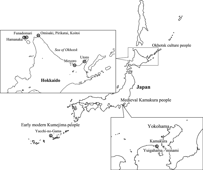 View Details | Figure 1. Location of the three skeletal series of the Okhotsk culture sites in Hokkaido, the Yuigahama-minami site of medieval Kamakura in Kanto, and the Yacchi-no-Gama site of the early-modern Kumejima, Okinawa, Japan. |
The second group of skeletal samples is from the medieval city of Kamakura, Kanto District, Central Japan (Figure 1). Medieval Kamakura was the capital of the Kamakura Shogunate in the period 1193–1333 AD. Huge numbers of human skeletal remains were recovered at the Yuigahama area, the coast of Kamakura, from the 1950s (Suzuki et al., 1956; Hirata et al., 2004). Because the samurai (military soldiers) were buried at a different place in Kamakura, the human remains from the Yuigahama area were considered to be those of common people. Mitochondrial DNA analysis revealed that the people of medieval Kamakura are very similar to the modern main-island Japanese (Shinoda, 2011).
Early-modern Kumejima samples from the Ryukyu Islands, the southernmost island chain of the Japanese archipelago, form the third group (Figure 1). One thousand human skeletal remains of the early-modern period were recovered from caves on Kumejima between 1998 and 2000 (Fukumine et al., 2001, 2006; Irei et al., 2008). These caves had been used as a mortuary during the 17th–19th centuries AD. The past existence of paddy fields in this area indicates that the skeletal series represent farmers (Fukumine et al., 2001). Isotopic analysis indicated that these people consumed both C3 plants, which fix carbon via the Calvin–Benson cycle (e.g. taro, rice and tree), and fish (Yoneda et al., 2004; Irei et al., 2008).
Moromizato et al. (2007) reported on degenerative changes of the spine for the Kumejima samples in Japanese. They found that the lumbar regions were most affected for both sexes, while severe degenerative changes of the lumbar region were more frequently seen in females. In addition, it was suggested that the greater frequency of degenerative changes seen in female cervical vertebrae may have been caused by traditional behavior, such as carrying items on the head.
On the basis of this report on Kumejima (Moromizato et al., 2007), we would like to present basic data on degenerative changes of the spine among three different peoples from the Japanese archipelago, and to compare rates of prevalence among them. Finally, we will mention causes of the different prevalence patterns of degenerative changes among them on the basis of their subsistence, nutrition, labor, and culture.
Table 1 and Figure 1 show numbers and locations of the materials used in this study. In our analysis of sex-based and age-related differences in degenerative changes, we determined the age and sex of the individuals from which the skeletal materials were derived. Sex was estimated by standard methods, such as pelvic and cranial morphology (White, 2000). Age was also estimated by standard methods using auricular surfaces (Nagaoka et al., 2006), sutural closure, and dental attrition (White, 2000). Because the age distribution of the Kamakura people is different to those of the other peoples owing to their relatively short life expectancy (Nagaoka et al., 2006), we interpreted their prevalence rates carefully.
Although we used individuals of known sex and age with more than 20% vertebral preservation in order to obtain relative large sample sizes of respective vertebrae, the preservation rates were high. That is, the averages of preservation rates were 83% (female) and 92% (male) in the Kumejima series, the averages were lower at 81% (female) and 85% (male) in the Kamakura series, and 62.9% (female) and 54.6% (male) in the Okhotsk series. No significant age-class differences were found.
For diagnosis of osteophytes on the vertebral body, the first author (Y.S.) examined osteophytes (bony outgrowth) on the body of vertebrae of the Okhotsk and Kamakura series, and the third author (K.M.) examined those of the Kumejima series (Moromizato et al., 2007). The osteophytes extend first horizontally and then in a vertical direction and finally bridge with the adjacent vertebrae (Resnick, 2002). We also examined progressive stages of osteophytes to evaluate severity (Figure 2). The normal situation is Grade 0; the next grade is when osteophytes grow horizontally (Grade 1); osteophytes can turn to grow in a vertical direction (Grade 2); osteophytes can then significantly grow in a vertical direction (Grade 3); eventually, osteophytes can bridge adjacent vertebrae (Grade 4), according to the diagnostic criteria of Rogers (1966) and Wada (1975). The grades were independently scored for the anterior, right and left sides, and posterior aspects of superior and inferior vertebral rims of the vertebral body. Differential diagnosis is very important but difficult because of lack of soft tissues, such as intervertebral discs. However, we could eliminate other bony outgrowths of the vertebral column that were clearly diagnosed as ankylosing spondylitis and psoriatic arthritis.
 View Details | Figure 2. Progressive stages of osteophytes on the body of vertebrae. Grade 0: normal condition; Grade 1: horizontal growth; Grade 2: growth in vertical direction; Grade 3: significant growth in vertical direction; Grade 4: bridging with adjacent vertebrae. Redrawn from Rogers (1966) and Wada (1975). |
For diagnosis of degenerative changes of apophyseal joints, degenerative changes of the articular surface of the articular process were also examined by the first (Y.S.) and third (K.M.) authors. The pathological characteristics of osteoarthritis of the apophyseal joint are concordant with those seen in other synovial joints. Owing to a lack of articular cartilage, bony eburnation and sclerosis, pitting and osteophytes were checked. We also judged progressive stages of degenerative change of the apophyseal joint to evaluate severity (Figure 3). The normal condition is Grade 0; the next stage is when osteophytes grow on the rim of articular surface without pitting on the surface (Grade 1); osteophytes can also grow on the rim of articular surface with lipping with slight pitting (Grade 2); osteophytes can then grow all around the rim of the articular surface with moderate pitting on the surface and the rims of articular surface tend to be broken (Grade 3); eventually, osteophytes can significantly grow on the rim of articular surface with severe pitting on the surface and the rim becomes unclear (Grade 4), according to the diagnostic criteria of Higuchi (1983). The grades were independently recorded for right and left sides of the superior and inferior articular processes of the respective vertebrae.
 View Details | Figure 3. Progressive stages of degenerative change of apophyseal joint surface. Grade 0: normal condition; Grade 1: osteophyte growing on the rim of articular surface without pitting; Grade 2: osteophyte growing on the rim of articular surface with lipping and slight pitting; Grade 3: osteophyte growing all around the rim of articular surface with moderate pitting; Grade 4: osteophyte significantly growing with severe pitting and unclear rim. Redrawn from Higuchi (1983). |
As a preliminary step toward analysis, the first author scored degenerative changes of Kumejima samples on independent occasions, and compared the data with the data scored by the third author to confirm that intra- and inter-observer errors were insignificant. We calculated crude prevalence rates of degenerative changes of the spine not per individual but per vertebra for comparisons of age-related changes, and sex-based and population-based differences because of the fragmentary condition of samples. We judged severity per vertebra using the most progressive stage of degenerative changes in the vertebral body and apophyseal joint. After that, we diagnosed greater than Grade 1 as a degenerative change and greater than Grade 3 as a severe degenerative change of the spine.
Fisher’s exact probability test, χ2 test and residual analysis were used to evaluate gender difference, age-related changes and regional differences. In addition, odds ratios (ORs) were calculated to evaluate age-related changes.
Appendix Tables 1–4 show detailed basic data of observed and affected numbers, and frequencies of osteophytes (more than Grade 1) on the respective vertebral rims of vertebral bodies from the Okhotsk culture remains and medieval Kamakura samples. Appendix Tables 5 and 6 also give basic data of observed and affected numbers and frequencies of degenerative changes of apophyseal joint (more than Grade 1) on the right and left sides of superior and inferior articular processes from the Okhotsk and Kamakura samples, respectively. Detailed data on the early-modern Kumejima samples have already been published (Moromizato et al., 2007).
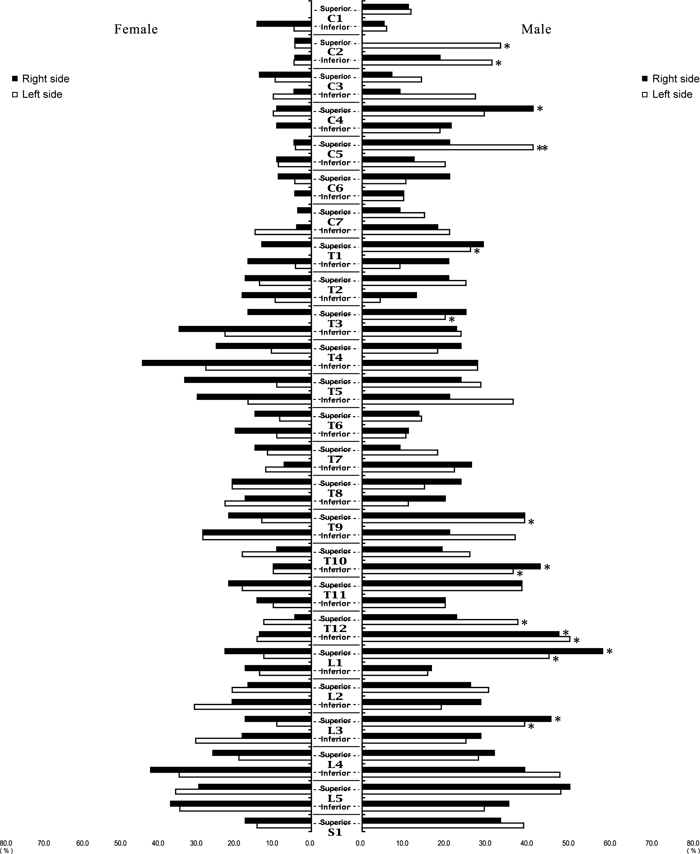
Osteophytes on the vertebral body of the thoracic and lumbar vertebrae of each sex generally grew more anteriorly than posteriorly in both Okhotsk and Kamakura samples (Figure 4, Figure 5), as well as in the Kumejima samples (Moromizato et al., 2007); interestingly, osteophytes on the inferior rims on the posterior side of the upper cervical vertebrae (C2–C4) were more frequent than those on the anterior side in both sexes of the Kamakura series (Figure 5). Osteophytes on the vertebral body were more frequent in the lower cervical spine and the lower lumbar spine. Degenerative changes of apophyseal joint tended to be more pronounced in men of both series (Figure 6, Figure 7), and were more frequent in the lumbar vertebrae, especially at the levels of Th12–L1 and L4–L5, for the Okhotsk series. These frequencies corresponded to previous clinical findings (Resnick, 2002).
 View Details | Figure 4. Frequencies of osteophytes on anterior and posterior sides of superior and inferior vertebral body surfaces: Okhotsk culture people. *Significantly different at the 0.05 level. |
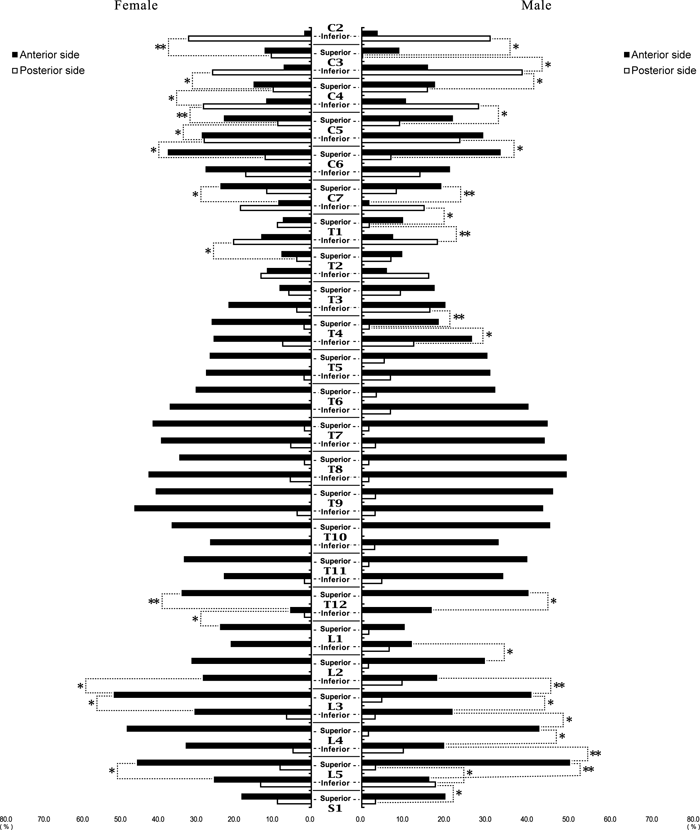 View Details | Figure 5. Frequencies of osteophytes on anterior and posterior sides of superior and inferior vertebral body surfaces: medieval Kamakura people. **,*Significantly different at the 0.01 or 0.05 level, respectively. |
 View Details | Figure 6. Frequencies of degenerative changes on right and left sides of superior and inferior apophyseal joints: Okhotsk culture people. **,*Significantly different between sexes at the 0.01 or 0.05 level, respectively. |
 View Details | Figure 7. Frequencies of degenerative changes on right and left sides of superior and inferior apophyseal joints: medieval Kamakura people. **,*Significantly different between sexes at the 0.01 or 0.05 level, respectively. |
In order to clarify the pattern of degenerative changes of joints, vertebrae were classified into four groups of cervical (C2–C7), upper thoracic (T1–T6), lower thoracic (T7–T12), and lumbar vertebrae (L1-L5, S1) to calculate crude prevalence rates of osteophytes on the vertebral body and degenerative changes of apophyseal joint per vertebra. Age-related changes and sex-based differences in degenerative changes of the spine were then analyzed using the Okhotsk and Kamakura samples.
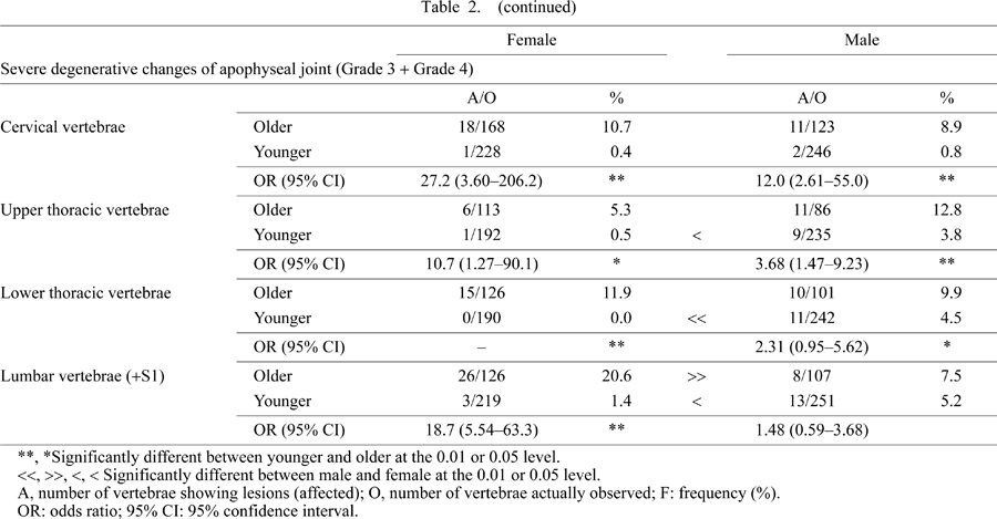
When the samples were divided into younger (under 40 years) and older groups (over 40 years), the older group of each sex had higher frequencies of osteophytes on the vertebral body and degenerative changes of apophyseal joint (more than Grade 1) than the younger group in the Kamakura sample (P < 0.01 or P < 0.05) except for degenerative changes of apophyseal joint of male cervical vertebrae, as shown in Table 2. Meanwhile, age-related differences of degenerative changes of apophyseal joint were not significant in males on the basis of the odds ratios. Severe degenerative changes (Grade 3 + Grade 4) were also more prevalent in the older group from Kamakura for each sex (P < 0.01 or P < 0.05) (Table 2).
In the Okhotsk series, on the other hand, age-related changes were not so clear, although more than half of the comparisons showed significant differences. For example, the differences in frequency between younger and older males were not statistically significant in all cases or in severe cases of degenerative changes of the apophyseal joint of lower thoracic vertebrae, and there were no significant differences between frequencies of all cases or in severe cases of degenerative changes of apophyseal joint of the upper thoracic region even in females (Table 3). In addition, severe osteophytes on the vertebral body of male cervical vertebrae were more pronounced in the younger group in the Okhotsk samples (P < 0.01). Thus, severe degenerative changes of the spine appeared even in the younger generation of the Okhotsk people.

As for sex-based differences, the older female group in the Kamakura samples had a higher frequency of osteophytes on the body of the cervical vertebra than the older males (P < 0.05), whereas the older females had a lower frequency in the lower thoracic region (P < 0.05). The frequencies in younger males were significantly higher in the cervical, upper, and lower thoracic vertebrae than those in younger females (P < 0.01 or P < 0.05). Severe osteophytes on the vertebral body showed a few differences between sexes. Degenerative changes of apophyseal joint of all vertebral groups were more pronounced in younger males from Kamakura than in younger females (P < 0.01), while the frequency for upper thoracic vertebrae was only significantly higher in older males than in older females (P < 0.05). Although severe degenerative changes of apophyseal joint of the younger generation also tended to be affected in the same pattern, the frequency for lumbar vertebrae was higher in older females than in older males (P < 0.01).
In the Okhotsk series, younger males had a higher frequency of osteophytes on the body of cervical, upper, and lower thoracic spine than younger females (P < 0.01 or P < 0.05). However, as for the older generation, the frequency of osteophytes on the body of the cervical vertebrae was significantly higher in females than in males (P < 0.05), as was the case for the Kamakura series, while osteophytes on the body of upper thoracic vertebrae were more pronounced in older males than in older females (P < 0.01). Severe osteophytes on the vertebral body in the older generation were more frequent in female cervical and male lumbar vertebrae (P < 0.01). Degenerative changes of apophyseal joint of cervical and lower thoracic spines were more pronounced in younger males than in younger females (P < 0.01), while the older generations exhibited no sex differences in this regard. Severe degenerative changes of apophyseal joint of lower thoracic vertebrae were more pronounced in younger males than in younger females (P < 0.01).
As a result, degenerative changes of the spine were generally more pronounced in males of both skeletal series, as described by Resnick (2002), while the degenerative changes of the spine of certain regions were more frequently seen in females. Because the younger males of both Kamakura and Okhotsk series tended to have high frequencies of degenerative changes of the spine, especially of apophyseal joint, age-related changes in males were obscure on the basis of the results of Fisher’s exact probability tests and odds ratios.
Next, we evaluated population differences of degenerative changes of the spine among the Okhotsk, medieval Kamakura, and early-modern Kumejima series (Table 4, Table 5). Although the three series were least affected in the upper thoracic vertebrae, with the exception of Okhotsk males (P < 0.01 or P < 0.05), they clearly showed different patterns of osteophytes on the vertebral body. For example, a significantly high frequency of affected cervical vertebrae was found for the Okhotsk people (P < 0.01 or P < 0.05), while the Kumejima samples had the highest frequency in the lumbar vertebrae (P < 0.01). As mentioned previously (Moromizato et al., 2007), the Kumejima females had a higher frequency in the cervical vertebrae than males (P < 0.01). In the Kamakura series, males were most affected in the lower thoracic vertebrae (P < 0.01). Among the three series, the Kumejima samples had the highest frequency in the lumbar vertebrae (P < 0.01).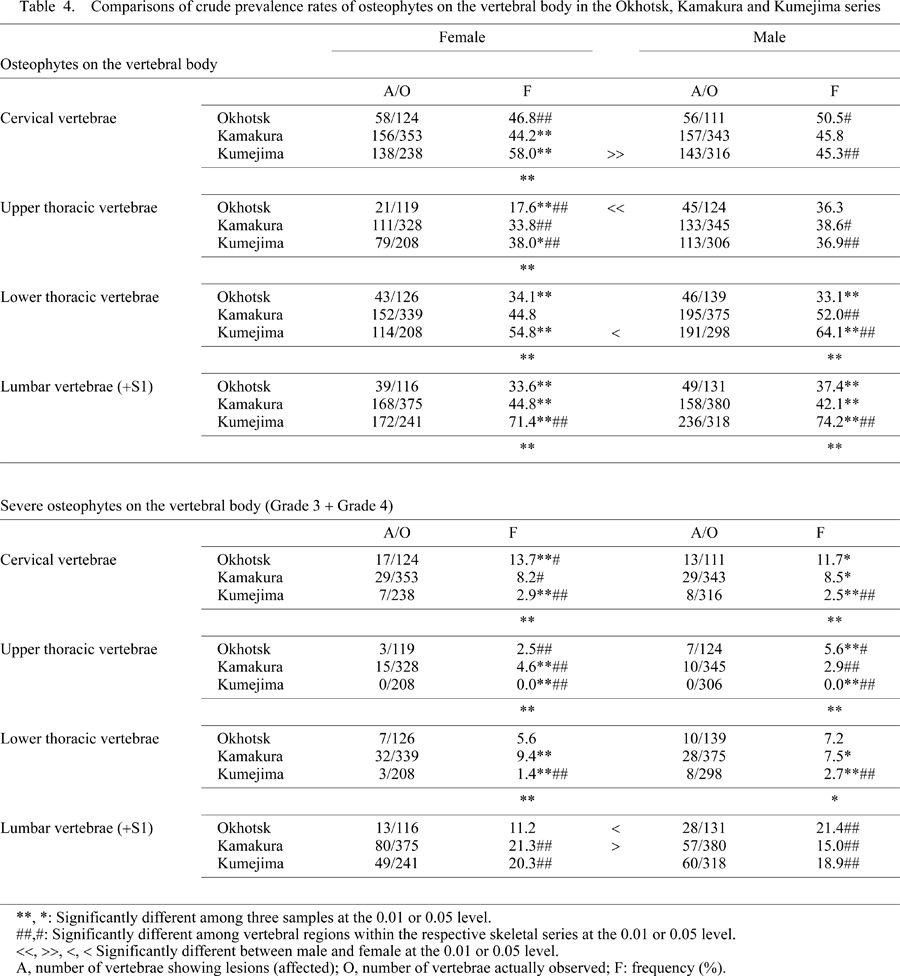
As for severe osteophytes on the vertebral body, all three series had the highest frequencies in the lumbar vertebrae, with the exception of the Okhotsk females (P < 0.01). Severe osteophytes on the body of other vertebral parts displayed markedly different patterns. The Okhotsk series had higher frequencies on the cervical vertebrae than the other two series (P < 0.01 or P < 0.05), while the Kamakura series generally had higher frequencies of severe osteophytes on the vertebral body, which was contrary to our expectations. The Kumejima series was the least affected in the cervical and thoracic vertebrae among the three series (P < 0.01).
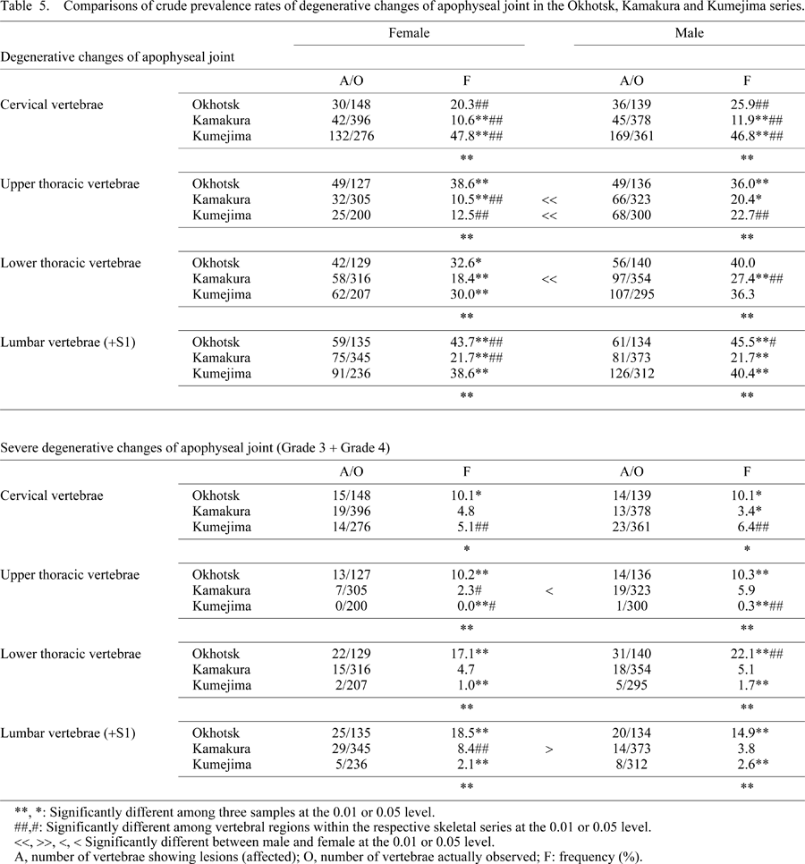
Affected patterns of degenerative changes of apophyseal joint within populations showed clearer differences. The Okhotsk series of each sex had the lowest frequencies in the cervical vertebrae and the highest frequencies in the lumbar vertebrae (P < 0.01 or P < 0.05), which contrasted with the highest frequency in the cervical vertebrae in the Kumejima series (P < 0.01). The Kamakura series of each sex had generally low frequencies of degenerative changes of apophyseal joint, especially in the cervical and upper thoracic regions, because of the relatively young age distribution.
Among the three series, severe degenerative changes of apophyseal joint dominantly affected the Okhotsk series of each sex. The Kumejima samples showed low frequencies (P < 0.01 or P < 0.05) although the relatively high frequencies in the cervical vertebrae remained (P < 0.01).
We confirmed that degenerative changes of the spine in ancient populations were more pronounced in men and older groups, as in contemporary populations. In addition, we revealed clear population-based and sex-based differences among the ancient groups from the Japanese archipelago.
Osteophytes developed more on the anterior side of the vertebral body in the three ancient peoples and recent Japanese (Wada, 1975). Age-related changes were clearly seen in the medieval Kamakura, early-modern Kumejima (Moromizato et al., 2007), and recent Japanese (Wada, 1975).
Osteophytes on the vertebral body tend to be more pronounced in men, leading to clinical cases (Resnick, 2002). However, older females had a higher frequency in the cervical vertebrae than the older males of both Kamakura and Okhotsk series. In addition, the Kumejima females of both younger and older groups combined had a higher frequency in the cervical vertebrae than males (Moromizato et al., 2007). Because there were similar frequencies between women and men and degenerative changes are more severe in men in clinical medicine (Resnick, 2002), this gender difference may be caused by some dynamic factors, including abnormal forces on the spinal column (Shedid and Benzel, 2007). Moromizato et al. (2007) mentioned that the Ryukyu women carry heavy loads on their head, called ‘Kasami.’ Although we do not know about the existence of such customs in the medieval Kamakura and Okhotsk periods, findings in other archeological cases suggest that older females followed this practice (Lovell, 1994).
Osteophytes on the body of the lumbar vertebrae were most frequent in the Kumejima samples, while those of the lower thoracic vertebrae and the cervical vertebrae were most frequent in the medieval Kamakura and Okhotsk series, respectively. Degenerative changes of the lumbar spine were found to be frequent in contemporary and archeological populations, including the Canadian Inuit (Merbs, 1983; Bridges, 1994; Larsen, 1997; Resnick, 2002; and many others). In Japan, the lumbar spine was found to be most affected in the Jomon, Yayoi, early-modern Edo and recent Japanese samples (Wada, 1975; Suzuki, 1978; Fukushima, 1988). Severe osteophytes on the body of the lumbar vertebrae were most frequent not only in the Kumejima series but also in the medieval Kamakura samples. Thus, the Okhotsk series had the peculiar characteristic of a higher frequency of osteophytes on the body of the cervical spine among populations from the Japanese archipelago, although the Kumejima females had the highest frequency, as mentioned above. The cervical vertebrae only support loads of or on the head, while the head, trunk and upper extremities produce loads on the lumbar vertebrae (Roh et al., 2005). Although we know of no relevant customs in prehistoric Okhotsk culture, some dynamic loads caused a high frequency of degenerative changes of cervical spine of clinical severity in other archeological cases (Shedid and Benzel, 2007; Lovell, 1994).
As mentioned above, the bodies of vertebrae are connected by intervertebral discs (symphyses), while the apophyseal joints are paired, true synovial joints. Therefore, the degenerative processes of the two joints may occur as independent phenomena (Resnick, 2002). However, intervertebral disc degeneration (intervertebral chondrosis and spondylosis deformans) causes narrowing of the intervertebral disc space, thus probably resulting in apophyseal articular alterations (Kalichman and Hunter, 2007). Although we could not analyze direct correlations between osteophytes on the vertebral body and degenerative changes of apophyseal joint within individuals because of the fragmented nature of skeletal materials, the frequency patterns of the samples were compared.
The lower lumbar vertebrae were most affected in the Okhotsk series, coinciding with findings for contemporary people (Fujiwara et al., 1999), whereas the Kamakura and Kumejima samples showed no clear peak in the lumbar vertebrae. Age-related differences in degenerative changes of apophyseal joint were more obscure than those in osteophytes on the vertebral body, especially in the Okhotsk males, whereas the recent Japanese showed clear age-related changes (Higuchi, 1983). This may be due to other factors, including labor and other activity-related causes.
Degenerative changes of the apophyseal joint also tend to be more pronounced in men, as in clinical cases (Resnick, 2002). It is interesting that the frequency of severe degenerative changes of apophyseal joint in the lumbar vertebrae of the Kamakura series was higher in older females than in older males. Because postmenopausal women had a high frequency of degenerative changes of lumbar apophyseal joint owing to increased expression of estrogen receptor (Ha et al., 2005), this high frequency seen in the Kamakura older females was possibly caused by age-related changes.
We found that there were marked differences in the pattern of effects exhibited between the Okhotsk and Kumejima series, i.e. the highest frequency for the lumbar spine in the Okhotsk series contrasts well with that of the cervical spine in the Kumejima series. However, an inverse pattern was exhibited for the frequencies of osteophytes on the vertebral body.
This may be explained by differences of anatomy and biomechanics between the apophyseal joints of cervical and lumbar vertebrae. The line of gravity runs just anterior to the apophyseal joints in the cervical vertebrae, while the line runs within the bodies of lumbar vertebrae, separate from the apophyseal joints (Standring, 2008). The superior and inferior flat articular facets of the cervical spine face posterosuperiorly and anteroinferiorly, respectively, at an angle of about 45° to the transverse plane at the upper cervical spine (Aiello and Dean, 1990; Kirpalani and Mitra, 2008). On the other hand, the facet joints of the lumbar vertebrae are very distinctive (Aiello and Dean, 1990; Kalichman and Hunter, 2007). The concave superior and convex inferior articular facets face medially and laterally, respectively, making an interlocking joint. Thus, the cervical apophyseal joint bears more weight and rotates easily, while the lumbar apophyseal joint has restricted rotation and prohibits anterior and lateral sliding.
The higher frequencies of degenerative changes of cervical apophyseal joint in the Kumejima series could be explained by additional loads on the head, especially in females (Moromizato et al., 2007). However, we would like to infer several dynamic reasons for the higher frequency of degenerative changes of lumbar apophyseal joint in the Okhotsk series because severe patterns were exclusively present at high frequency in this series among the three samples.
The Okhotsk culture appeared in southern Sakhalin and spread to northeastern Hokkaido and the Kuril Islands from the 5th to 12th century AD (Yamaura and Ushiro, 1999; Amano, 2003). The average annual temperature today is about 7°C, with −5°C in January, at Wakkanai City, the northernmost city on Hokkaido. Sea drift ice occupies the coast of the Okhotsk Sea from January to March. The Okhotsk culture developed considerable sea-mammal hunting, in both shallow and deep water, unlike the native population of Hokkaido (Hudson, 2004). These sea mammals consisted of fur seal, whales, sea lions and others. For hunting and fishing, the Okhotsk people used bone tools, including hooks and harpoons, as well as iron tools (Amano, 2003). In addition, they traveled in boats and probably fished using nets based on findings of stone weights. The nature of subsistence of the Okhotsk culture was recently confirmed using stable isotope analysis (Naito et al., 2010).
There has been little bioarcheological research on the Okhotsk people; Kodama (1948) mentioned severe dental wear and Yamaguchi (1995) identified a wound caused by a stone arrowhead in the right hip bone of a male specimen. Recently, oral health has attracted attention (Ishida et al., 1994; Hudson, 2004; Fukumoto et al., 2007; Oxenham and Matsumura, 2008; Hoover and Matsumura, 2008). However, only vertebral compression fractures were reported with regard to the skeletal system of the Okhotsk people (Ishida et al., 1994; Ishida and Matsumura, 2000).
Degenerative changes were found even in the younger Okhotsk individuals. As mentioned above, degenerative changes of lumbar apophyseal joint were more pronounced in the Okhotsk people. This was possibly caused by travel by boat and net fishing, as suggested by archeological evidence, because of the associated need for much rotation, extension and flexion of the lumbar spine.
Intensive paleodemographic research has been performed and has revealed that the medieval Kamakura people were not long lived (Nagaoka et al., 2006; Nagaoka and Hirata, 2008). In addition, many weapon-related traumas were found among the skeletal remains from medieval Kamakura (Suzuki et al., 1956; Hirata et al., 2004; Nagaoka et al., 2009, 2010). However, there have been few studies on the life activity patterns of these people. As mentioned above, the skeletal remains from medieval Kamakura were thought to be from common people (Hirata et al., 2004; Nagaoka et al., 2006). Residents in the medieval city of Kamakura had many different occupations, such as merchants, civil servants, workmen and housekeepers. Although they had no specific labor patterns, age-related changes of degenerative diseases were apparent, as in contemporary and clinical cases (Wada, 1975).
Regarding the Ryukyu Islands, there have been only a few bioarcheological studies. Dental disease, including dental caries and linear enamel hypoplasia, was investigated by Oyamada et al. (1996) and Hudson and Takamiya (2003). Degenerative changes in the elbow joints and lumbar vertebrae were found in early-modern samples on Ishigaki Island, in the southernmost island group of the Ryukyu Islands (Zukeran et al., 2002). As for the early-modern Kumejima samples, dental diseases were reported to have been found with a high rate of dental caries in adult females, suggesting differences in food preference may have led to this sex difference, as suggested by isotopic analysis (Irei et al., 2008). We have previously reported that the pattern of a high frequency of degenerative changes in the lumbar spine coincided with that of farming peoples, including the prehistoric northern Thailand people from the Ban Chiang site (Pietrusewsky and Douglas, 2002), which is due to lasting flexion posture during farming, and that the gender difference seen in the cervical spine may have been caused by labor specific to women, such as carrying items on the head (Moromizato et al., 2007).
Degenerative joint diseases have been investigated in clinical medicine (Mukai et al., 2009a, b). These established epidemiological studies using very large populations and exact diagnosis revealed many risk factors and the etiology of degenerative joint diseases. Because archeological human skeletal remains are imperfect for age and sex determination, in terms of preservation, population size, and especially lack of soft tissue, methods and results of analysis cannot guarantee definitive conclusions.
For example, there are some limitations to this study. First, because the age distribution in the Okhotsk females tended to be biased towards an older age than in the males (P = 0.15), male dominant sex differences almost disappeared when conjoined with age groups. Second, the age distribution of the Kumejima people was different from those of the other peoples owing to their relatively long life expectancy. Therefore, there was a good possibility that the high frequencies of degenerative changes seen in the Kumejima series were caused not by population-specific events but were merely aging effects.
However, in this study using relatively large populations of three ancient human groups, we could reveal age-related, sex-based, and population-based patterns of degenerative joint changes among peoples of the Japanese archipelago. We will continue to investigate such skeletal changes of many other ancient peoples in order to obtain important new information on their quality of life.
This study was supported in part by Grants-in-aid for Scientific Research from the Japan Society for the Promotion of Science (Nos. 18370099, 22370087). We are deeply grateful to two anonymous reviewers for their valuable comments.
|