| * Correspondence to: Cláudia Umbelino, CIAS—Centro de Investigação em Antropologia e Saúde, Department of Life Sciences, University of Coimbra, Apartado 3046, 3001-401 Coimbra, Portugal. E-mail: umbelino@antrop.uc.pt Published online 28 December 2011 in J-STAGE (www.jstage.jst.go.jp) DOI: 10.1537/ase.101130 |
For those interested in diseases of past populations, human identified osteological collections can be very helpful, especially when complete documentary records are available. This is the case of the Coimbra Identified Skeletal Collection (Rocha, 1995; Santos, 2000; Cunha and Wasterlain, 2007). For a total of 505 individuals, a wide range of biographical information is available: birthplace, sex, age at death, occupation, marital status, individual’s and parents’ names, cause of death, date and place of death, and place of inhumation. Although the value of documented collections is unquestionable, the presence of bone lesions not reported on the documentary records is an important matter to consider in the paleopathological analysis. The medical knowledge and complementary techniques available at the time of the individual’s death must have to be considered, since it could lead to a misdiagnosis (Matos and Santos, 2006) or to a diagnosis not accepted nowadays (Brickley and Ives, 2008). Additionally, the individual may have suffered from multiple pathological conditions, visible or unrecognizable on the skeleton, but not related to the cause of death and thus omitted from the death records (Santos and Roberts, 2001; Matos and Santos, 2006; Brickley and Ives, 2008). This is the case of the skeleton 470 from the Coimbra Identified Collection that will be portrayed in the present paper. Thus, the main goals are: (i) to describe the signs of pathology perceptible on the skeleton; and (ii) to consider the possible aetiologies.
The skeleton recorded in the Coimbra Identified Skeletal Collection book with the number 470 (Sk. 470) is from a female, single, born in the city of Coimbra. This individual’s occupation was ‘doméstica,’ which can mean either a housekeeper or a housewife. This woman died on 25 May 1933, aged 27 years old, at the Sanatorium of Celas, the female sanatorium of the town; pulmonary tuberculosis was recorded as the cause of death. Regarding the paleopathological analysis, the skeleton was examined macroscopically with the naked eye and with the help of a magnifying (10×) lens and the affected bones were also studied by conventional radiography. This analysis was followed by a detailed description of the bone changes, according to their morphology and distribution, and concludes with a differential diagnosis.
Sk. 470 is well preserved and all the bones are present. The macroscopic and radiological analysis showed several bony abnormalities.
In the skull, the occipital condyles and the superior articular facets of the atlas have sharp edges. The articularis eminence near to the glenoid fossa shows a flat surface, with associated microporosity. However, no corresponding changes were seen in the mandibular condyles. At the external occipital protuberance, an asymmetrical superior curved line running downward from the left side is observed.
Thirteen teeth are present—four incisors, two canine, four premolars, one molar, and two roots—seven of them with caries. Both maxilla and mandible exhibit signs of alveolar infections and two apical cysts. The dentition is in poor condition with dental defects such as enamel hypoplasias.
The study of the axial skeleton revealed agenesis of the last pair of ribs and of the 12th thoracic vertebra (T12). Periosteal bone deposition is present on the sternal end of two left ribs. Additionally, a slight bone growth is observed in the vertebral end, near the articular tubercle from the 3rd to the 8th right ribs. These can be the result of ossification of the costotransverse ligament.
Besides the vertebral agenesis, there is an abnormal curvature between the vertebrae T2 and T6, compatible with kyphoscoliosis (Figure 1). This deformity, more conspicuous in T3 and T4, is characterized by an asymmetric reduction of the body height, observable on the right side in T2 and T3, and on the left between T4 and T6. Radiographically, there is a decrease in radio-opacity in the affected spinal elements (Figure 2). Nevertheless, the contours of the vertebral body are well defined. The spine misalignment also introduced changes in the geometry of the spinous and transverse processes from both thoracic and lumbar vertebrae. At the lumbar (L3–L5) and sacral apparatus a slight depression in the upper vertebral plateau is seen (Figure 3).
 View Details | Figure 1. Abnormal curvature between the T2 and T6 vertebrae visible in an anterolateral view. |
 View Details | Figure 2. Radiographic image in a lateral view showing a decrease radiopacity in the affected vertebrae. |
 View Details | Figure 3. Lumbar vertebrae presenting changes in the spinous and transverse processes and slight depression of the end plates. The left is in superior and the right in inferior view. |
The right innominate is higher (168 mm) than the left (161 mm), presenting a flat ilium. The ischiopubic portion is oriented upward and laterally. In opposition, the left bone exhibits a broader and S-shaped morphology, accentuated by the iliac fossa concavity. Differences were also found in the volume and configuration of the acetabulum, with the right one being wider and almost circular, while the left one presents a lesion that closed and constricted anterolaterally the hip joint (Figure 4); the affected area reveals a marked porosity with bone outgrowth. A digital impression is located on the superomedial edge of the acetabulum ring. Bone resorptive lesions were perceived in the acetabular fossa of both innominate, more strikingly in the left one. In the medial view, the swollen aspect of the ischium body at the area of fusion of the triradiate cartilage should also be noted.
 View Details | Figure 4. Left and right innominate bone in lateral view showing differences in the acetabulum morphology. The left bone exhibits a lesion that closed and constricted anterolaterally the hip joint, while the right one is wider and almost circular. |
The apendicular skeleton presents long bone deformities, namely in the femurs and fibulae. The femurs have thin and gracile shafts and severe abnormalities in the epiphyseal portion. In the right bone a reduced neck angle (c. 90°), classified as coxa vara (Shapiro, 2001), and formed by the horizontal insertion of the femoral head in the collodiaphyseal axis is seen (Figure 5). The head presents a forward rotation, showing multiple vascular holes at the upper epiphyseal line. Furthermore, its medial surface is crossed by a transverse depression and the fovea capitis is absent. The left femur exhibits a head displacement as well as the absence of the neck (Figure 6). The detached head is small, eroded, and with irregular contours.
 View Details | Figure 5. Right femur with a reduced neck angle (c. 90°) formed by the horizontal insertion of the femoral head in the collodiaphyseal axis is seen. |
 View Details | Figure 6. Left femur in medial view showing bone outgrowth. |
Two types of bone changes are seen in the intertrochanteric platform. The posteromedial side is markedly vascularized, showing signs of bone resorption and remodelling, whereas the anterior portion exhibits bone outgrowth only. The small trochanter is abnormally salient, ‘squeezed,’ and dislocated downward. The radiographic spectrum reveals an increased radio-opacity along the right femoral shaft with increased thickness of the cortical bone and a reduced bony mass in the trochanters. Radiographically, there is an amplified radioluscency, mainly in the proximal segment of the left femur. Comparing both femurs, there is a loss of bone tissue in the head, trochanters and shaft of the left femur (Figure 7). For all bones radiographed a generalized osteopenia is present. An acute fibular bowing is observed (Figure 8), more pronounced in the right bone. In this particular case the bending deformity lies medially down.
 View Details | Figure 7. Radiographic image of both femurs, showing an increase radiopacity along the femoral shaft with increased thickness of the cortical bone and a reduced bony mass in the trochanters. |
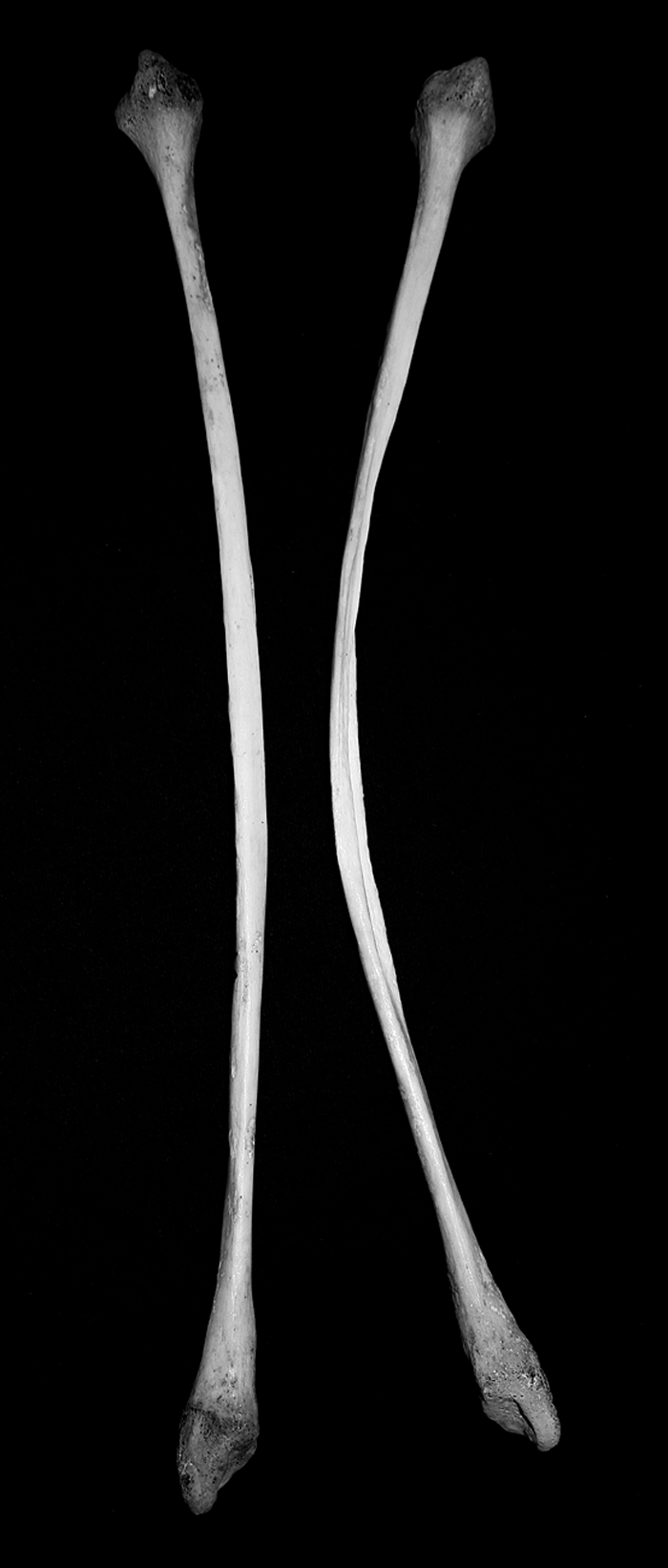 View Details | Figure 8. Left and right fibulae with an acute bowing, more pronounced in the right bone. |
The remaining bones do not show any evidence of pathological lesions.
Sk. 470 belongs to an identified collection with a complete record; therefore the individual’s cause of death, pulmonary tuberculosis, is known. This condition can be traced by the macroscopic observations of the rib cage that shows new bone formation on the visceral surface. This type of bone alteration is commonly associated with pulmonary tuberculosis (Kelley and Micozzi, 1984; Roberts et al., 1994; Santos and Roberts, 2001; Matos and Santos, 2006), and will not be explored in this work since it has already been discussed in other publications (Santos, 2000; Santos and Roberts, 2006).
Regarding the bone changes observed on the vertebral column and on the left lower limb, a causal relation with the cause of death was investigated. Although kyphoscoliosis of the thoracic vertebrae was considered, the absence of massive bone destruction, abscess formation and collapse of the vertebral body, spinal features known as Pott’s disease (Roberts and Buikstra, 2003), excluded this hypothesis. The same was considered regarding the left femur, since none of the most distinctive feature of hip tuberculosis was observed, such as the resorpting grooving of the articulating bone, the formation of sequestra and/or cavitation with incipient bone remodelling and the unifocal joint fusion present in advanced cases (Ortner, 2003; Waldron, 2009). Thus, it is possible to discard bone tuberculosis from the present diagnosis.
Beyond the cause of death, other conditions can be pointed out for this individual. Rib and vertebral agenesis can result from developmental defects during the growth and ossification processes (Glass et al., 2002; Castriota-Scanderbeg and Dallapiccola, 2005). Bilateral aplasia of ribs is a common occurrence in normal individuals as an isolated finding. Nevertheless, it can also reveal the existence of a systemic disorder, such as Edwards syndrome (trisomy 18), occurring in association with other skeletal abnormalities (Glass et al., 2002; Castriota-Scanderbeg and Dallapiccola, 2005; Hudgins and Vaux, 2006). According to Olivier (1960), this spine variation affects 1.5% of individuals without major consequences.
Abnormal curvatures of the spine are common in human pathology, and can have a congenital or idiopathic aetiology (Zimmerman and Kelley, 1982; Castriota-Scanderbeg and Dallapiccola, 2005). Congenital kyphoscoliosis includes hemivertebrae, unilateral block vertebrae, pediculate bars, and neural arch fusion, among others (Williams et al., 1982 after Castriota-Scanderbeg and Dallapiccola, 2005: 211). Sk. 470 does not present any kind of the above-mentioned conditions linked to a congenital/genetic origin (e.g. Turner syndrome, trisomy 18, neurofibromatosis, osteogenesis imperfecta), so it appears that the kyphoscoliosis observed has an idiopathic origin. According to Ortner (2003: 397), “the vertebral bodies may have decreased height due to compression, often combined with a deeper scalloping of the endplate.” This misalignment caused by kyphoscoliosis could explain the asymmetry of the spinous process from the lower spine, the slight degenerative signs at the atlantoccipital joint as well as the marked oblique line for muscle attachment at the occipital base.
The congenital dislocation of the hip, albeit plausible for left innominate bone, because of the new joint formation for the femoral head, does not exhibit the typical signs of hip dislocation already described, namely its bilateral occurrence. Alternatively, a subcapital or transcervical neck fracture that occurred at early ages can be proposed as a possible diagnosis. This fracture, induced by acute trauma or accumulated stress, may have been responsible for a disruption of the bone supply to the femoral head, resulting in the tissue death (Ortner, 2003). This aseptic necrosis may underlie the detachment of the left femoral head and the formation of a new articulation in the innominate bone.
For the right femur of Sk. 470 the differential diagnosis of the epiphyseal alterations observed included slipped femoral capital epiphysis (SFCE), Legg–Calvé–Perthes disease and congenital dislocation of the hip. The first condition is an adolescent hip disorder, characterized by a posteroinferior displacement of the femoral head in relation to the neck at the level of the growth plate (Szőke et al., 2009; Bullough, 2010), being more frequent in males and on the left side (Aufderheide and Rodríguez-Martín, 1998). Although certain similarities with SFCE exist, the forward orientation of the femoral head of Sk. 470, and the absence of the ligamentum teres depression, usually well defined in SFCE, allow us to exclude this condition. Legg–Calvé–Perthes disease, an avascular necrosis of the epiphyses (Shapiro, 2001; Ortner, 2003; Szőke et al., 2009), was also rejected since the femoral head does not present the typical ‘mushroom-like’ appearance, porosity, and exuberant bony growth that characterize this disorder (Aufderheide and Rodríguez-Martín, 1998; Ortner, 2003). A developmental pathology was also pondered to explain the congenital dislocation of the hip. This condition manifests through the complete or partial dislocation of the femoral head in relation to the acetabulum, leading to severe pseudoarthrosis, shallow hip concavity, and progressive deformation of the femoral epiphyses that may appear flattened or distorted (Zimmerman and Kelley, 1982; Hudgins and Vaux, 2006; Waldron, 2009). The normal appearance of the acetabular fossa and the absence of degenerative lesions at the femoral head make this diagnosis less probable.
It is clear that this individual suffered from a more systemic condition that predisposed the skeleton to structural weakness and failure during growth and development, such as residual rickets. One of the primary causes of rickets is Vitamin D deficiency (in fact cholecalciferol is a steroid hormone: see DeLuca and Schnoes, 1983), since it is involved in calcium metabolism (Zimmerman and Kelley, 1982; Ortner, 2003; Pettifor, 2003; Mays et al., 2007; Brickley and Ives, 2008; Mays et al., 2009). A deficient calcium supply leads to an accumulation of unmineralized osteoid, resulting in a failure or delay of the endochondral ossification of long bones (Parsons, 1980; Ortner and Mays, 1998; Pettifor, 2003; Brickley and Ives, 2008; Mays, 2008; Mays et al., 2009). In long-standing rickets, biomechanical forces can induce limb deformities that persist into adulthood, a condition that is called residual rickets (RR) or healed rickets (Ortner, 2003; Brickley and Ives, 2008; Mays, 2008; Waldron, 2009; Brickley et al., 2010). Other features of RR includes kyphosis or scoliosis, changes in the morphology of the ribs and sternum, lateral narrowing of pelvis, abnormal shape of ilia, thickening in the concave face of long bones and mediolateral widening of proximal femora (Brickley et al., 2005; Brickley and Ives, 2008).
Sk. 470 exhibited kyphoscoliosis, wedged vertebral bodies, an abnormal size and asymmetric pelvis with cortical thinning, particularly in the central region of the ilia, and symphyseal faces projected forward instead of being normally articulated at the ventral aspect, and dental enamel defects—all features that can be ascribed to RR. Additionally, the bone fragility imposed by rickets may also explain the right coxa vara deformity and subcapital neck fracture observed in the left femur. In addition to those features the presence of osteopenia, perceptible in the majority of bones, agrees with the possibility of rickets. Furthermore, the lateral and exaggerated bending of both fibulae, more accentuated in the right bone, gave more strength to this diagnosis. Assuming that this condition is responsible for some bone fragility, with an onset after the infant begins to walk, it can produce bending deformities. According to Ortner (2003: 396), when this happens, “bending abnormalities may be limited to the long bones of the lower extremity,” this explains the absence of bent bones among the upper limbs. Many of the above-mentioned lesions are common to osteomalacia. However, the absence of the most distinctive features such as buckling of scapular body and pubic ramus, rib curvature and sternum angulation, vertebral body compression, also known as ‘codfish vertebra’ and Looser–Milkman’s zones of radiolucency associated with bilateral and symmetrical pseudofractures considered as pathognomonic (Parsons, 1980; Sittampalam and Rosenberg, 2001; Ortner, 2003; Kamath et al., 2005; Brickley and Ives, 2008; Waldron, 2009) led to the exclusion of osteomalacia.
Another pathology that should be considered in the diagnosis is juvenile osteoporosis. Though a rare condition, it may have associated with it kyphosis, scoliosis, traumatic vertebral compression fractures, and fragility fractures (Langman, 2005). Apart from kyphoscoliosis and vertebral compression Sk. 470 does not present any of the other distinctive features of juvenile osteoporosis.
From the above-mentioned differential diagnosis, RR seems the most probable aetiology for the majority of the lesions observed in this individual. RR coexisted with pulmonary tuberculosis, her cause of death, and with morphological variation of congenital origin. Moreover, if this individual was retrieved from an assemblage of commingled bones the pathological conditions described would probably lead to the diagnosis of different pathologies depending on the observed anatomical area, and it would be difficult to diagnose a probable case of rickets.
This individual presents several pathological lesions perceptible on the ribs, vertebral column, pelvic bones, femurs, and fibulae, some of them clearly not related to the cause of death, which was recorded as pulmonary tuberculosis. Concerning the differential diagnosis it is clear that this young female suffered from a more generalized systemic disorder instead of isolated pathological conditions. A case of residual or healed rickets can be put forward to explain the changes on the skeleton, as well as the coxa vara, the femoral head displacement and fibular bending. At some point in this woman’s life she contracted pulmonary tuberculosis, causing her death during adulthood.
This study also indicates that even when dealing with individuals with known causes of death, cautious analysis of the skeletons should always be done since various conditions might exist apart from the cause of death recorded.
We would like to thank Carina Marques, Carmina Gomes Silva, and the Clínica Universitária de Imagiologia at the Coimbra University Hospital, and the former Museum of Anthropology at the University of Coimbra. This paper was developed from an oral presentation at the 36th Annual Meeting of the Paleopathology Association (Chicago, IL, USA) in 2009.