Abstract
Biomacromolecules (>40 kDa) have been developed as drug delivery system (DDS) carriers of low-molecular weight drugs to promote these drugs’ uptake by cancer tissues via enhanced permeability and retention (EPR) effects. Human serum albumin (HSA) has been found to accumulate in cancer tissues via this EPR effect. HSA is the most abundant protein in serum, which performs essential physiological functions such as the transportation of many endogenous and exogenous ligands. Nitric oxide (NO) is a very small ligand of HSA; it is a unique and diffusible molecular messenger that plays a central role in mammalian physiology. Although the in vivo half-life of NO is extremely short, HSA could prolong the half-life of NO via S-nitrosation at the position of Cys-34. S-Nitrosated HSA (mono-SNO-HSA) is called an ‘Endogenous NO traffic protein,’ due to the highly stable S-nitroso form in circulating blood, and to the efficiency of S-transnitrosation in cells that require NO. Mono-SNO-HSA possesses a very strong cytoprotective action via the induction of heme oxygenase-1. On the other hand, HSA reinforced with approximately seven NO molecules (poly-SNO-HSA), which we developed by means of chemical modification, possesses multiple anticancer activities. Our previous data clarified that the high expression of protein disulfide isomerase on the surface of cancer cells plays a very important role in the anticancer action of poly-SNO-HSA. In this review, we focus on the advantage of poly-SNO-HSA in treating intractable cancers from the viewpoint of drug delivery systems and drug resistance.
1. HUMAN SERUM ALBUMIN AS THE KEY PLAYER IN THE NITRIC OXIDE TRAFFIC SYSTEM IN HUMANS
A gas modulator, nitric oxide (NO), plays an important role in the human body.1–7) A small amount of NO is synthesized by endothelial and neuronal NO synthases. Its actions, including the relaxation of vascular smooth muscle, are pleiotropic.8,9) However, NO can sometimes be cytotoxic. For instance, a large amount of NO inhibits the growth of cancer cells and induces cell death via apoptosis.10,11) Many studies have identified that the apoptosis of cancer cells (directly) and the inhibition of cancer progression (indirectly) have relevance to NO.12) Unfortunately, the T1/2 of NO is so extremely short (<5 s) that NO without a carrier cannot be applied as a therapeutic biological agent. Thus, NO releasing compounds with pharmacological activity have been synthesized around the world.
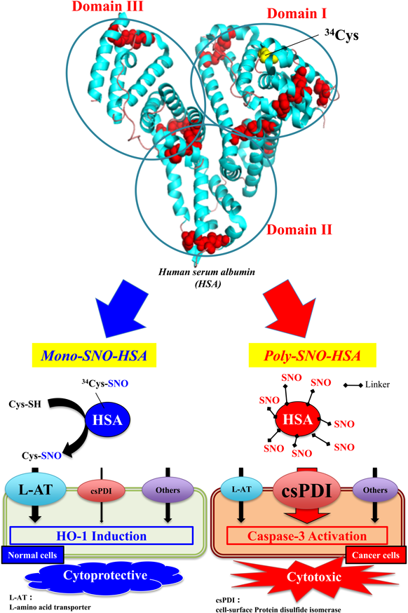
For a long-acting and safe NO donor, we examined the possibility of developing a ‘NO traffic protein’ as a superior NO carrier. This ‘NO traffic protein’ would include (1) a NO binding protein with the superior efficiency of S-nitrosation, (2) superior stability of the S-nitroso form in human blood, and (3) the superior efficiency of NO transfer in selected cells which require NO. As a candidate for creating such a NO traffic protein, we selected human serum albumin (HSA), because HSA has high biocompatibility and good biodegradability, and because ‘endogenous’ S-nitrosothiols in serum are largely related to HSA.13,14) Endogenous HSA has one S-nitrosated site at Cys-34 thiol. The endogenous S-nitrosated HSA (SNO-HSA) was named ‘Mono-SNO-HSA,’ which has significantly superior stability compared with low molecular weight (LMW) S-nitrosothiols.
2. POLY-SNO-HSA AS A CARRIER FOR THE TARGETED DELIVERY OF NO TO CANCER CELLS
In many animal models, it is well established that Mono-SNO-HSA possesses many beneficial and cytoprotective actions. For example, Mono-SNO-HSA can inhibit the apoptosis of hepatic cells and bacteria growth. In an attempt to create highly effective SNO-HSA preparations, we synthesized a SNO-HSA that contained approximately 7 (S-nitroso mol/mol HSA) S-nitroso groups by chemical reaction with lysine residues on the surface of HSA, and named this ‘poly-SNO-HSA.’ Poly-SNO-HSA characteristics were compared in detail with those of a mono-SNO-HSA preparation, per their effectiveness against murine colon carcinoma (C26) and Human hepatoma (HepG2) cells.15) In the case of mono-SNO-HSA, the NO uptake of cells partly takes place via a LMW thiol, such as glutathione or cysteine, which cytoprotectively results in the development of actions via heme oxygenase-1 induction. In contrast to mono-SNO-HSA, the NO transfer from poly-SNO-HSA was more rapid and more pronounced. The cytoprotective action mainly occurs via the reaction of a protein disulfide isomerase (PDI) on the cell surface (Fig. 1). Thus, the excessive NO inflow resulted in apoptotic cell death caused by the induction of reactive oxygen species (ROS) production, caspase-3 activation and DNA fragmentation. We concluded that the site of S-nitroso groups on a carrier such as HSA is more important than an increased number of S-nitroso groups, and that it is difficult to enhance cellular responses to NO without inducing opposite actions. Here, we wish to focus on the possibility of using poly-SNO-HSA as a secure and effective multifunctional anticancer agent in biological systems.
3. THE DIRECT ANTICANCER ACTION OF POLY-SNO-HSA VIA APOPTOSIS
Previously, poly-SNO-HSA was incubated with various cancer cells, and we observed that this HSA form had the ability to induce the apoptosis of cancer cells.16–18) Via activation of the intrinsic apoptosis pathway, apoptosis occurred in various cancer cells such as murine colon cancer, C26 cells, and a variant of Yoshida sarcoma, LY-80 cells, both in vivo and in vitro.
The apoptotic pathway was induced as follows. First, poly-SNO-HSA increased intracellular NO via PDI on the surface of the cancer cells. Subsequently, the elevated NO concentration induced ROS. Then, mitochondrial function and membrane potential were damaged by ROS. Finally, caspase-3, which is a critical instigator of apoptosis, was activated, causing cells to undergo morphological changes, such as chromatin condensation and the engulfment of apoptotic bodies.
Next, the anticancer actions of poly-SNO-HSA were investigated in vivo: C26-bearing mouse and LY-80-bearing rat were given an intravenous injection of poly-SNO-HSA. Interestingly, cancer growth in the poly-SNO-HSA treated animals was reduced to 30% of that observed in the control or HSA-treated animals. These findings indicate that poly-SNO-HSA has anticancer actions both in vitro and in vivo by its ability to induce apoptosis. Similar results were observed when we performed the same experiments using mice bearing other cell types, such as SW480 cells, indicating that poly-SNO-HSA has anticancer action against various cancers.
4. POLY-SNO-HSA CAN OVERCOME MULTIDRUG RESISTANCE
One barrier in various cancer therapies has been well characterized: the development of multidrug resistance by cancer cells. To overcome this problem, various methods have been performed, including the use of agents that inhibit P-glycoprotein (P-gp) through targeted drug delivery, and by changing the cell membrane. However, while some approaches have succeeded in mouse and rat models, clinical applications remain limited. Hence, we propose that NO is an interesting and promising compound in the search for treatments to combat the multidrug resistance of cancer.
Besides inducing apoptosis, poly-SNO-HSA could affect the multidrug resistance of cancer. In practice, we reported that poly-SNO-HSA (0.5–10 µM) significantly reduced the resistance to doxorubicin in human leukemia multidrug resistant (K562/doxorubicin) cells. The inhibitory actions of doxorubicin and poly-SNO-HSA on the growth of K562/doxorubicin cells were synergistic. These data strongly indicate that poly-SNO-HSA possesses not only the ability to inhibit cancer cell growth, but also to attenuate resistance to doxorubicin in vitro.19)
To evaluate the action of poly-SNO-HSA on chemo-resistance in vivo, we investigated the role of poly-SNO-HSA using K562/doxorubicin-bearing mice. After the cancer tissues had reached the size of about 150 mm3, doxorubicin and/or poly-SNO-HSA was injected to the mice biweekly. As a result, in the K562/doxorubicin mice, doxorubicin had no significant action on cancer growth. Intriguingly, a combination treatment of both doxorubicin and poly-SNO-HSA in the K562/doxorubicin mice resulted in a decrease in cancer volume, to 30% that of the mice treated with each alone. This result indicates that poly-SNO-HSA attenuates doxorubicin resistance in vivo. Our findings demonstrate that poly-SNO-HSA has potential for use as a secure and powerful multifunctional anticancer agent. Concerning clinical application, a limitation of the current nanoparticles used for targeted drug delivery is that these carriers also accumulate in non-target organs, inducing serious side actions and side effects. In the case of our carrier, HSA has good biodegradability and low accumulation in non-target organs; therefore, serious side effects would not be encountered. Additionally, our carrier presents a unique opportunity for treating cancers without significant side effects because almost no NO is released from poly-SNO-HSA near cancer tissues. The current tool of drug delivery to cancers, based on nanotechnology, can provide significant therapeutic benefit through understanding the basic mechanisms of drug actions, including that of therapeutic NO.20)
5. POLY-SNO-HSA AS AN AUTOPHAGY INHIBITOR
Recent clinical trials support the hypothesis that the use of antiangiogenic drugs, which target abnormal blood vessels in cancer, can inhibit cancer progression.21) However, longer term clinical responses to angiogenesis inhibitors do not always lead to success. For instance, in clinical trials, approximately 50% of cancers progressed after an initially successful treatment using bevacizumab in glioblastomas,22) consistent with the development of resistance to antiangiogenic therapy, indictating a low prognosis and low response to valuable treatments developed thus far.23) Some clinical data indicate that cancer hypoxia and hypoxia-mediated resistance to antiangiogenic therapy are enhanced via the devascularization induced by antiangiogenic treatment.24) Importantly, hypoxia activates autophagy, which is a lysosomal degradation pathway which may accelerate the survival of cancer cells.25,26) One possible mechanism has been proposed in which BCL2/adenovirus E1B 19 kDa protein-interacting protein 3, a hypoxia inducible factor (HIF)-1α downstream target gene, is essential to hypoxia-mediated autophagy.27,28) Hence, autophagy inhibitors could be candidates for the prevention of resistance to antiangiogenic therapy. In our previous studies, we demonstrated that poly-SNO-HSA has anti-autophagy activity via the inhibition of HIF-1α in cancer cells in vitro and in vivo.29) We are conducting further studies to clarify the issue of whether poly-SNO-HSA can also be used as a specific and potent autophagy inhibitor.
6. POLY-SNO-HSA AS A MHC CLASS I-RELATED CHAIN A (MICA) SHEDDING INHIBITOR
Cancer rejection via binding to natural killer group 2, member D (NKG2D), can be highly effective during early phases of cancer proliferation.30–33) However, the expression of MICA on the surface of cancer cells, and the shedding of soluble MICA by late phase cancer progression, negatively imprints on the systemic immune response. Thus, cancer cells facilitate the cancer immune escape response.34–36)
MICA on the surface of cancer cells is accompanied by endoplasmic reticulum protein 5, which, identically to PDI, usually helps in the folding of many proteins in the endoplasmic reticulum.37) Interestingly, a previous report demonstrated that the function of cell-surface endoplasmic reticulum protein 5 is required for MICA shedding.37) The shedding of MICA is regarded as an important step in human cancer progression and metastasis in order to escape the immunosurveillance mediated by NKG2D. MICA shedding not only decreases the density of MICA on the surface of cancer cells, but also produces soluble MICA, which has been determined to down-regulate systemic NKG2D on cytotoxic effector cells, thus expanding the immunosuppressive microenvironment in the cancerous region.34,36,38) Therefore, inhibition of MICA shedding is a promising strategy to therapeutically improve anticancer immunity.
Our previous reports clearly showed that the transfer of NO from poly-SNO-HSA into cancer cells was induced by cell-surface PDI. Although it is well known that PDI is similar to ERp5, the exact action of poly-SNO-HSA on MICA shedding by ERp5 remains unknown. To clarify this, the human breast cancer cell line MCF-7, which expressed MICA, was incubated with poly-SNO-HSA. As a result, poly-SNO-HSA significantly increased the expression of MICA on the surface of MCF-7 cancer cells, and the soluble MICA in medium was decreased compared with the control. On the other hand, Phorbol 12-myristate 13-acetate (PMA), which stimulates the shedding of MICA, actually decreased the expression of MICA on the surface of MCF-7 and increased the soluble MICA in medium (Fig. 2). These data indicate that poly-SNO-HSA plays an important role as an inhibitor of immune escape mechanisms of cancers (Fig. 3). Therefore, poly-SNO-HSA is a promising component in the development of targeted tumor immunotherapy.
7. PERSPECTIVES OF POLY-SNO-HSA AS AN ANTICANCER DRUG
As described above, poly-SNO-HSA functions as an apoptosis inducer, a multidrug resistance suppressor, an autophagy inhibitor and a MICA-shedding inhibitor (Fig. 4). However, the in vivo half-life of S-nitroso in poly-SNO-HSA was approximately 20 min in cancer-bearing models. These data suggest that poly-SNO-HSA is not a reliably long-lasting NO donor.39) To construct a superior NO delivery carrier, we investigated the factors involved in the stability of S-nitroso on poly-SNO-HSA. Previous findings demonstrated that a polyethylene glycol (PEG) elevated the stability of S-nitroso compounds, because the PEG acts as a stabilizer of S-nitroso bonding.40) We confirmed that this action can be applied to construct more useful poly-SNO-HSA preparations.41) Furthermore, the problem of drug targeting to cancers should be addressed, in addition to the in vivo stability of S-nitroso. From the point of view of cancer targeting, the enhanced permeability and retention (EPR) effect is very important. This effect is a unique and well-known phenomenon in the solid cancer-selective delivery of nanoparticles, liposomes or large proteins, including antibodies.42) Matsumura and Maeda demonstrated that various long-lasting pharmaceutical nanocarriers larger than 40 kDa are able to spontaneously accumulate in solid cancers.43) Additionally, Kataoka’s group indicated that approximately 30 nm micelles, but not 60 nm micelles, are able to enter very low permeable pancreatic cancers.44) These reports strongly indicate that nanoparticle therapeutics ranging in size from 40 kDa to less than 30 nm can serve as efficient drug delivery carriers to even poorly permeable cancers, such as pancreatic cancers. To clarify this possibility, we constructed a HSA dimer (20 nm) produced by Pichia pastoris using a method of genetic engineering. In our previous study, we reported that HSA dimer has a longer blood circulation time than HSA monomer in rats and mice.45) Therefore, we speculated that HSA dimer could be promising clinically as a novel DDS material with superior serum retention as well as increased cancer-specific accumulation. And in fact, the dimerization of poly-SNO-HSA did result in improved anticancer activity via more efficient delivery of NO in C26-bearing mice.41) The PEGylation product of poly-SNO-HSA also possesses good biocompatibility and biodegradability. Furthermore, nanoparticles of PEGylated poly-SNO-HSA-dimer (about 30 nm) are potentially more efficient carriers of anticancer drugs. In the near future, we hope that the promising PEGylated poly-SNO-HSA dimer and its nanoparticles will be developed as a superior anticancer drug with reduced side effects (Fig. 5).
This review of the author’s work was written by the author upon receiving the 2016 Pharmaceutical Society of Japan Award for Young Scientists.
Acknowledgments
I would like to acknowledge Dr. Masaki Otagiri, Dr. Toru Maruyama and current laboratory members at the Department of Biopharmaceutics, Graduate School of Pharmaceutical Sciences, Kumamoto University, for their valuable contributions to this work. I wish to thank Dr. H. Maeda, Institute of DDS Research, Sojo University and Dr. T. Akaike, Graduate School of Medicine, Tohoku University, for their valuable advice and kind consideration. I also thank Dr. Victor T. G. Chuang for editing the manuscript.
Conflict of Interest
The author declares no conflict of interest.
REFERENCES
- 1) Moncada S, Palmer R, Higgs E. Nitric oxide: physiology, pathophysiology, and pharmacology. Pharmacol. Rev., 43, 109–142 (1991).
- 2) Mizutani T, Layon A. Clinical applications of nitric oxide. Chest, 110, 506–524 (1996).
- 3) Stamler JS, Singel D, Loscalzo J. Biochemistry of nitric oxide and its redox-activated forms. Science, 258, 1898–1902 (1992).
- 4) Nathan C. Nitric oxide as a secretory product of mammalian cells. FASEB J., 6, 3051–3064 (1992).
- 5) Lancaster JR Jr. Simulation of the diffusion and reaction of endogenously produced nitric oxide. Proc. Natl. Acad. Sci. U.S.A., 91, 8137–8141 (1994).
- 6) Gaston B, Reilly J, Drazen J, Fackler J, Ramdev P, Arnelle D, Mullins M, Sugarbaker D, Chee C, Singel D. Endogenous nitrogen oxides and bronchodilator S-nitrosothiols in human airways. Proc. Natl. Acad. Sci. U.S.A., 90, 10957–10961 (1993).
- 7) Clancy RM, Levartovsky D, Leszczynska-Piziak J, Yegudin J, Abramson S. Nitric oxide reacts with intracellular glutathione and activates the hexose monophosphate shunt in human neutrophils: evidence for S-nitrosoglutathione as a bioactive intermediary. Proc. Natl. Acad. Sci. U.S.A., 91, 3680–3684 (1994).
- 8) Furchgott RF, Zawadzki JV. The obligatory role of endothelial cells in the relaxation of arterial smooth muscle by acetylcholine. Nature, 288, 373–376 (1980).
- 9) Palmer RM, Ferrige AG, Moncada S. Nitric oxide release accounts for the biological activity of endothelium-derived relaxing factor. Nature, 327, 524–526 (1987).
- 10) Cui S, Reichner JS, Mateo RB, Albina JE. Activated murine macrophages induce apoptosis in tumor cells through nitric oxide-dependent or -independent mechanisms. Cancer Res., 54, 2462–2467 (1994).
- 11) Tendler DS, Bao C, Wang T, Huang EL, Ratovitski EA, Pardoll DA, Lowenstein CJ. Intersection of interferon and hypoxia signal transduction pathways in nitric oxide-induced tumor apoptosis. Cancer Res., 61, 3682–3688 (2001).
- 12) Fukumura D, Kashiwagi S, Jain RK. The role of nitric oxide in tumour progression. Nat. Rev. Cancer, 6, 521–534 (2006).
- 13) Stamler JS, Jaraki O, Osborne J, Simon DI, Keaney J, Vita J, Singel D, Valeri CR, Loscalzo J. Nitric oxide circulates in mammalian plasma primarily as an S-nitroso adduct of serum albumin. Proc. Natl. Acad. Sci. U.S.A., 89, 7674–7677 (1992).
- 14) Marley R, Feelisch M, Holt S, Moore K. A chemiluminescense-based assay for S-nitrosoalbumin and other plasma S-nitrosothiols. Free Radic. Res., 32, 1–9 (2000).
- 15) Ishima Y, Kragh-Hansen U, Maruyama T, Otagiri M. Poly-S-nitrosated albumin as a safe and effective multifunctional antitumor agent: characterization, biochemistry and possible future therapeutic applications. Biomed. Res. Int., 2013, 353892 (2013).
- 16) Ishima Y, Yoshida F, Kragh-Hansen U, Watanabe K, Katayama N, Nakajou K, Akaike T, Kai T, Maruyama T, Otagiri M. Cellular uptake mechanisms and responses to NO transferred from mono- and poly-S-nitrosated human serum albumin. Free Radic. Res., 45, 1196–1206 (2011).
- 17) Katayama N, Nakajou K, Komori H, Uchida K, Yokoe J, Yasui N, Yamamoto H, Kai T, Sato M, Nakagawa T, Takeya M, Maruyama T, Otagiri M. Design and evaluation of S-nitrosylated human serum albumin as a novel anticancer drug. J. Pharmacol. Exp. Ther., 325, 69–76 (2008).
- 18) Katayama N, Nakajou K, Ishima Y, Ikuta S, Yokoe J, Yoshida F, Suenaga A, Maruyama T, Kai T, Otagiri M. Nitrosylated human serum albumin (SNO-HSA) induces apoptosis in tumor cells. Nitric Oxide, 22, 259–265 (2010).
- 19) Ishima Y, Hara M, Kragh-Hansen U, Inoue A, Suenaga A, Kai T, Watanabe H, Otagiri M, Maruyama T. Elucidation of the therapeutic enhancer mechanism of poly-S-nitrosated human serum albumin against multidrug-resistant tumor in animal models. J. Control. Release, 164, 1–7 (2012).
- 20) Otagiri M, Chuang VT, Maruyama T, Kragh-Hansen U. Human Serum Albumin: New insights on its structural dynamics, functional impacts and pharmaceutical applications. Sojo University Publishing Center, Kumamoto (2013).
- 21) Jain RK. Lessons from multidisciplinary translational trials on anti-angiogenic therapy of cancer. Nat. Rev. Cancer, 8, 309–316 (2008).
- 22) Vredenburgh JJ, Desjardins A, Herndon JE 2nd, Dowell JM, Reardon DA, Quinn JA, Rich JN, Sathornsumetee S, Gururangan S, Wagner M, Bigner DD, Friedman AH, Friedman HS. Phase II trial of bevacizumab and irinotecan in recurrent malignant glioma. Clin. Cancer Res., 13, 1253–1259 (2007).
- 23) Clark AJ, Lamborn KR, Butowski NA, Chang SM, Prados MD, Clarke JL, McDermott MW, Parsa AT, Berger MS, Aghi MK. Neurosurgical management and prognosis of patients with glioblastoma that progresses during bevacizumab treatment. Neurosurgery, 70, 361–370 (2012).
- 24) Hu YL, DeLay M, Jahangiri A, Molinaro AM, Rose SD, Carbonell WS, Aghi MK. Hypoxia-induced autophagy promotes tumor cell survival and adaptation to antiangiogenic treatment in glioblastoma. Cancer Res., 72, 1773–1783 (2012).
- 25) Mazure NM, Pouysségur J. Hypoxia-induced autophagy: cell death or cell survival? Curr. Opin. Cell Biol., 22, 177–180 (2010).
- 26) Leone RD, Amaravadi RK. Autophagy: a targetable linchpin of cancer cell metabolism. Trends Endocrinol. Metab., 24, 209–217 (2013).
- 27) Tracy K, Dibling BC, Spike BT, Knabb JR, Schumacker P, Macleod KF. BNIP3 is an RB/E2F target gene required for hypoxia-induced autophagy. Mol. Cell. Biol., 27, 6229–6242 (2007).
- 28) Hu YL, Jahangiri A, Delay M, Aghi MK. Tumor cell autophagy as an adaptive response mediating resistance to treatments such as antiangiogenic therapy. Cancer Res., 72, 4294–4299 (2012).
- 29) Ishima Y, Inoue A, Fang J, Kinoshita R, Ikeda M, Watanabe H, Maeda H, Otagiri M, Maruyama T. Poly-S-nitrosated human albumin enhances the antitumor and antimetastasis effect of bevacizumab, partly by inhibiting autophagy through the generation of nitric oxide. Cancer Sci., 106, 194–200 (2015).
- 30) Cerwenka A, Baron JL, Lanier LL. Ectopic expression of retinoic acid early inducible-1 gene (RAE-1) permits natural killer cell-mediated rejection of a MHC class I-bearing tumor in vivo. Proc. Natl. Acad. Sci. U.S.A., 98, 11521–11526 (2001).
- 31) Diefenbach A, Jensen ER, Jamieson AM, Raulet DH. Rae1 and H60 ligands of the NKG2D receptor stimulate tumor immunity. Nature, 413, 165–171 (2001).
- 32) Girardi M, Oppenheim DE, Steele CR, Lewis JM, Glusac E, Filler R, Hobby P, Sutton B, Tigelaar RE, Hayday AC. Regulation of cutaneous malignancy by gammadelta T cells. Science, 294, 605–609 (2001).
- 33) Smyth MJ, Swann J, Cretney E, Zerafa N, Yokoyama WM, Hayakawa Y. NKG2D function protects the host from tumor initiation. J. Exp. Med., 202, 583–588 (2005).
- 34) Groh V, Wu J, Yee C, Spies T. Tumour-derived soluble MIC ligands impair expression of NKG2D and T-cell activation. Nature, 419, 734–738 (2002).
- 35) Doubrovina ES, Doubrovin MM, Vider E, Sisson RB, O’Reilly RJ, Dupont B, Vyas YM. Evasion from NK cell immunity by MHC class I chain-related molecules expressing colon adenocarcinoma. J. Immunol., 171, 6891–6899 (2003).
- 36) Groh V, Smythe K, Dai Z, Spies T. Fas ligand-mediated paracrine T cell regulation by the receptor NKG2D in tumor immunity. Nat. Immunol., 7, 755–762 (2006).
- 37) Ellgaard L, Ruddock LW. The human protein disulphide isomerase family: substrate interactions and functional properties. EMBO Rep., 6, 28–32 (2005).
- 38) Salih HR, Antropius H, Gieseke F, Lutz SZ, Kanz L, Rammensee HG, Steinle A. Functional expression and release of ligands for the activating immunoreceptor NKG2D in leukemia. Blood, 102, 1389–1396 (2003).
- 39) Katayama N, Nakajou K, Ishima Y, Ikuta S, Yokoe J, Yoshida F, Suenaga A, Maruyama T, Kai T, Otagiri M. Nitrosylated human serum albumin (SNO-HSA) induces apoptosis in tumor cells. Nitric Oxide, 22, 259–265 (2010).
- 40) Shishido SM, de Oliveira MG. Polyethylene glycol matrix reduces the rates of photochemical and thermal release of nitric oxide from S-nitroso-N-acetylcysteine. Photochem. Photobiol., 71, 273–280 (2000).
- 41) Ishima Y, Fang J, Kragh-Hansen U, Yin H, Liao L, Katayama N, Watanabe H, Kai T, Suenaga A, Maeda H, Otagiri M, Maruyama T. Tuning of poly-S-nitrosated human serum albumin as superior antitumor nanomedicine. J. Pharm. Sci., 103, 2184–2188 (2014).
- 42) Maeda H, Sawa T, Konno T. Mechanism of tumor-targeted delivery of macromolecular drugs, including the EPR effect in solid tumor, and clinical overview of the prototype polymeric drug SMANCS. J. Control. Release, 74, 47–61 (2001).
- 43) Matsumura Y, Maeda H. A new concept for macromolecular therapeutics in cancer chemotherapy: mechanism of tumoritropic accumulation of proteins and the antitumor agent SMANCS. Cancer Res., 46, 6387–6392 (1986).
- 44) Cabral H, Matsumoto Y, Mizuno K, Chen Q, Murakami M, Kimura M, Terada Y, Kano MR, Miyazono K, Uesaka M, Nishiyama N, Kataoka K. Accumulation of sub-100 nm polymeric micelles in poorly permeable tumours depends on size. Nat. Nanotechnol., 6, 815–823 (2011).
- 45) Matsushita S, Chuang VT, Kanazawa M, Tanase S, Kawai K, Maruyama T, Suenaga A, Otagiri M. Recombinant human serum albumin dimer has high blood circulation activity and low vascular permeability in comparison with native human serum albumin. Pharm. Res., 23, 882–891 (2006).