A 30-year-old asymptomatic male with a giant coronary aneurysm attended our hospital for a follow-up examination. The patient also had a childhood history of Kawasaki disease. Non-contrast-enhanced magnetic resonance imaging (MRI) was performed to assess the coronary aneurysm and investigate the presence of aneurysms in other blood vessels. The known coronary aneurysm had not increased in size since the previous examination. However, concomitantly, a huge aneurysm was detected in the right upper arm (Figure A). Furthermore, a high-intensity plaque was observed at the same site (Figure B). When the site was examined further by arterial ultrasound (Figure C), a fresh floating thrombus was found and, consequently, warfarin therapy was initiated.
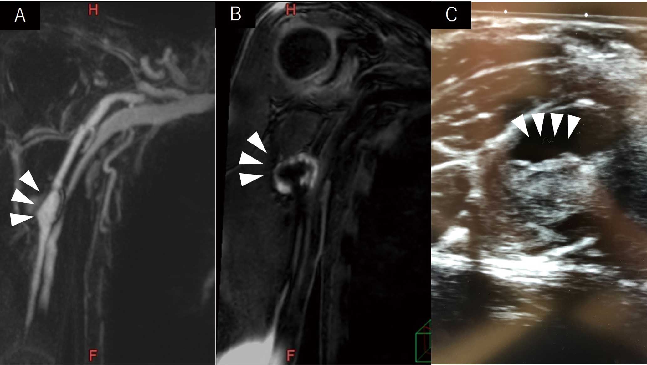
A previous study reported that a high-intensity plaque seen on MRI reflects thrombus and arteriosclerosis.1
Because Kawasaki disease is systemic vasculitis, the inflammation may spread not only to the coronary arteries, but also to systemic arteries throughout the body. In Kawasaki disease, which often occurs in young patients, non-contrast MRI without exposure can be effective for evaluation of the entire systemic vasculature, including the detection of coronary aneurysms.
Reference
- 1.
Ehara S, Matsumoto K, Shimada K. The clinical value of high-intensity signals on the coronary atherosclerotic plaques: Noncontrast T1-weighted magnetic resonance imaging. Int J Mol Sci 2016; 17: 1187.
