| To whom correspondence should be addressed: Takashi Muramatsu, Department of Biochemistry, Nagoya University Graduate School of Medicine, 65 Tsurumai-cho, Showa-ku, Nagoya 466-8550, Japan. Tel: +81–52–744–2059, Fax: +81–52–744–2065 Abbreviations: BCHS, blue cheese; E, embryomic day. |
The BEACH domain has been defined as a sequence motif of 282 amino acids present in the mouse beige protein and its human homologue, the CHS protein (Nagle et al., 1996; Spritz, 1998). The CHS gene is mutated in patients with Chediak-Higashi syndrome (CHS) (Nagle et al., 1996; Spritz, 1998). All proteins with the BEACH domain also possess several WD40 motifs in their C-terminal portions (Wang et al., 2001; Wang et al., 2000). Proteins with the BEACH-WD40 domain are implicated in protein sorting between endosomes, lysosomes and the plasma membrane. WD40 repeats seem to be a functional motif that facilitates defined protein-protein interaction (Wang et al., 2001; Adam-Klages et al., 1996; Neer et al., 1994; Tsukazaki et al., 1998). Recently, the FYVE (Fab1p/YOTB/Vac1p/EEA1) domain has been shown to bind phosphatidyl inositol-3-phosphate (PtdIns(3)P) with high specificity and thus represents a novel signaling module that can mediate protein interaction with membranes (Gaullier et al., 1998; Patki et al., 1998).
Here, we report the cloning, genomic structure, and mode of expression of a protein containing the BEACH, WD40 and FYVE domains that we have designated as BWF1. After completion of the present study, the Drosophila protein, blue cheese (BCHS), was reported by another team (Finley et al., 2003). The loss of function of the bchs gene leads to degeneration in the nervous tissue. The bchs gene might be important for prevention of neurodegeneration. BCHS has 45% homology with BWF1 at the amino acid level, and has been detected in the cytoplasm of neuronal cells (Finley et al., 2003). The loss of function of the bchs gene leads to formation of protein aggregates, probably due to failure in vesicle transport (Finley et al., 2003). To date, however, nothing has been reported on the expression profile of the mammalian homolog.
Full-length BWF1 cDNA was obtained by rapid amplification of 5'-cDNA ends (5'-RACE) polymerase chain reaction (PCR) using ICR mouse embryonic brain cDNA and liver cDNA as templates (Chen et al., 2001). Two nested primers (BWF1-R1, 5'-GAGTACCTTGTTCTGCTCCAC-3' and BWF1-R2, 5'-AGGGAAGGCCTCAGGTTGTCTAGG-3') were designed based on the partial cDNA sequence (GenBank accession number D37791). The PCR products were sequenced directly to avoid detecting the mutations introduced during PCR. Both strands of each template were sequenced, and the sequence was confirmed by analysis of the PCR products both from ICR mouse brain and liver mRNA. New primers were designed from these sequences, and further 5'-RACE PCR was conducted to extend the cDNA sequence in the 5' direction. The following primers were used: BWF1-R3, 5'-GCACCTCCTTGATGTCTTC-3', BWF1-R4, 5'-CCTTGCCTGGCACCTCGAGG-3', BWF1-R5, 5'-CGCAGCAGCTCGCACTCGATCTGAC-3', BWF1-R6, 5'-TCTGACACCACTCCTCGGTCACGTAC-3', BWF1-R7, 5'-GCAGGCTGACCTTGAAGAGCT-3', BWF1-R8, 5'-GTGGATCAGCTCGGTCCAGACT-3', BWF1-R9, 5'-AACTGCATCAGGGTCACTGGA-3', BWF1-R10, 5'-AAGTAGTGGCGACAGGCTTAGGGA-3', BWF1-R11, 5'-CATGAAGTCTGATGTCATGAG-3', BWF1-R12, 5'-CACAGTGTGCAGGAGCTCCAACAC-3', BWF1-R13 and 5'-CAGCCGGCTTCAGCTCGCTGA-3', BWF1-R14, 5'-TCCCTTGGACAGCTTGTTCTGAGT-3'. PCR was performed for 35 cycles at 94°C for 30 sec, 55°C for 30 sec, and 72°C for 2 min. Mouse genomic DNA sequence was extracted from available public (NCBI) database using the BLAST program.
Northern blotting analysis was performed as described previously (Chen et al., 2000). Poly (A) RNA (2 μg) was prepared from 129/Sv mouse tissues. Multiple tissue Northern blot (Clontech, Palo Alto, CA) was also used for hybridization analysis. A mouse cDNA fragment was amplified by PCR with the primers: 5'-CCCATCGTGAGTGTGAACACA-3' and 5'-GCAGTTCCGAGGTCCATCTT-3'. The PCR products were labeled with [α-32P]dCTP using a Rediprime II DNA labeling system (Amersham Biosciences) and used as the probe. The filters were hybridized with the probe in 2×SSC, 0.5% SDS, 5×Denhardt’s solution containing 100 μg/ml heat denatured salmon sperm DNA at 42°C overnight. Filters were washed twice for 5 min at room temperature (RT) in 2×SSC, 0.5% SDS and twice for 30 min at 68°C in 0.1×SSC, 0.1% SDS. Hybridization signals were detected with a BAS 2000 radioimage analyzer (Fuji Film, Tokyo, Japan). A β-actin cDNA (Clontech, Palo Alto, CA) probe was used as a control.
In situ hybridization was performed as previously described (Kurosawa et al., 1999). Briefly, sense and anti-sense RNA probes for BWF1 were prepared using T7 and T3 RNA polymerase in the presence of digoxigenin-11-UTP (Roche Applied Science). A mouse BWF1 cDNA fragment was amplified by PCR with the primers: 5'-AACTGACTATGCACACAGCC-3', 5'-GCAGTTCCGAGGTCCATCTT-3', and the mouse embryonic brain cDNA as a template. The amplified DNA fragment was subcloned into pBluescript II SK+, and used as a template in the transcription reaction. Hybridization was performed at 65°C for 16 h, followed by washing in 0.2×SSC at 68°C for 2 h. Samples were incubated overnight with alkaline phosphatase-conjugated goat anti-digoxigenin Fab fragment (Roche Applied Science) at 1:1000 dilution, washed and detected using 4-nitroblue tetrazolium chloride (NBT) (Boehringer Mannheim) and X-phosphate/5-bromo-4-chloro-3-indolyl-phosphate (BCIP) (Roche Applied Science).
To generate the expression vector pBWF-C (Fig. 5A), containing the FYVE domain (amino acids, 3409–3508), PCR was performed. The primers used for amplification were: 5'-AACTGACTATGCACACAGCC-3',5'-GAGGTCGACTCAGCAGTTCCGAGGTCCATCTT-3' (the underline shows the SalI site) and mouse liver cDNA as the template. The PCR products were digested with BamHI/SalI and then ligated in-frame into the mammalian expression vector pCMV-Tag2C (Sigma), resulting in the expression of an N-terminal Flag-tagged fusion protein. The pBWF-B expression vector, containing the BEACH domain and FYVE domain (amino acids, 2714–3508), was generated by insertion of the BamHI-digested PCR product into the pBWF-C vector. The pBWF-A expression vector (amino acids, 2190–3508) was generated by inserting a HincII-digested PCR product into SmaI-digested pBWF-C, and then by the insertion of a BamHI fragment from pBWF-B. The single insertion in the correct orientation was finally confirmed by restriction enzyme analysis and DNA sequencing.
COS7 cells were cultured in Dulbecco’s modified Eagle’s Medium (DMEM) containing 10% fetal calf serum under 5% CO2 at 37°C. When the cells reached a confluence of 70% they were transiently transfected with 4 μg DNA using Lipofectamine plus (Invitrogen). After 24 h, the cells were recovered.
After 24 h of incubation, the transfected COS7 cells were washed thoroughly with Dulbecco’s phosphate-buffered saline (PBS) and fixed for 20 min at room temperature (RT) in 4% phosphate-buffered formalin freshly prepared from paraformaldehyde. After extensive washing with 1×PBS, the cells were incubated in 0.1% Triton X-100/PBS for 10 min at RT. The cells were blocked in 1% bovine serum albumin for 20 min at RT and then incubated with 1:500 dilution of the anti-Flag M2 monoclonal antibody (Sigma) for 1 h at RT. After washing, the cells were incubated with 1:100 dilution of the secondary antibody, fluorescein isothiocyanate (FITC)-conjugated goat anti-mouse IgG (Sigma), in the dark for 30 min at RT. After washing, immunolocalization of the BWF1 fusion protein was detected by a laser scanning confocal imaging system (Bio-Rad Microscience).
A partial cDNA sequence, which we reported previously as a β-1, 4-galactosyltransferase gene (Uehara and Muramatsu, 1997), was used to design PCR primers to amplify the upstream regions from the mouse embryonic brain cDNA library. Initially, we obtained a 1.5-kb cDNA sequence by this strategy. 5'-RACE was performed to obtain the full-length cDNA of BWF1. We eventually obtained a full-length cDNA sequence which encodes a protein consisting of 3508 amino acids with a predicted molecular weight of 385 kDa. Homology search indicated that the BWF1 gene has many interesting features. It contains a BEACH domain, 5 WD40 repeats and a FYVE domain; hence we called the protein BWF1 (Fig. 2A). A 282 amino acid sequence of BWF1 has approximately 70% homology with the BEACH domain in other proteins (Fig. 2B). The BEACH domain has been defined as a sequence motif of 282 amino acids present in the mouse beige protein and its human homologue, the CHS protein (Nagle et al., 1996; Spritz, 1998). The CHS gene is mutated in patients with Chediak-Higashi syndrome (CHS), an inherited disease characterized by hypopigmentation, severe immunological deficiency, a bleeding tendency, and neuronal abnormalities (Nagle et al., 1996; Spritz et al., 1998). BEACH is also present in other proteins (Wang et al., 2001; Wang et al., 2000; Adam-Klages et al., 1996; Wang et al., 2002; Jogl et al., 2002), but the function of the BEACH domain is still unknown. All proteins with BEACH domains also possess several WD40 motifs in their C-terminus portions (Wang et al., 2001; Wang et al., 2000). WD40 repeats seem to be a functional motif that facilitates defined protein-protein interaction (Wang et al., 2001; Adam-Klages et al., 1996; Neer et al., 1994; Tsukazaki et al., 1998). All the WD40-repeat proteins described so far seem to provide regulatory functions in various cellular processes; none of them contain intrinsic enzymatic function. FAN, which contains a BEACH-WD40 domain, interacts with the cytoplasmic domain of the 55 kDa tumor necrosis factor receptor through its C-terminal cluster of WD40 repeats and signals the activation of neutral sphingomyelinase (N-Smase) (Adam-Klages et al., 1996). BWF1 contains 5 WD40 repeats in its C-terminus portion (Fig. 1 and 2C). Therefore, BWF1 may interact with other proteins through its C-terminal cluster of WD40 repeats. More interestingly, BWF1 has 47% homology with the FYVE domain in other proteins (Fig. 1 and 2D). Recently, analysis of a number of FYVE domains from yeast and mammals has revealed that this motif binds phosphatidyl inositol-3-phosphate with high specificity and thus represents a novel signaling module that can mediate protein interaction with membranes (Gaullier et al., 1998; Patki et al., 1998; Patki et al., 1997). The BWF1 gene contains the three motifs, therefore the cloned BWF1 provided a good molecular model to study the function of the three motifs. The partial sequence of BWF1 was considered to encode a β-1,4-galactosyltransferase (Uehara and Muramatsu, 1997). The low specific activity of the recombinant enzyme (Uehara and Muramatsu, 1997) was inferred to be due to expression in E. coli system. However, after extensive purification the BWF1 protein did not show any galactosyltransferase activity (unpublished results). Most probably protein-binding motifs in the truncated protein (Uehara and Muramatsu, 1997) recruited whatever endogenous β-1,4-galactosyltransferase was present in E. coli.
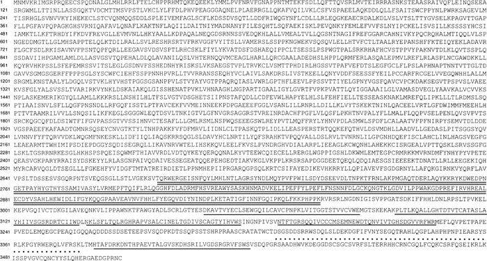 View Details | Fig. 1. Amino acid sequences of mouse BWF1. The BEACH domain sequence is doubly underlined (=); the 5 WD40 motifs are underlined (–); the FYVE domain is dotted (.). |
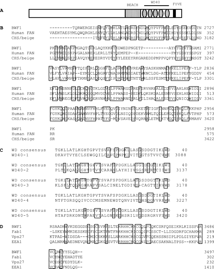 View Details | Fig. 2. Structural characterization of BWF1. (A) Schematic map showing the motif of the BWF1 protein: the gray box indicates the BEACH domain; the flat gray circles indicate the WD40 motifs; the black box indicates the FYVE domain. (B) Alignment of the BEACH domain from BWF1, human Chediak-Higashi syndrome protein (GenBankTM accession number U67615), and human FAN protein (GenBankTM accession number Q92636). (C) Alignment of the WD40 domain consensus sequence (GenBankTM Conserved Domain Database, accession number Smart00320) with the BWF1 WD40 domain. (D) Alignment of the FYVE domain from BWF1, Fab1 (GenBankTM accession number U01017), Vps27 (GenBankTM accession number U24218) and EEA1 (GenBankTM accession number L40157). Identical residues are boxed. |
BLAST analysis against mouse genomic databases showed that the gene is located on chromosome 5 and contains 67 exons, which span more than 270 kb of genomic DNA. The splice junctions were predicted based on cDNA BLAST alignment to mouse genome DNA and all adhered to the characteristic GT-AG configuration (Breathnach and Chambon, 1981). The complete sequence of the BWF1 cDNA has been deposited within the GenBank Database (accession number AY336569), and the partial cDNA sequence which contains all the three motifs was deposited on August 2001 (accession number D37791).
Northern blot analysis revealed approximately 10 kb mRNA of BWF1; the BWF1 mRNA size was consistent with the cDNA size. The mRNA was strongly expressed in the liver, moderately in the kidney and testis, and weakly expressed in the brain (Fig. 3). We noted that BWF1 is mainly expressed in organs with high activity of protein synthesis. Smear of the mRNA bands might be due to differential splicing of the large BWF1 transcript. During development of the mouse brain, BWF1 mRNA was abundant at embryonic day (E)14–16; after birth, the level of BWF1 mRNA expression decreased markedly to reach the adult level at postnatal day 3 (Fig. 3). To obtain precise information on the distribution of BWF1 mRNA in the developing mouse brain, in situ hybridization analysis was performed (Fig. 4). The signal was detected mainly in the marginal region both in E14 and E16 embryonic brain; after birth, the mRNA expression became diffuse. Subcellular distribution of BWF1 was studied by transfection of Flag-tagged BEACH-WD40-FYVE domain fusion protein (Fig. 5A). The protein was diffusely distributed in the cytoplasm (Fig. 5B). Two kinds of truncated fusion proteins (Fig. 5A) showed similar distribution patterns (Fig. 5B). Therefore, BWF1 is considered to be a cytoplasmic protein. Together with the high expression in organs with high activity of protein synthesis, it is possible that BWF1 functions in some stages of protein synthesis including vesicle transport. Identification of the protein that binds to BWF1 is required as the next step of study on this interesting protein.
 View Details | Fig. 3. Northern blot analysis of the expression of mouse BWF1. The membrane hybridized with the BWF1 probe was deprobed and re-hybridized with a β-actin cDNA probe. (A) Analysis of mouse BWF1 expression in adult organs. (B) Analysis of mouse BWF1 expression in the developing mouse brain: E14, embryonic brain day 14; E16, embryonic brain day 16; P0, postnatal day 0; P3, postnatal day 3. |
 View Details | Fig. 4. In situ hybridization studies on the expression of BWF1 mRNA. Sections were hybridized with the digoxigenin-labeled sense and antisense RNA probes for BWF1. The signals were detected using NBT and BCIP. (A) The developing mouse brain. Sagittal sections of E14, E16 and P0 mouse brain were analyzed. Scale bar, 1 mm. (B) Higher magnification of the boxed area in A. Scale bar, 100 μm. |
 View Details | Fig. 5. Localization of the Flag-BWF1 fusion protein in COS7 cells. COS7 cells were transfected with Flag-BWF1 expression plasmid, fixed and incubated with mouse monoclonal anti-FLAG antibody, followed by FITC-conjugated goat anti-mouse IgG antibody. A: A schematic diagram of the constructs used for transfection, B:(I) Cells transfected with the BWF-A expression plasmid; (II) Cells transfected with BWF-B expression plasmid; (III) Cells transfected with BWF-C expression plasmid; (IV) Untransfected cells. |
During manuscript preparation a report on Drosophila BCHS appeared (Finley et al., 2003). BCHS has 45% sequence identify with BWF1. A sequence of a human homolog has also appeared (ALFY, GenBankTM accession number AAN15137). ALFY and bchs have 46% sequence identity, and ALFY and BWF1 93% identity. BCHS is expressed only in the nervous tissue, and nothing is reported on the expression profile of ALFY. The present paper indicates that subcellular localization of BWF1 is similar to that of BCHS. However, BWF1 in mammals is more broadly located than BCHS, hence is likely that BWF1 plays a broader role than BCHS.
We thank H. Inoue and T. Adachi for secretarial assistance. This work was supported in part by grants from the Ministry of Education, Culture, Sports, Science and Technology of Japan.
|