| To whom correspondence should be addressed: Yoshikazu Ohya, Laboratory of Signal Transduction, Department of Integrated Biosciences, Graduate School of Frontier Sciences, University of Tokyo, 5-1-5 Kashiwanoha, Kashiwa, Chiba 277-8562, Japan. Tel: +81–4–7136–3650, Fax: +81–4–7136–3651 E-mail: ohya@k.u-tokyo.ac.jp |
Cell cycle progression is regulated by several checkpoint mechanisms that monitor cell cycle landmarks. When a cell cycle event is perturbed, progression of the cell cycle to subsequent events is halted until the completion of the former event. Well-known checkpoint targets are DNA damage, DNA replication (Weinert et al., 1994), kinetochore attachment to the mitotic spindle (Rudner and Murray, 1996), and bud morphogenesis (Lew and Reed, 1995).
We recently discovered a new cell cycle checkpoint, termed the cell wall integrity checkpoint (Suzuki et al., 2004). In response to a defect in cell wall synthesis, cells of the yeast Saccharomyces cerevisiae arrest cell growth at the small bud before entry into mitosis. The cell wall of S. cerevisiae consists predominantly of 1,3-β-glucan (Cid et al., 1995). Mutants defective in 1,3-β-glucan synthesis cannot form the medium or large bud, and these mutants arrest the cell cycle progression before nuclear division without forming a bipolar spindle (Suzuki et al., 2004). This observation indicates that the perturbation of 1,3-β-glucan synthesis triggers the cell wall integrity checkpoint, leading to cell cycle arrest before the separation of the spindle pole body (SPB). An analysis of the mutant defective in the cell wall integrity checkpoint, wac1 (wall checkpoint defective), revealed that this mutant accumulates cells with a bipolar spindle after 1,3-β-glucan synthesis has stopped (Suzuki et al., 2004). WAC1 is identical to ARP1, and the wac1 product possesses a single amino acid replacement (P268L). These findings suggest that Arp1p (Wac1p) is required to arrest the cell cycle at G2 phase when cell wall synthesis is perturbed, but little is known about how Arp1p actually functions in the cell wall integrity checkpoint.
Biochemical and microscopic studies have revealed that Arp1p is the core subunit of the dynactin complex. Nine or 10 Arp1p monomers form the actin-like short filament backbone of the dynactin complex, as well as multiple species of subunits assemble along this filament (Eckley et al., 1999; Schafer et al., 1994). The dynactin complex of the budding yeast contains at least three species of polypeptides: Arp1p, Nip100p, and Jnm1p (Geiser et al., 1997; Kahana et al., 1998; McMillan and Tatchell, 1994). In addition to these proteins, comprehensive two-hybrid screening has isolated three Arp1p binding proteins: Arp10p, Jsn1p, and Yjr008wp (Ito et al., 2001; Uetz et al., 2000). Genetic studies have shown that the dynactin complex is required for the nuclear migration process after anaphase. The deletion of the ARP1 gene causes the defect in dynein-mediated nuclear migration, and cells with two nuclei in the mother cell are frequently observed (Muhua et al., 1994). The dynactin complex in S. cerevisiae is proposed to associate with the cytoplasmic dynein motor, resulting in the pulling force to promote nuclear migration (Hildebrandt and Hoyt, 2000). In addition, components of the dynactin complex, namely Arp1p, Nip100p, and Jnm1p, are required for the cell wall integrity checkpoint. Surprisingly, the dynactin-related proteins Dyn1p, Pac11p, Num1p, Pac1p, and Bik1p are not required for the checkpoint (Suzuki et al., 2004). Thus Arp1p functions in the cell wall integrity checkpoint as well as in nuclear migration, although the relationship between these two functions remains unclear.
In this study, we used a genetic approach to determine whether the cell wall integrity checkpoint and nuclear migration functions of Arp1p are based on independent molecular functions. We constructed 32 mutated arp1 alleles with alanine mutations at the clusters of charged amino acids that are predicted to be on the surface of the Arp1p ultrastructure. We then examined the effects of the mutations on the cell wall integrity checkpoint and nuclear migration functions of Arp1p in yeast cells.
Standard molecular biological techniques for yeast and bacteria were used (Sambrook et al., 1989; Sherman et al., 1986).
The yeast strains used in this study are listed in Table I. All strains are derivatives of YPH499 and YPH500 (Sikorski and Hieter, 1989). The FKS1 or fks1-1154 mutant strains used in this study harbored an FKS1 or fks1-1154 temperature-sensitive allele integrated into the Δfks1 Δfks2 background, in order to avoid the effect of Fks2p that has overlapping functions with Fks1p. The FKS1 wac1 (YOC2857) and fks1-1154 wac1 (YOC2858) strains have the wac1 mutation (arp1P268L) in ARP1 of the FKS1 (YOC1001) and fks1-1154 (YOC1087) strains, respectively.
The genomic ARP1 was replaced with the gene Cg-LEU2 (Candida glablata LEU2 gene compatible with Saccharomyces cerevisiae LEU2) in strains FKS1 and fks1-1154 to generate the FKS1 Δarp1 (YOC2888) and fks1-1154 Δarp1 (YOC2889) strains, respectively.
The arp1-x:URA3 strains (YOC3032-3099) were constructed by replacing the Δarp1::LEU2 locus of the FKS1Δarp1 (YOC2888) and fks1-1154Δarp1 (YOC2889) strains with the HindIII-XbaI fragment containing arp1-x:URA3 prepared from the pBS-arp1-x plasmid. In the wac1:URA3 strains, the genomic Δarp1::LEU2 region was replaced with a HindIII-DraI fragment, because wac1 was digested with XbaI. The integrants were selected on SD-uracil plates, and the correct integrations were verified by PCR.
The YOC4106-4112 were constructed by transforming the plasmid pYO2763, centromeric vector containing KAR9 and URA3, into FKS1 (YOC1001), FKS1 wac1 (YOC2857), FKS1 Δarp1:: KanMX (YOC4113), FKS arp1-31 (YOC3035), FKS1 arp1-236 (YOC3052), FKS1 arp1-266 (YOC3056) and FKS1 arp1-374 (YOC3065) strains (URA3 marker of genomic YOC3035, 3052, 3056 and 3065 were deleted). The genomic KAR9 gene of the resultant strains was then replaced with the Cg-LEU2 marker.
The plasmids used in this study are listed in Table II. The plasmid pRS315-ARP1 (pYO2521) contains the 2.1-kb HindIII-XbaI fragment, including the entire ORF of ARP1, inserted into pRS315. For pBS-ARP1-URA3 (pYO2522), the 2.1-kb HindIII-XbaI fragment, including the entire ORF of ARP1, was prepared from pRS315-ARP1 and was inserted into pBluescript II SK(+). The yeast URA3 fragment amplified from pRS316 by PCR was digested with SmaI and was inserted at the blunt-ended EcoT22I site located 200 bp upstream from the XbaI site.
For pBS-wac1-URA3 (pYO2523), a fragment including the wac1 allele was amplified from the FKS1 wac1 genome by PCR, digested with HindIII and DraI, and used to replace the HindIII-DraI region of pBS-ARP1-URA3. Positive clones were selected based on XbaI digestion, because an additional XbaI site was generated by the wac1 mutation.
To construct pBS-arp1-x-URA3 (pYO2524-pYO2555), the NruI-SphI fragment of pBS-ARP1-URA3 (or, in the case of arp1-294 to -374, the SphI-EcoT22I fragment) was replaced with EGFP. Next, the EGFP fragment inserted in pBS-ARP1-URA3 was replaced with the NruI-SphI fragment (or, in the case of arp1-294-arp1-374, the SphI-EcoT22I fragment), which included a part of the ORF of arp1-x containing an alanine mutation, prepared from mutated pRS315-ARP1. Positive clones were selected based on plasmid digestion with HindIII and Bsp1407I.
A QuickChange site-directed mutagenesis kit (Stratagene, La Jolla, CA, USA) was used to make point mutations. We used 32 synthetic oligonucleotides to change selected charged amino acids in yeast ARP1 to alanine. The designed mutations were placed in the middle of each oligonucleotide, with 15–20 bases of correct sequence in each flanking region. Using pRS315-ARP1, which included intact ARP1 as a template, the mutagenic oligonucleotide primers complementary to opposite strands of the plasmid were extended by PCR. Following the PCR, the products were treated with DpnI (target sequence: 5'-Gm6ATC-3'), which is specific for methylated and hemimethylated DNA, to digest the parental DNA template and to select for the mutation-containing synthesized DNA. Template plasmid isolated from Escherichia coli is methylated and therefore susceptible to DpnI digestion. The nicked plasmids incorporating the desired mutation were then transformed into E. coli XL-1-Blue, and the mutations were confirmed by DNA sequencing.
Cells were cultured to a density of 107 cells/ml at 25°C in YPD medium, and 100 ml of cells were centrifuged and resuspended in 10 ml of aqueous medium. The cells were sonicated and loaded into an elutriator (CR20E centrifuge with R5E rotor, Hitachi, Tokyo, Japan). Centrifugation was carried out at 870×g at 4°C. The flow rate was adjusted to 10 ml/min and was shifted to 12 ml/min before collecting 200 ml of G1 cells to be used for synchronized culture. The collected cells were concentrated by centrifugation, resuspended in fresh medium, and incubated at the restrictive temperature (37°C). Cells were collected every 30 min and fixed with 3.7% formaldehyde. The fixed cells were washed three times with SP buffer (0.5 mg/ml BSA, 150 mM NaCl, 50 mM HEPES, 0.1% Tween-20, and 1 mM NaN3 at pH 7.5) and treated with 28 μg/ml zymolyase 100T (Seikagaku Corporation, Tokyo, Japan), for 30 min at 30°C. The cells were washed with SP buffer and incubated in SP buffer containing 0.1% Triton X-100 for 10 min at room temperature. The cells were then washed twice and suspended in an appropriate volume of SP buffer. To visualize spindles, the cells were spread on a poly-L-lysine-coated slide and stained with rat monoclonal anti-yeast-tubulin antibody (1/34 YOL; Oxford Biotechnology, Oxford, UK) and Alexa488-conjugated goat-anti-rat antibody (Molecular Probes, Eugene, OR) as first and second antibodies, respectively. After the antibody treatment, the cells were mounted in mounting buffer (90% glycerol containing 1 mg/ml p-phenylenediamine) containing 200 ng/ml DAPI, overlaid with a coverslip, and sealed. The stained cells were observed and photographed with a fluorescence microscope (DMRE, Leica, Bannockburn, IL) using a 100×objective. At least 200 cells were counted.
Cells were cultured to mid-log phase in YPD medium at 25°C. Synchronized G1 cells prepared by centrifugal elutriation were incubated in YPD medium at 37°C for 4 h; cells were collected every 30 min and fixed in 3.7% formaldehyde. The fixed cells were stained with DAPI and observed with a DMRE microscope (Leica). Only cells that had completed nuclear division were counted, and the percentage of cells with two nuclei in the mother cell after the first cell cycle was determined. At least 200 cells were counted.
To investigate the structure-function relationship of Arp1p, we used a comprehensive mutagenesis method called “charged cluster-to-alanine scanning mutagenesis,” in which a cluster of charged amino acids in a target protein is replaced with alanines. In this procedure, it is expected that the surfaces of functional domains are altered with minimal misfolding or structural alterations at a distance from the mutated site. The algorithm for selecting the target clusters was two or three charged amino acids (Glu, Asp, Arg, Lys, or His) in a window of five. As a result, 32 clusters of charged amino acids were identified in the amino acid sequence of Arp1p (Fig. 1). Using a PCR-based mutagenesis method, we constructed a series of mutated arp1 alleles (designated arp1-x, where x indicates the number of the first amino acid mutated in a charged cluster) that have alanine mutations at the clusters of charged amino acids, and then integrated the mutated alleles in the yeast chromosome (Fig. 2).
 View Details | Fig. 1. Amino acid sequence of the yeast Arp1p. Charged clusters are underlined (with allele numbers below), and the residues that are replaced with alanine are in boldface. A previously identified checkpoint-defective mutation is shown with a white letter: wac1, P268L. |
 View Details | Fig. 2. Diagram of the strategy for integrating the URA3-linked arp1-x allele into the yeast genome at the ARP1 locus. (1) Genomic ARP1 was deleted via homologous recombination with a PCR product composed of Cg-LEU2 with 45 bp of the sequence flanking the coding region. (2) Homologous recombination between the HindIII-XbaI fragment of pBS-arp1-x-URA3 and the chromosome containing a Cg-LEU2 replacement of ARP1 (Δarp1::LEU2) results in a strain with URA3-linked arp1-x (arp1-x:URA3) at the ARP1 locus. |
We examined the effect of alanine mutations in Arp1p on the cell wall integrity checkpoint in the fks1-1154 arp1-x cells (YOC3066-YOC3099). Synchronized G1 cells of each strain were prepared by centrifugal elutriation and were incubated in YPD medium at the restrictive temperature (37°C). Cell cycle progression was monitored at 30-min intervals by observing the assembly of the bipolar spindle. To activate the cell wall integrity checkpoint, we used the fks1-1154 mutant, a temperature-sensitive mutant defective in 1,3-β-glucan synthase activity. In a control strain, which contained URA3-linked wac1 at the ARP1 locus, about 80% of the cells assembled bipolar spindles at 180 min after the temperature shift, as did checkpoint-defective wac1 cells (Fig. 3). In nine of the 32 mutant strains (fks1-1154 arp1-73, -84, -162, -214, -222, -233, -236, -263, and -266), over 50% of the cells did not arrest at G2 phase and assembled bipolar spindles. The remaining mutant strains exhibited slight or almost no effect on the cell wall integrity checkpoint. These results imply that the amino acids mutated in the arp1-73, -84, -162, -214, -222, -233, -236, -263, and -266 alleles are required for the cell wall integrity checkpoint function of the Arp1p molecule.
 View Details | Fig. 3. Effect of the alanine-replaced mutations on the cell wall integrity checkpoint. (A) Synchronized G1 cells of fks1-1154 (YOC1087), fks1-1154 wac1 (YOC2858), fks1-1154 Δarp1 (YOC2889), fks1-1154 arp1-31 (YOC3069), fks1-1154 arp1-236 (YOC3086), fks1-1154 arp1-266 (YOC3090), and fks1-1154 arp1-374 (YOC3099) were prepared by centrifugal elutriation and then incubated at the restrictive temperature (37°C). The percentage of cells with bipolar spindles was examined every 30 min after the temperature shift. At least 200 cells were counted. (B) Fluorescence microscopy of tubulin, the spindle polar body, and the nucleus. Photographs were taken at 180 min after the temperature shift. |
We examined the effect of alanine mutations in Arp1p on nuclear migration by observing the accumulation of cells that did not segregate a nucleus into the daughter cell in FKS1 arp1-x cells (YOC3032-YOC3065). In 14 of the 32 mutant strains (FKS1 arp1-46, -73, -84, -108, -162, -214, -222, -251, -266, -294, -326, -344, -368, and -374), over 20% of the cells accumulated as mother cells with two nuclei, as in the nuclear migration-defective FKS1 Δarp1 cells (Fig. 4). In the remaining mutant strains, including the wac1 mutant, most of cells properly segregated one nucleus into each daughter cell (Fig. 4). These results suggest that the amino acids mutated in arp1-46, -73, -84, -108, -162, -214, -222, -251, -266, -294, -326, -344, -368, and -374 are required for the nuclear migration function of the Arp1p molecule.
 View Details | Fig. 4. Effect of the alanine-replaced mutations on nuclear migration. (A) Synchronized G1 cells of FKS1 ARP1:URA3 (YOC3032), FKS1 wac1:URA3 (YOC3033), FKS1 Δarp1 (YOC2888), FKS1 arp1-31 (YOC3035), FKS1 arp1-236 (YOC3052), FKS1 arp1-266 (YOC3056), and FKS1 arp1-374 (YOC3065) were prepared by centrifugal elutriation and then incubated at 37°C. The percentage of cells with two nuclei in the mother cell was examined after anaphase. At least 200 cells that had completed nuclear division were counted. (B) Fluorescence microscopy of the nucleus. Photographs were taken at the time when most of the cells had completed nuclear division. (C) The indicated cells were streaked on a plate containing 1 μg/ml 5-FOA and were incubated for 2 days at 25°C. |
Most of the arp1-x alleles could be classified into one of four groups based on these phenotypes (Table III). The first group of mutants (arp1-2, -31, -35, -79, -133, -185, -192, -202, -256, -282, -300, and -317) shows almost no difference from wild type. The second group of mutants (arp1-233, -236, and -263) has a defect only in the cell wall integrity checkpoint and shows an intact nuclear migration function, as do cells with the wac1 allele. The third mutant group (arp1-46, -251, -294, -326, -344, -368, and -374) has a defect only in nuclear migration and has an intact cell wall integrity checkpoint function. The fourth group of mutants (arp1-73, -84, -162, -214, -222, and -266) is defective in both cell wall integrity checkpoint and nuclear migration functions, as is the deletion mutant of ARP1.
KAR9 is a component of the kinesin-mediated nuclear migration pathway. A mutation in KAR9 can cause a nuclear migration defect that conveys synthetic lethality in conjunction with mutations in genes of the dynein/dynactin pathway (Miller and Rose, 1998). To confirm whether mutants representative of each of the four mutant groups (arp1-31, -236, -266, and -374) have defects in dynein/dynactin-mediated nuclear migration, we tested for synthetic lethality between arp1 mutants and Δkar9. Cells of the strains YOC4106, YOC4106, YOC4108, YOC4109, YOC4110, YOC4111, and YOC4112 were streaked on a plate containing 1 μg/ml 5-FOA and were incubated for 2 days at 25°C. The mutation combinations of Δarp1 Δkar9, arp1-266 Δkar9, and arp1-374 Δkar9 showed obvious growth defects, whereas the ARP1 Δkar9, wac1 Δkar9, arp1-31 Δkar9, and arp1-236 Δkar9 strains formed colonies (Fig. 4C). These results further indicate that strains containing arp1-266 and arp1-374 have defects in dynein/dynactin-mediated nuclear migration, and that strains containing wac1, arp1-31, and arp1-236 do not.
To clarify the structure-function relationship of the Arp1p molecule, the three-dimensional structure of the Arp1p monomer was predicted using the homology modeling software MODELLER (Marti-Renom et al., 2000; Sali and Blundell, 1993). The prediction was performed with the three-dimensional structure of yeast conventional actin as a template, because Arp1p has about 50% amino acid identity and 70% similarity with yeast conventional actin and is considered to share a conserved ultrastructure (Goodson and Hawse, 2002; Kabsch and Holmes, 1995; Poch and Winsor, 1997). The putative three-dimensional structure of Arp1p was visualized with RasMol software (Sayle and Milner-White, 1995), and the positions of the corresponding alanine-replaced mutations were labeled on the structure (Fig. 5A and B). The amino acids labeled in red are essential only for the cell wall integrity checkpoint function (arp1-233, -236, -263, and wac1). The positions of these amino acids are concentrated in one domain and are on the surface of the Arp1p molecule, with the exception of the wac1 mutation, which is buried in the core of the structure. The amino acids essential for only nuclear migration are shown in green (arp1-46, -251, -294, -326, -344, -368, and -374) and are located on the overall surface of the Arp1p molecule. The positions of the mutations suggest that these residues exposed on the surface of the molecule may be required for physical interactions with Arp1p binding proteins. These physical interactions may determine the functional specificity of Arp1p related to the cell wall integrity checkpoint or nuclear migration. The amino acids colored yellow are essential for both the cell wall integrity checkpoint and nuclear migration functions (arp1-73, -84, -162, -214, -222, and -266). Several of these residues are concentrated close to the ATP binding cleft that is required for Arp1p monomer polymerization (Bingham and Schroer, 1999). Therefore, the mutation of these amino acids may inhibit Arp1p polymerization, resulting in the destruction of the dynactin complex.
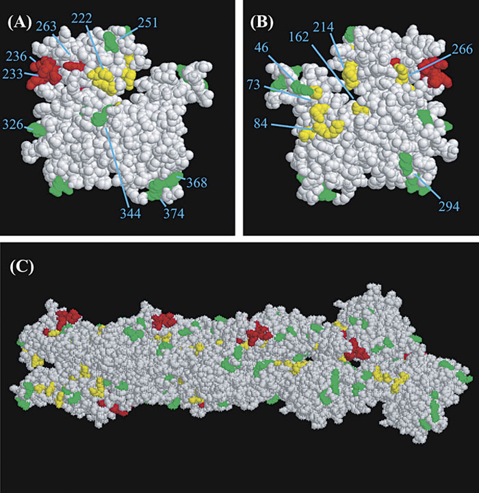 View Details | Fig. 5. Positions of the functional residues on the monomer and polymer forms of Act1p. (A) The three-dimensional structure of the yeast Act1p structure is presented with RasMol molecular visualization software (Sayle and Milner-White, 1995). This view is designated as the “front view” of Act1p. The alanine mutations associated with the different effects are indicated according to the following color scheme: red, cell wall integrity checkpoint defective (over 50% cells formed bipolar spindle at 180 min); green, nuclear migration defective (over 20% cells have two nuclei in mother cell); and yellow, both cell wall integrity checkpoint and nuclear migration defective. Alleles are represented by number. (B) Same as A, except back view. (C) Ultrastructure of the rabbit muscle actin 8-mer (Lorenz et al., 1993). The colored residues correspond to the equivalent residues in yeast Act1p. Color scheme is the same as A and B. |
Arp1p forms an actin-like short filament composed of nine or ten Arp1p monomers (Bingham and Schroer, 1999; Schafer et al., 1994). Therefore, the ultrastructure of the rabbit muscle actin 8-mer was used for predicting the positions of the mutations on the filamentous form of Arp1p (Fig. 5C) (Lorenz et al., 1993). Most of the amino acids colored red and green are located on the surface of the Arp1p polymer. Thus, mutations in these amino acids are unlikely to inhibit the formation of the Arp1p filament. On the other hand, the amino acids colored yellow tend to lie on the hidden surface of the filament. This supports the possibility that mutations of these residues may cause the destruction of the Arp1p filament, thereby abolishing both the cell wall integrity checkpoint and dynein-mediated nuclear migration functions in these cells. Alternatively, these mutated Arp1p molecules may be rapidly degraded, producing the same phenotype as that of the arp1 deletion.
Recently, Clark and Rose (Clark and Rose, 2005) reported that an arp1 mutant with the same mutations as our arp1-214 mutant (E214A, R215A, E216A) has a defect in the polymerization of the Arp1p filament. In our study, we showed that the arp1-214 mutant is defective in both cell wall integrity checkpoint and nuclear migration. These data suggest that the precise formation of the Arp1p filament is essential not only for nuclear migration but also for the cell wall integrity checkpoint. Clark and Rose (Clark and Rose, 2005) proposed that residues K369, D371, E374, and D375 play an important role in the interaction with Jnm1p. We showed that the arp1-368 (K368A, K369A, D371A) and arp1-374 (E374A, D375, R378A) mutants were defective only in nuclear migration, suggesting that the interaction of Arp1p with Jnm1p is not critical for its checkpoint function.
In summary, the deletion of arp1 causes defects in both cell wall integrity checkpoint and nuclear migration functions, whereas the wac1 mutation causes a defect only in the cell wall integrity checkpoint. One possible explanation for the phenotypic difference between wac1 and arp1 deletion mutants is that the cell wall integrity checkpoint and nuclear migration are based on distinct molecular functions of Arp1p, and that these two functions are separable in the Arp1p molecule. These results, together with our previous results showing that dynein-related proteins, except dynactin subunits, are not required for the cell wall integrity checkpoint, indicate that the molecular function of the cell wall integrity checkpoint may be independent of the dynein-based nuclear migration mechanism.
This work was supported by a grant (#16026205) for Scientific Research from the Ministry of Education, Science, Sports, and Culture of Japan.
|