Japanese Journal of Medical Technology
Online ISSN : 2188-5346
Print ISSN : 0915-8669
ISSN-L : 0915-8669
Part 3
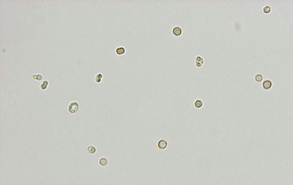
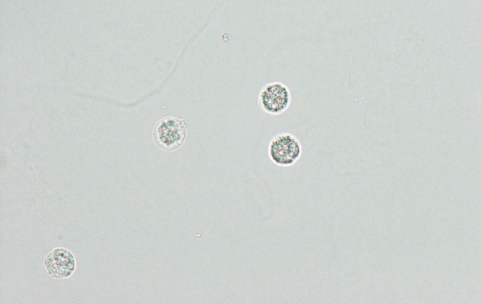
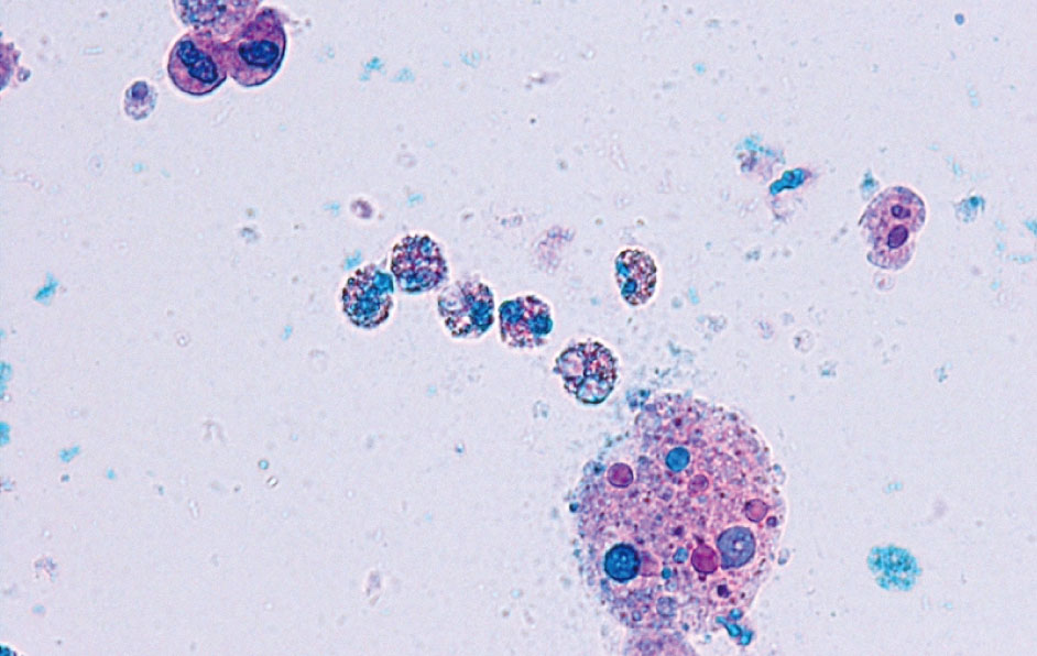
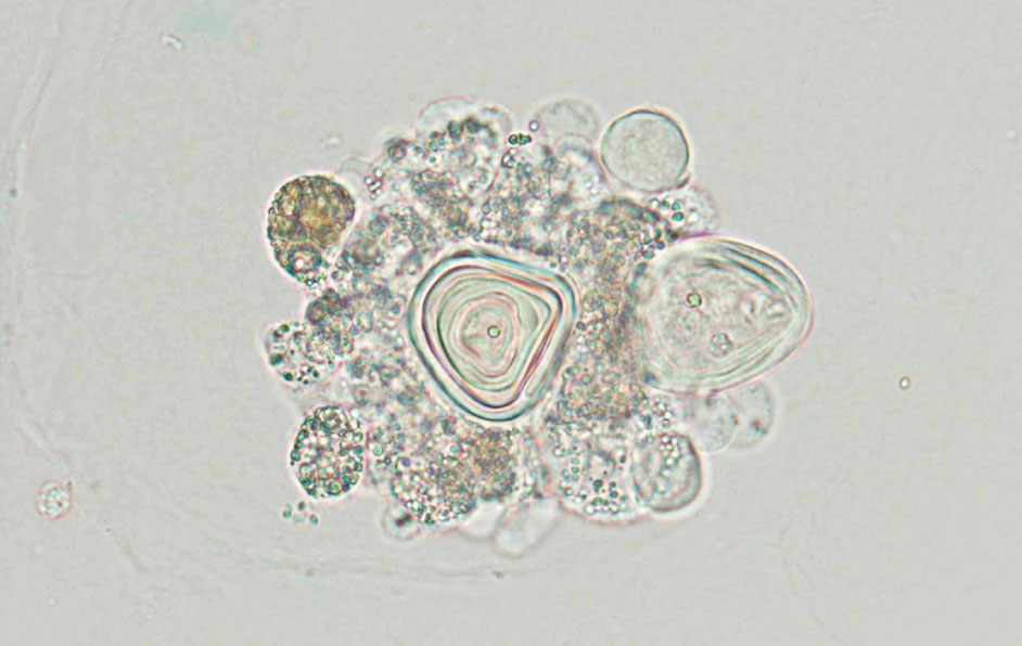
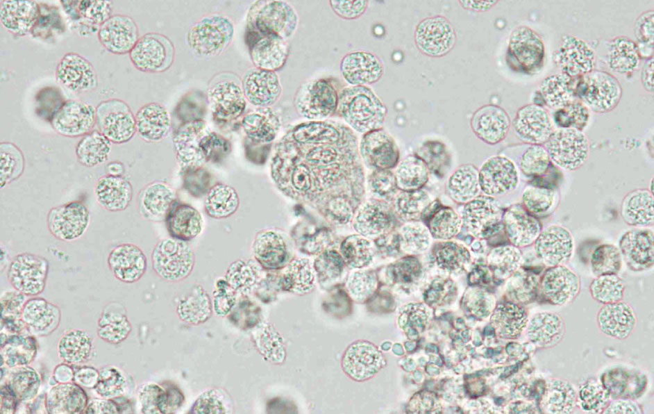
Atlas of Urinary Sediment: I Non-Epithelial Cells: Blood Cells, etc.
Japanese Association of Medical Technologists; Editorial Committee of the Special Issue: Urinary Sediment
Author information
JOURNAL FREE ACCESS FULL-TEXT HTML

2017 Volume 66 Issue J-STAGE-1 Pages 86-94

Details
Abstract

尿沈渣検査は,尿中に出現する成分を尿の遠心操作にて得られた沈殿物を観察する検査である。尿沈渣の標本作成における操作が単純であるにもかかわらず,尿沈渣に出現する成分は多種多様であるため,鑑別が非常に複雑である。その要因としては,尿沈渣に出現する尿中有形成分が,ひとつの成分においても様々な形態で存在することがある。たとえばシュウ酸カルシウム結晶では正八面体型とビスケット型,コマ型などが存在し,尿細管上皮細胞に至っては基本型,特殊型と細胞形態のバリエーションが多岐にわたる。このように尿沈渣検査では成分を正しく鑑別するための知識と技術が必要である。この部では,尿沈渣に出現する基本的な尿中有形成分を鑑別する知識を習得するために,最も基本となる成分の写真について「尿沈渣検査法2010」の尿沈渣アトラスを引用(一部改編)し掲載する。また「*」でマークした写真は,尿沈渣成分の新たな情報として追加したものである.この尿沈渣アトラスを利用し,各成分の特徴を捉えることをしっかりと身につけ,今後遭遇するであろう鑑別困難な成分に対しても対処できるよう,基礎知識を学習することを目的とする。

I  非上皮細胞類 ―血球類,その他―   Non-Epithelial Cells: Blood Cells, etc.
Figure 3.1 

非糸球体型赤血球 40× 無染色

Non–glomerular-type red blood cells 40× No staining

中央がくぼんだ典型的な円盤状赤血球である。

Typical discocytes with a dimple in the center.

Figure 3.2 

非糸球体型赤血球 40× 無染色

Non–glomerular-type red blood cells  40× No staining

金平糖状を示す萎縮・円盤状赤血球である。

Atrophic discocyte exhibiting a confetti-like shape.

Figure 3.3 

非糸球体型赤血球 40× 無染色

Non–glomerular-type red blood cells 40× No staining

pHや浸透圧の影響を受け膨化状や脱ヘモグロビン状を示す膨化・円盤状赤血球である。

Swollen and discotic red blood cells showing expansion and a dehemoglobinized state under the influence of pH and osmotic pressure.

Figure 3.4 

非糸球体型赤血球 40× 無染色

Non–glomerular-type red blood cells 40× No staining

コブを有しヘモグロビン色素を豊富に含んだコブ・球状赤血球である。これらは典型的な非糸球体型赤血球である。

Humped spherocytes that have humps and contain an abundance of the hemoglobin pigment. These are typical non-glomerular red blood cells.

Figure 3.5 

非糸球体型赤血球 40× 無染色

Non–glomerular-type red blood cells  40× No staining

典型的な円盤状赤血球と球状赤血球がみられる。中央にはコブ・球状赤血球が確認できる(矢印)。

Typical discocytes and spherocytes. Humped spherocytes are confirmed in the center (arrow).

Figure 3.6 

非糸球体型赤血球 40× S染色

Non–glomerular-type red blood cells  40× S staining

Figure 3.5と同様の赤血球のS染色像である。

The red blood cells shown in Figure 3.5 with S staining.

Figure 3.7 

非糸球体型赤血球 40× 無染色

Non–glomerular-type red blood cells 40× No staining

コブ・球状赤血球のコブ部分がみられる。これらは赤血球の断片として判断し,計数しない。

Hump regions of humped spherocytes are shown. These are identified as fragments of red blood cells and are not counted.

Figure 3.8 

非糸球体型赤血球 40× 無染色

Non–glomerular-type red blood cells  40× No staining

やや萎縮状を示す赤血球である。高浸透圧尿または低pH 尿ではこのような像を示すことがある。

Red blood cells exhibiting a somewhat atrophic shape. Such an image may appear in hyperosmotic or low pH urine.

Figure 3.9 

非糸球体型赤血球 40× 無染色

Non–glomerular-type red blood cells 40× No staining

膜部顆粒成分凝集状脱ヘモグロビン赤血球である。前立腺生検後や多発性嚢(のう)胞腎の患者尿にみられることが多い。糸球体型赤血球との鑑別は重要である。

Dehemoglobinized red blood cells with granular components aggregated in the membrane area are shown. These are often found in urine after a prostate biopsy or from patients with a polycystic kidney. Differentiation from the glomerular-type red blood cells is important.

Figure 3.10 

糸球体型赤血球 40× 無染色

Glomerular-type red blood cells 40× No staining

糸球体型赤血球のドーナツ不均一赤血球である。特にコブ・ドーナツ状不均一赤血球が多く確認できる。 コブのみは赤血球の断片として判断し,計数しない。

Doughnut-like dysmorphic red blood cells of glomerular-type. In particular, many humped doughnut-like dysmorphic red blood cells are confirmed. Humps only are identified as fragments of red blood cells and are not counted.

Figure 3.11 

糸球体型赤血球 40× 無染色

Glomerular-type red blood cells 40× No staining

大小不同をともなった多彩な形態を示す糸球体型赤血球が多数みられる。

Many glomerular-type red blood cells exhibiting various forms with large and small dissimilarities are shown.

Figure 3.12 

糸球体型赤血球 40× 無染色

Glomerular-type red blood cells 40× No staining

大小不同のドーナツ状不均一赤血球が多数みられる。

Many doughnut-like dysmorphic red blood cells with various sizes are shown.

Figure 3.13 

糸球体型赤血球 40× 無染色

Glomerular-type red blood cells 40× No staining

ドーナツ状不均一赤血球に大小不同が確認できる。

Doughnut-like dysmorphic red blood cells with various sizes can be identified.

Figure 3.14 

糸球体型赤血球 40× 無染色

Glomerular-type red blood cells 40× No staining

やや大型のドーナツ状不均一赤血球や標的・ドーナツ状不均一赤血球がみられる。

Slightly large doughnut-like dysmorphic red blood cells and codocyte/doughnut-like dysmorphic red blood cells.

Figure 3.15 

糸球体型赤血球 40× 無染色

Glomerular-type red blood cells 40× No staining

大型のドーナツ状不均一赤血球が多くみられる。

Many large doughnut-like dysmorphic red blood cells can be seen.

Figure 3.16 

糸球体型赤血球 40× 無染色

Glomerular-type red blood cells 40× No staining

小球性の赤血球の中には,コブ・ドーナツ状不均一赤血球がみられる。

Humped doughnut-like dysmorphic red blood cells are found in microcytic red cells.

Figure 3.17 

糸球体型赤血球 40× 無染色

Glomerular-type red blood cells 40× No staining

多彩性がなく大部分が2~4 μmの小球状を示している。赤血球の中には,詳細に観察すると不均一構造が確認できる。

There is no variety, and most cells are small and spherical in shape (2–4 μm). In the red blood cells, when observed in detail, a heterogeneous structure can be confirmed.

Figure 3.18 

糸球体型赤血球 40× 無染色

Glomerular-type red blood cells 40× No staining

左上に赤血球円柱がみられる。糸球体型赤血球の出現時には,同時に赤血球円柱を伴うことが多い。

A red blood cell cast is seen in the upper left. When glomerular-type red blood cells appear, they often simultaneously accompany red blood cell casts.

Figure 3.19 

白血球(好中球) 40× 無染色

White blood cells (neutrophils) 40× No staining

無染色での色調は灰白調から灰色調を呈し,なかには分葉核が確認できるものもある。

The color tones without staining are grayish white to gray, and some of the white blood cells show a segmented nucleus.

Figure 3.20 

白血球(好中球) 40× S染色

White blood cells (neutrophils) 40× S staining

S染色により輝細胞,淡染細胞,濃染細胞が確認できる。

Glitter cells, pale-stained cells, and dark-stained cells can be confirmed with S staining.

Figure 3.21 

白血球(好中球) 40× 無染色

White blood cells (neutrophils) 40× No staining

小突起を有する白血球像である。これは浸透圧の影響による生細胞の形態変化のひとつと考えられる。

A white blood cell image with small protrusions. This appears to be a morphological change of viable cells caused by osmotic pressure.

Figure 3.22 

白血球(好中球) 40× 無染色

White blood cells (neutrophils) 40× No staining

生細胞は,低浸透圧尿下ではアメーバ状の形態を呈する。

Viable cells exhibit an amoeboid form in hypoosmotic urine.

Figure 3.23 

白血球(好中球) 40× 無染色

White blood cells (neutrophils) 40× No staining

低浸透圧尿では細胞は膨化状を示し,核は明瞭となり単核様のものもみられる。

In urine with a low osmotic pressure, the cells become swollen and the nucleus becomes clear. Mononuclear-like cells are also visible.

Figure 3.24 

白血球(好中球) 40× 無染色

White blood cells (neutrophils) 40× No staining

好中球が粘液中で伸びた状態で崩壊したときにみられる。核が明瞭のため死細胞であることが示唆される。

This is seen when neutrophils become elongated and collapse in mucus. The nucleus is clearly shown, suggesting that these are dead cells.

Figure 3.25 

白血球(リンパ球) 40× 無染色

White blood cells (lymphocytes) 40× No staining

リンパ球は単核でN/C比が高く,核形は円形,類円形を呈する。好中球と比較して小型で,顆粒成分がほとんどみられない。

Lymphocytes are mononuclear and have a high N/C ratio, and the nucleus is circular or near-circular in shape. Compared with neutrophils, lymphocytes are smaller and contain less granule components.

Figure 3.26 

白血球(リンパ球) 40× S染色

White blood cells (lymphocytes) 40× S staining

Figure 3.25同様のリンパ球である。S染色では核は青色~青紫色を呈し,N/C比が高い。核小体がみられるものもある。細胞質は淡桃色を呈しわずかにみられる。

These are lymphocytes, similar as shown in Figure 3.25. With S staining, the nucleus exhibits blue or bluish purple color and the N/C ratio is high. Some show nuclear bodies. The cytoplasm appears pale pink, it is slightly observed.

Figure 3.27 

白血球(好酸球) 40× 無染色

White blood cells (eosinophils) 40× No staining

好酸球は光沢があり均一で黒色調の顆粒が細胞質内にみられる。

Eosinophils are shiny and homogeneous. Blackish granules are found in the cytoplasm.

Figure 3.28 

白血球(好酸球) 40× 無染色

White blood cells (eosinophils) 40× No staining

好酸球の核は2分葉を呈することが多く,丸みを帯びている。細胞質は好中球同様にアメーバ状の変化も有する。

The nucleus of eosinophils is often bifurcated and rounded. The cytoplasm has amoeboid-like changes, as observed in neutrophils.

Figure 3.29 

白血球(好酸球) 40×  Hansel染色

White blood cells (eosinophils)  40× Hansel staining

Hansel染色により,好酸球の顆粒は赤色調に染色される。

By Hansel staining, the granules of eosinophils are stained red.

Figure 3.30 

白血球(好酸球) 40× S染色

White blood cells (eosinophils) 40× S staining

S染色では核は青色,細胞質は赤紫色に染色されるが,好酸性顆粒は染まらない。

By S staining, the nucleus is stained blue, the cytoplasm is stained reddish purple, but the eosinophilic granules remain unstained.

Figure 3.31 

白血球(単球) 40× 無染色

White blood cells (monocytes) 40× No staining

単球の色調は灰白色調を呈する。表面構造は細顆粒状で,辺縁構造は不明瞭である。

Monocytes exhibits a grayish white tone. The surface structure is fine granular, and the marginal structure is unclear.

Figure 3.32 

白血球(単球) 40× 無染色

White blood cells (monocytes) 40× No staining

核形は馬蹄形やくびれ状を呈し,大きさは大小不同を認める。

The nuclei exhibit the shape of a horseshoe and are constricted in shape as well as irregular in size.

Figure 3.33 

白血球(単球) 40× 無染色

White blood cells (monocytes) 40× No staining

細胞質は透明感が強く,無染色でも核の観察が容易であることが多い。 また細胞境界が不明瞭な集塊状を形成することがある。

The cytoplasm is transparent and makes the observation of the nucleus often easy, even when unstained. Cell boundaries can be formed unclear conglomerates.

Figure 3.34 

白血球(単球) 40× S染色

White blood cells (monocytes) 40× S staining

核形は切れ込み状やくびれ状など種々の形状を示す。S染色での染色性は,青紫色または赤紫色調に染め出される。

The nuclear form exhibits various shapes, such as a cleaved shape and a constricted shape. By S staining, the nuclear is stained bluish purple or reddish purple.

Figure 3.35 

大食細胞 40× 無染色

Macrophages 40× No staining

たくさんの精子や大小の顆粒を貪食している。

Macrophages are shown to be phagocytizing many sperm and various sized granules.

Figure 3.36 

大食細胞 40× S染色

Macrophages 40× S staining

核は変性濃縮状を呈し,青色に染色されている。Figure 3.35同様に精子や顆粒を貪食している。

The nucleus is in a denatured concentrated state and stained blue. As shown in Figure 3.35, it is phagocytosing sperm and granules.

Figure 3.37 

大食細胞 40× 無染色

Macrophages 40× No staining

類でんぷん小体を取り囲むように出現している像である。性腺由来の成分を取り囲んでいることから前立腺由来の大食細胞であることが推定できる。

This is an image of macrophages surrounding an amyloid body. They are presumed to be prostate-derived macrophage cells because they surround a gonadal component.

Figure 3.38 

大食細胞 40× S染色

Macrophages 40× S staining

Figure 3.37同様の類でんぷん小体を取り囲んでいる。 細胞質は淡く,不明瞭な辺縁構造を示している。

Macrophages surround an amyloid body as seen in Figure 3.37. The cytoplasm is pale in color and has unclear marginal structures.

Figure 3.39 

大食細胞 40× 無染色

Macrophages 40× No staining

細胞質の辺縁構造は不明瞭で多核を示している。

The marginal structures of the cytoplasm are unclear and multinuclear.

Figure 3.40 

大食細胞 40× S染色

Macrophage 40× S staining

大型の大食細胞のS染色像である。不明瞭な辺縁構造を示し,細胞質は淡く赤紫色に染まる。

This is an S-stained image of a large macrophage. It shows unclear marginal structures, and its cytoplasm is stained pale reddish purple.

Figure 3.41 

子宮内膜間質細胞 40× 無染色

Endometrial stromal cells 40× No staining

集塊の外側を形成する細胞は子宮内膜円柱上皮細胞であり,中央部の集塊は子宮内膜間質細胞である。子宮内膜間質細胞を取り巻くように子宮内膜円柱上皮細胞がみられる。

The cells that form the outside of the conglomerate are endometrial columnar epithelial cells, and the center of the conglomerate is endometrial stromal cells. Endometrial columnar epithelial cells surround endometrial stromal cells.

Figure 3.42 

子宮内膜間質細胞 40× S染色

Endometrial stromal cells 40× S staining

Figure 3.41と同一例。子宮内膜円柱上皮細胞と子宮内膜間質細胞は区別が困難なため,円柱上皮細胞(子宮内膜由来)などとして報告する。

The same example as Figure 3.41. Because it is difficult to distinguish endometrial columnar epithelial cells from endometrial stromal cells, they are reported as, for example, columnar epithelial cells (derived from the endometrium).

Figure 3.43 

中皮細胞 40× 無染色

Mesothelial cells 40× No staining

核は中心性で厚い細胞質を有し,細胞結合部分は直線的である。

They have a central nucleus, thick cytoplasm, and a linear cell-binding region.

Figure 3.44 

中皮細胞 40× S染色

Mesothelial cells 40× S staining

細胞質は厚く,2核を有している。

A thick cytoplasm and two nuclei.

Figure 3.45 

中皮細胞 40× Papanicolaou染色

Mesothelial cells 40× Papanicolaou staining

Papanicolaou染色像においても中皮細胞は厚い細胞質を有し,核は中心性~偏在性である。中央の3核の上皮は尿路上皮細胞である(矢印)。

Papanicolaou staining also shows that mesothelial cells have a thick cytoplasm. The nuclei are central or eccentrically located. The epithelia with three nuclei in the middle are urothelial cells (arrow).

Figure 3.46 

中皮細胞 20× Calretinin染色(酵素抗体法)

Mesothelial cells 20× Calretinin staining (enzyme antibody technique)

標本中に出現している細胞のうち,中皮細胞のマーカーであるCalretininに陽性を示している。

Among the cells appearing in the specimen, they are positive for Calretinin, a marker for mesothelial cells.

 
© 2017 Japanese Association of Medical Technologists
feedback
Top