第三部
尿沈渣アトラス:V 微生物類・寄生虫類
2017 年 66 巻 J-STAGE-1 号 p. 142-147
詳細
2017 年 66 巻 J-STAGE-1 号 p. 142-147
尿沈渣検査は,尿中に出現する成分を尿の遠心操作にて得られた沈殿物を観察する検査である。尿沈渣の標本作成における操作が単純であるにもかかわらず,尿沈渣に出現する成分は多種多様であるため,鑑別が非常に複雑である。その要因としては,尿沈渣に出現する尿中有形成分が,ひとつの成分においても様々な形態で存在することがある。たとえばシュウ酸カルシウム結晶では正八面体型とビスケット型,コマ型などが存在し,尿細管上皮細胞に至っては基本型,特殊型と細胞形態のバリエーションが多岐にわたる。このように尿沈渣検査では成分を正しく鑑別するための知識と技術が必要である。この部では,尿沈渣に出現する基本的な尿中有形成分を鑑別する知識を習得するために,最も基本となる成分の写真について「尿沈渣検査法2010」の尿沈渣アトラスを引用(一部改編)し掲載する。また「*」でマークした写真は,尿沈渣成分の新たな情報として追加したものである.この尿沈渣アトラスを利用し,各成分の特徴を捉えることをしっかりと身につけ,今後遭遇するであろう鑑別困難な成分に対しても対処できるよう,基礎知識を学習することを目的とする。

細菌 40× 無染色
Bacteria 40× No staining
尿路感染症では,多数の細菌と白血球がみられる。単純性尿路感染症ではEscherichia coliなどのグラム陰性桿菌が大部分であるが,複雑性尿路感染症ではPseudomonas aeruginosaや球菌もみられる。
In urinary tract infections, numerous bacteria and white blood cells are found. In simple urinary tract infections, gram-negative bacilli, such as Escherichia coli, are primarily found, whereas in complex urinary tract infections, Pseudomonas aeruginosa and cocci may also be present.
細菌 40× S染色
Bacteria 40× S staining
尿路に炎症がある場合,細菌と同時に多数の白血球(好中球)がみられる。
When there is inflammation in the urinary tract, numerous white blood cells (neutrophils) are simultaneously observed with bacteria.

細菌 40× S染色
Bacteria 40× S staining
写真の中央に白血球(好中球)と背景に青~黒色に染まった細菌が散在性にみられる。好中球は多形核が観察できる。
White blood cells (neutrophils) in the center and bacteria stained blue to black on the background are observed in a scattered manner. Polymorphonuclear neutrophils can also be observed.

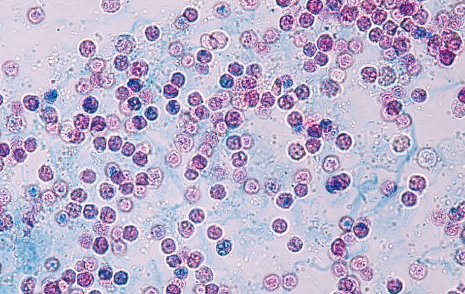
細菌 40× S染色
Bacteria 40× S staining
多数の細菌と白血球がみられる。尿路感染症では出血を伴うことが多く,背景に赤血球がみられる。
Numerous bacteria and leukocytes are found. Urinary tract infections often involve hemorrhaging; thus, red blood cells can be found in the background.

細菌 40× S染色
Bacteria 40× S staining
多数の細菌と数個の白血球(矢印)および赤血球がみられる。図のように桿菌の場合,一般に鑑別は容易でるが,球菌は大きさが小さく判定に戸惑うこともしばしばある。
A number of bacteria, several leukocytes (arrows), and red blood cells are observed. In the case of bacilli, as shown in the figure, it is generally easy to differentiate, whereas cocci are small in size and are often difficult to identify.

細菌 40× S染色
Bacteria 40× S staining
菌塊状にみられた細菌である。S染色に染まり,一見,尿細管上皮細胞に類似する。顕微鏡の微動を動かし菌塊の詳細を観察し鑑別を行う。
Bacteria found in a bacterial conglomerate. These are stained with S stain and resemble renal tubular epithelial cells in appearance. Adjust the micromotion of the microscope to observe the details of the bacterial conglomerate and perform differentiation.

細菌(球菌) 40× S染色
Bacteria (cocci) 40× S staining
多数の細菌(球菌)がみられる。尿中にみられる細菌はグラム陰性桿菌が多いが,StaphylococcusやEnterococcusなどの球菌がみられる場合も多い。
A number of bacteria (cocci) are visible. In urine, gram-negative bacilli are often found, but cocci such as Staphylococcus and Enterococcus may also be seen.

細菌(球菌) 40× S染色
Bacteria (cocci) 40× S staining
球菌はサイズが小さく判定に戸惑うことが多い。とくに無晶性塩類との鑑別が困難な場合があるが,球菌は形状,大きさが整っていることが多い。
Because the cocci are small, they are difficult to differentiate. Although it is particularly difficult to differentiate cocci from amorphous salts, cocci typically have a consistent shape and size.

細菌 40× S染色
Bacteria 40× S staining
右の扁平上皮細胞に多数の細菌がみられる。大部分は女性から検出され,膣内に常在する乳酸桿菌が扁平上皮細胞の表面を被ったものである。
Numerous bacteria are found on the squamous epithelial cells on the right. Most cases are identified in females, and this is a lactobacillus resident in the vagina covering the surface of squamous epithelial cells.

細菌 40× S染色
Bacteria 40× S staining
菌体がフィラメント状に細長く伸びた細菌(変形細菌)である。細胞壁合成阻害剤(β‐ラクタム系抗菌薬)使用例で,しばしば観察される。
Bacteria (deformed bacteria) in which the bacterial cells are elongated in a filament form. This is frequently observed in cases in which inhibitors of cell wall synthesis (β-lactam antibiotics) are used.

細菌 40× S染色
Bacteria 40× S staining
フィラメント状に細長く伸びた細菌(変形細菌)である。抗菌薬により細菌の細胞壁合成阻害が生じると形態保持,隔壁合成などが阻害され菌体に変化を生じる。
Bacteria elongated in a filament form (deformed bacteria). Inhibition of bacterial cell wall synthesis by antibacterial drugs inhibits morphological retention and partition wall synthesis, resulting in a change in the bacterial cells.

細菌 40× S染色
Bacteria 40× S staining
フィラメント状に細長く伸びた細菌(変形細菌)である。菌塊状にみられる。
These bacteria elongated in a filament form (deformed bacteria) and constitute bacterial conglomerates.

細菌 40× 無染色
Bacteria 40× No staining
菌体がフィラメント状に細長く伸び,一部がコブ状に膨らんでいる細菌(変形細菌)である。この形状を一般にスフェロプラスト型という。
A bacterium in which the bacterial cell elongates in a filament form and partly swells into a hump shape (deformed bacterium). This shape is generally referred to as a spheroplast.

細菌 40× S染色
Bacteria 40× S staining
スフェロプラスト型の変形細菌で,グラム陰性菌が抗菌薬により細胞壁の一部が失われることにより菌体に変形を生じる。
A deformed bacterium of the spheroplast type. Bacterial cells of gram-negative bacteria become deformed due to the loss of a partial cell wall by antibacterial agents.
細菌 40× S染色
Bacteria 40× S staining
スフェロプラスト型の変形細菌でβ‐ラクタム系抗菌薬のなかでカルバペネム系抗菌薬使用時にしばしば観察される。白血球に類似するが大小不同が著しい。
Spheroplast-type deformed bacteria are frequently observed when using carbapenem antibiotics, one of the β-lactam antibacterial agents. These deformed bacteria are similar to leukocytes, but appear in significantly irregular sizes.

真菌 40× 無染色
Fungi 40× No staining
真菌は,灰白色の楕円形で大小不同がある。
Fungi are light gray, oval shape, and appear in irregular sizes.

真菌 40× 無染色
Fungi 40× No staining
菌糸を長く伸ばした状態の真菌である。
Fungi with long stretched mycelial threads.

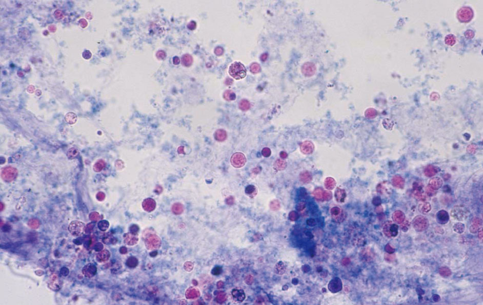
真菌 40× S染色
Fungi 40× S staining
矢印の白血球は真菌を貪食しており,細胞質内に多数の真菌がみられる。
Leukocytes identified by the arrows phagocytose fungi, and numerous fungi are found in the cytoplasm.

白血球の裸核 40× S染色
Naked nuclei of white blood cells 40× S staining
白血球の膜が壊れ,核が残存している状態(裸核)である(矢印)。女性で外陰部などからの混入時などでしばしば観察される。大部分は好中球分葉核球を認める。
The leukocyte membrane is broken, but the nuclei remain (naked nucleus) (arrows). These are frequently observed during contamination by white blood cells from the vulva in female patients. The majority exhibit neutrophilic segmented nuclei.

フサリウム 40× S染色
Fusarium 40× S staining
フサリウムは土壌真菌で田畑の土壌から分離され,本来は植物の病原菌である。大部分は外部からの混入であるが,感染防御能の低下した患者にもみられる。
Fusarium is a soil fungus. It is isolated from the soil of fields and is originally a pathogenic bacterium of plants. It usually appears due to external contamination but may also be seen in immunocompromised patients.

真菌 40× 無染色
Fungi 40× No staining
抗真菌薬投与中の患者尿で,真菌薬の影響により菌体が大きく変形している。図はポリエン系抗菌薬(アムホテリシンB)服用患者例で観察された。
In the urine of a patient who is receiving antifungal medicine, the bacterial bodies were greatly deformed due to the influence of the fungal drug. These were observed in patients taking polyene antibiotics (amphotericin B).

真菌 40× S染色
Fungi 40× S staining
Figure 3.341のS染色像である。低比重尿でしばしば観察される膨化した赤血球と類似する場合がある。潜血反応や酢酸添加による溶血の有無などで鑑別する。
S staining image of Figure 3.341. It may be similar to the swollen red blood cells often observed in low-density urine. The occult blood reaction and the presence of hemolysis by adding acetic acid are used for differentiation.

膣トリコモナス 40× 無染色
Trichomonas vaginalis 40× No staining
新鮮尿では活発に鞭毛や波動膜を動かして観察される。採尿後,経過時間が長いと(尿温の低下など)活動を停止し,白血球との鑑別が困難な場合がある。
In fresh urine, Trichomonas vaginalis is observed actively moving flagella and undulating membranes. When a long time passes after taking a urine sample (i.e., decrease in urine temperature), the bacteria cease activity. Thus, it can become difficult to differentiate these cells from white blood cells.

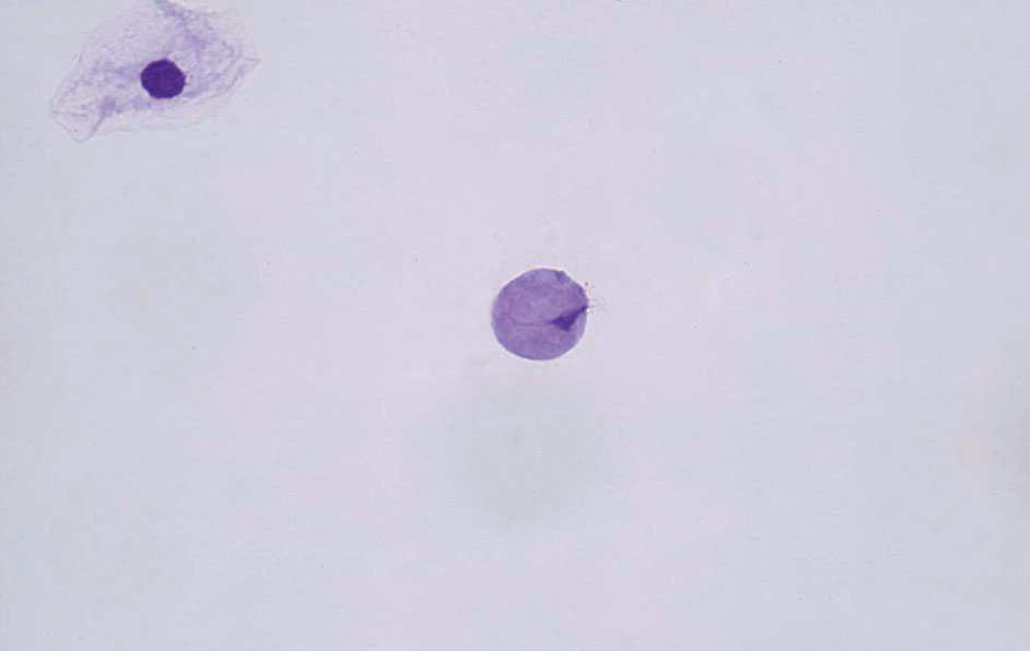
膣トリコモナス 40× S染色
Trichomonas vaginalis 40× S staining
矢印が膣トリコモナスで活動を停止すると白血球との鑑別が重要である。酢酸を添加すると白血球の核が明瞭に描出され鑑別が容易となる。
When Trichomonas vaginalis (arrows) stop their activity, differentiation from leukocytes become important. Adding acetic acid, the nuclei of leukocytes are clearly depicted, it makes differentiation easy.
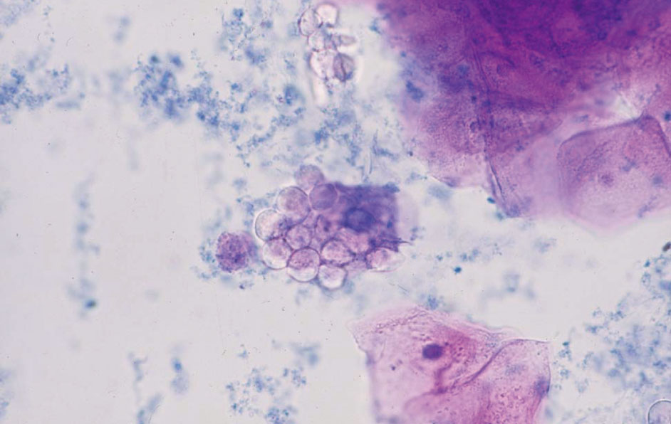
膣トリコモナス 40× S染色
Trichomonas vaginalis 40× S staining
扁平上皮細胞の表面(ブドウの房状)に寄生している膣トリコモナスで,扁平上皮細胞のグリコーゲンを栄養源としている。
Trichomonas vaginalis are parasitic to the surface of squamous epithelial cells (grape cluster-shaped). Their nutrient source is the glycogen of squamous cells.
膣トリコモナス 40× MG染色
Trichomonas vaginalis 40× MG staining
白血球との鑑別が困難な場合,塗抹標本を作成後,MG染色などを施すと白血球との鑑別が容易である。膣トリコモナスは細長い核と鞭毛が観察される。
When it is difficult to distinguish from white blood cells, MG staining for smear sample makes differentiation easier. Trichomonas vaginalis has an elongated nucleus and flagella.

ビルハルツ住血吸虫卵 40× 無染色
Schistosoma haematobium egg 40× No staining
ビルハルツ住血吸虫卵は,長径110~170 μm,短径40~70 μmで尾端に大きな棘を有する虫卵である。血尿を伴うことが多い。
Schistosoma haematobium egg has a size of major axis: 110–170 μm, minor axis: 40–70 μm, and has a large spine at the tail end. It is often accompanied by hematuria.

ビルハルツ住血吸虫(ミラシジウム) 40× 無染色
Schistosoma haematobium (miracidium) 40× No staining
ビルハルツ住血吸虫に感染した患者尿中には,虫卵のほか卵から生まれた幼虫(有毛幼虫;ミラシジウム)も同時に観察されることがある。
In the urine of patients infected with the causative agent of Schistosoma haematobium, larvae (hairy larvae; miracidium) born from eggs as well as the eggs may be simultaneously observed.

蟯虫卵 40× 無染色
Enterobius vermicularis egg 40× No staining
蟯虫卵は,尿路に産卵されるものではなく,肛門周囲に産卵された虫卵が,採尿の際に尿中へ混入したものである。色調は透明で柿の種状の形状を示す。
Enterobius vermicularis eggs are not laid in the urinary tract. Rather, eggs laid around the anus are mixed into the urine during urine collection. The color tone is transparent, and the eggs are shaped like persimmon seeds.

糞線虫(フィラリア型幼虫) 20× 無染色
Strongyloides stercoralis (filariform larva)
20× No staining
糞線虫は,本来は小腸の粘膜内に寄生するが,免疫力が低下している患者では尿中から糞線虫がみられる場合がある。
Although Strongyloides stercoralis normally infest the mucosa of the small intestine, they may be detected in the urine of patients with a weakened immune system.