抄録
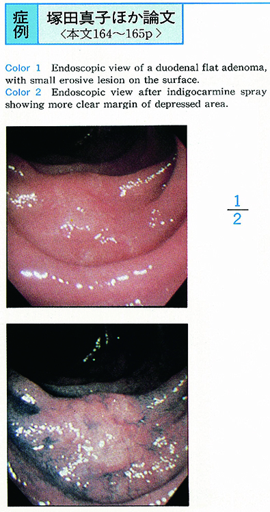
A 60-year-old man was admitted to our hospital because of a flat lesion at the anal side of supra duodenal angle. This lesion was pointed out by screening upper GI endoscopy. The follow-up examination showed the adenoma with severe atypia.
Tumor was 15×10mm in size with eroded surface. After spraying indigocalmin, this lesion was clearly identified. Endoscopic ultrasonograpy revealed the adenoma was located within duodenal mucosa.
The endoscopic mucosal resection (EMR) was successfully done, the resected specimen was 18×15×3mm in size and histological examination revealed 7×3mm flat type of adenoma. The over expression of p53 by using immunohistochemistry was not observed in the tissue.
Reported case of flat adenoma was relatively rare. There was 6 cases in Japanese literature (during 1956-1993 period) .