Biophysics and Physicobiology
Online ISSN : 2189-4779
ISSN-L : 2189-4779
Volume 20, Issue 1
Displaying 1-14 of 14 articles from this issue
Database and Computer Program
  • Kento Yonezawa, Masatsuyo Takahashi, Keiko Yatabe, Yasuko Nagatani, No ...
    Article type: Database and Computer Program
    2023 Volume 20 Issue 1 Article ID: e200001
    Published: 2023
    Released on J-STAGE: February 04, 2023
    Advance online publication: January 07, 2023
    JOURNAL OPEN ACCESS FULL-TEXT HTML
    Supplementary material

    Recent small-angle X-ray scattering (SAXS) for biological macromolecules (BioSAXS) is generally combined with size-exclusion chromatography (SEC-SAXS) at synchrotron facilities worldwide. For SEC-SAXS analysis, the final scattering profile for the target molecule is calculated from a large volume of continuously collected data. It would be ideal to automate this process; however, several complex problems exist regarding data measurement and analysis that have prevented automation. Here, we developed the analytical software MOLASS (Matrix Optimization with Low-rank factorization for Automated analysis of SEC-SAXS) to automatically calculate the final scattering profiles for solution structure analysis of target molecules. In this paper, the strategies for automatic analysis of SEC-SAXS data are described, including correction of baseline-drift using a low percentile method, optimization of peak decompositions composed of multiple scattering components using modified Gaussian fitting against the chromatogram, and rank determination for extrapolation to infinite dilution. In order to easily calculate each scattering component, the Moore-Penrose pseudo-inverse matrix is adopted as a basic calculation. Furthermore, this analysis method, in combination with UV–visible spectroscopy, led to better results in terms of accuracy in peak decomposition. Therefore, MOLASS will be able to smoothly suggest to users an accurate scattering profile for the subsequent structural analysis.

Commentary and Perspective (Invited)
Regular Article
  • Kanta Fujimoto, Kosuke Inada, Kotaro Oka, Etsuro Ito
    Article type: Regular Article
    2023 Volume 20 Issue 1 Article ID: e200003
    Published: 2023
    Released on J-STAGE: February 04, 2023
    Advance online publication: January 14, 2023
    JOURNAL OPEN ACCESS FULL-TEXT HTML

    Some evidence suggests that oxytocin, which is a neuropeptide conventionally thought to be synthesized in the hypothalamus and released by the posterior pituitary, is generated in peripheral keratinocytes, but the details are lacking and the mRNA analysis is further required. Oxytocin and neurophysin I are generated together as cleavage products after splitting the precursor molecule, preprooxyphysin. To confirm that oxytocin and neurophysin I are also generated in the peripheral keratinocytes, it must first be clarified that these molecules contained in peripheral keratinocytes did not originate in the posterior pituitary gland and then the expression of oxytocin and neurophysin I mRNAs must be established in keratinocytes. Therefore, we attempted to quantify preprooxyphysin mRNA in keratinocytes using various primers. Using real-time PCR, we observed that the mRNAs of both oxytocin and neurophysin I were located in keratinocytes. However, the mRNA amounts of oxytocin, neurophysin I, and preprooxyphysin were too small to confirm their co-existence in keratinocytes. Thus, we had to further determine whether the PCR-amplified sequence was identical to preprooxyphysin. The PCR products analyzed by DNA sequencing were identical to preprooxyphysin, finally determining the co-existence of both oxytocin and neurophysin I mRNAs in keratinocytes. In addition, the immunocytochemical experiments showed that oxytocin and neurophysin I proteins were located in keratinocytes. These results of the present study provided further support indicating that oxytocin and neurophysin I are generated in peripheral keratinocytes.

Review Article (Invited)
  • Akiko Yamada, Akira Watanabe, Takenori Yamamoto
    Article type: Review Article (Invited)
    2023 Volume 20 Issue 1 Article ID: e200004
    Published: 2023
    Released on J-STAGE: February 04, 2023
    Advance online publication: January 19, 2023
    JOURNAL OPEN ACCESS FULL-TEXT HTML

    Mitochondria play an important role in energy conversion as well as in intracellular calcium (Ca2+) storage. Ca2+ uptake from the cytosol to the mitochondria is mediated by the calcium uniporter, which functions as a Ca2+ ion channel. However, the molecular composition of this uniporter has remained unclear until recently. The Ca2+ ion channel consists of seven subunits. The yeast reconstitution technique revealed that the mitochondrial calcium uniporter (MCU) and essential MCU regulatory element (EMRE) are the core subunits of the complex. Furthermore, detailed structure-function analyses of the core subunits (MCU and EMRE) were performed. In this review, the regulatory mechanism of mitochondrial Ca2+ uptake is discussed.

Regular Article
  • Shintaroh Kubo, Kenichi Umeda, Noriyuki Kodera, Shoji Takada
    Article type: Regular Article
    2023 Volume 20 Issue 1 Article ID: e200006
    Published: 2023
    Released on J-STAGE: February 14, 2023
    Advance online publication: February 02, 2023
    JOURNAL OPEN ACCESS FULL-TEXT HTML
    Supplementary material

    The high-speed atomic force microscopy (HS-AFM) is a unique and prominent method to observe structural dynamics of biomolecules at single molecule level at near-physiological condition. To achieve high temporal resolution, the probe tip scans the stage at high speed which can cause the so-called parachuting artifact in the HS-AFM images. Here, we develop a computational method to detect and remove the parachuting artifact in HS-AFM images using the two-way scanning data. To merge the two-way scanning images, we employed a method to infer the piezo hysteresis effect and to align the forward- and backward-scanning images. We then tested our method for HS-AFM videos of actin filaments, molecular chaperone, and duplex DNA. Together, our method can remove the parachuting artifact from the raw HS-AFM video containing two-way scanning data and make the processed video free from the parachuting artifact. The method is general and fast so that it can easily be applied to any HS-AFM videos with two-way scanning data.

Review Article (Invited)
  • Hidekazu Hiroaki
    Article type: Review Article (Invited)
    2023 Volume 20 Issue 1 Article ID: e200007
    Published: 2023
    Released on J-STAGE: March 04, 2023
    Advance online publication: February 03, 2023
    JOURNAL OPEN ACCESS FULL-TEXT HTML

    To completely treat and ultimately prevent dementia, it is essential to elucidate its pathogenic mechanisms in detail. There are two major hypotheses for the pathogenesis of Alzheimer’s dementia: the β-amyloid (Aβ) hypothesis and the tau hypothesis. The modified amyloid hypothesis, which proposes that toxic oligomers rather than amyloid fibrils are the essential cause, has recently emerged. Aβ peptides [Aβ(1–40) and Aβ(1–42)] form highly insoluble aggregates in vivo and in vitro. These Aβ aggregates contain many polymorphisms, whereas Aβ peptides are intrinsically disordered in physiological aqueous solutions without any compact conformers. Over the last three decades, solid-state nuclear magnetic resonance (NMR) has greatly contributed to elucidating the structure of each polymorph, while solution NMR has revealed the dynamic nature of the transient conformations of the monomer. Moreover, several methods to investigate the aggregation process based on the observation of magnetization saturation transfer have also been developed. The complementary use of NMR methods with cryo-electron microscopy, which has rapidly matured, is expected to clarify the relationship between the amyloid and molecular pathology of Alzheimer’s dementia in the near future. This review article is an extended version of the Japanese article, Insights into the Mechanisms of Oligomerization/Fibrilization of Amyloid β Peptide from Nuclear Magnetic Resonance, published in SEIBUTSU BUTSURI Vol. 62, p. 39–42 (2022).

  • Toshiki Yagi, Akiyuki Toda, Muneyoshi Ichikawa, Genji Kurisu
    Article type: Review Article (Invited)
    2023 Volume 20 Issue 1 Article ID: e200008
    Published: 2023
    Released on J-STAGE: March 04, 2023
    Advance online publication: February 08, 2023
    JOURNAL OPEN ACCESS FULL-TEXT HTML
    Supplementary material

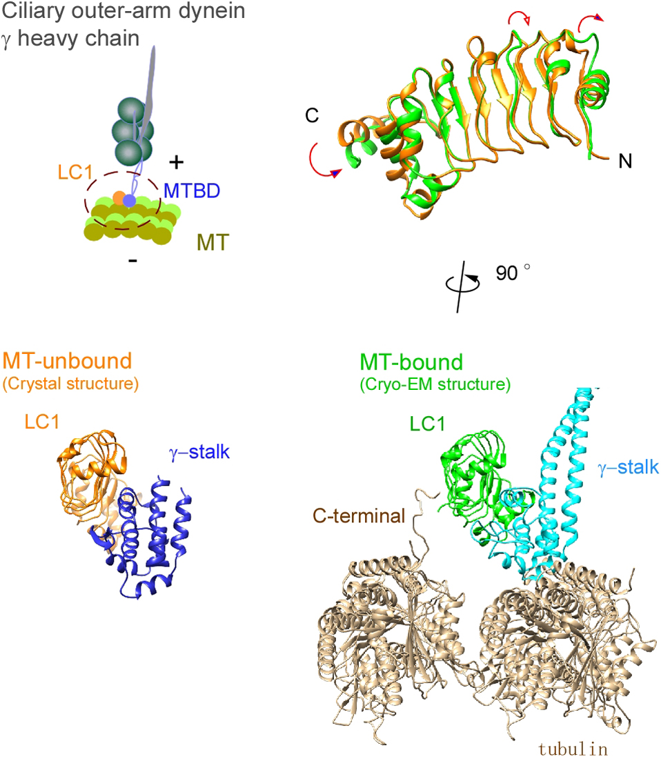
    Ciliary bending movements are powered by motor protein axonemal dyneins. They are largely classified into two groups, inner-arm dynein and outer-arm dynein. Outer-arm dynein, which is important for the elevation of ciliary beat frequency, has three heavy chains (α, β, and γ), two intermediate chains, and more than 10 light chains in green algae, Chlamydomonas. Most of intermediate chains and light chains bind to the tail regions of heavy chains. In contrast, the light chain LC1 was found to bind to the ATP-dependent microtubule-binding domain of outer-arm dynein γ-heavy chain. Interestingly, LC1 was also found to interact with microtubules directly, but it reduces the affinity of the microtubule-binding domain of γ-heavy chain for microtubules, suggesting the possibility that LC1 may control ciliary movement by regulating the affinity of outer-arm dyneins for microtubules. This hypothesis is supported by the LC1 mutant studies in Chlamydomonas and Planaria showing that ciliary movements in LC1 mutants were disordered with low coordination of beating and low beat frequency. To understand the molecular mechanism of the regulation of outer-arm dynein motor activity by LC1, X-ray crystallography and cryo-electron microscopy have been used to determine the structure of the light chain bound to the microtubule-binding domain of γ-heavy chain. In this review article, we show the recent progress of structural studies of LC1, and suggest the regulatory role of LC1 in the motor activity of outer-arm dyneins. This review article is an extended version of the Japanese article, The Complex of Outer-arm Dynein Light Chain-1 and the Microtubule-binding Domain of the Heavy Chain Shows How Axonemal Dynein Tunes Ciliary Beating, published in SEIBUTSU BUTSURI Vol. 61, p. 20–22 (2021).

  • Kohei Otomo, Hirokazu Ishii, Tomomi Nemoto
    Article type: Review Article (Invited)
    2023 Volume 20 Issue 1 Article ID: e200009
    Published: 2023
    Released on J-STAGE: February 22, 2023
    Advance online publication: February 08, 2023
    JOURNAL OPEN ACCESS FULL-TEXT HTML

    Two-photon excitation laser scanning microscopy (TPLSM) has provided many insights into the life sciences, especially for thick biological specimens, because of its superior penetration depth and less invasiveness owing to the near-infrared wavelength of its excitation laser light. This paper introduces our four kinds of studies to improve TPLSM by utilizing several optical technologies as follows:

    (1) A high numerical aperture objective lens significantly deteriorates the focal spot size in deeper regions of specimens. Thus, approaches to adaptive optics were proposed to compensate for optical aberrations for deeper and sharper intravital brain imaging.

    (2) TPLSM spatial resolution has been improved by applying super-resolution microscopic techniques. We also developed a compact stimulated emission depletion (STED) TPLSM that utilizes electrically controllable components, transmissive liquid crystal devices, and laser diode-based light sources. The spatial resolution of the developed system was five times higher than conventional TPLSM.

    (3) Most TPLSM systems adopt moving mirrors for single-point laser beam scanning, resulting in the temporal resolution caused by the limited physical speed of these mirrors. For high-speed TPLSM imaging, a confocal spinning-disk scanner and newly-developed high-peak-power laser light sources enabled approximately 200 foci scanning.

    (4) Several researchers have proposed various volumetric imaging technologies. However, most technologies require large‐scale and complicated optical setups based on deep expertise for microscopic technologies, resulting in a high threshold for biologists. Recently, an easy‐to‐use light‐needle-creating device was proposed for conventional TPLSM systems to achieve one-touch volumetric imaging.

Commentary and Perspective
Review Article (Invited)
  • Takayuki Umakoshi
    Article type: Review Article (Invited)
    2023 Volume 20 Issue 1 Article ID: e200011
    Published: 2023
    Released on J-STAGE: March 04, 2023
    Advance online publication: February 14, 2023
    JOURNAL OPEN ACCESS FULL-TEXT HTML

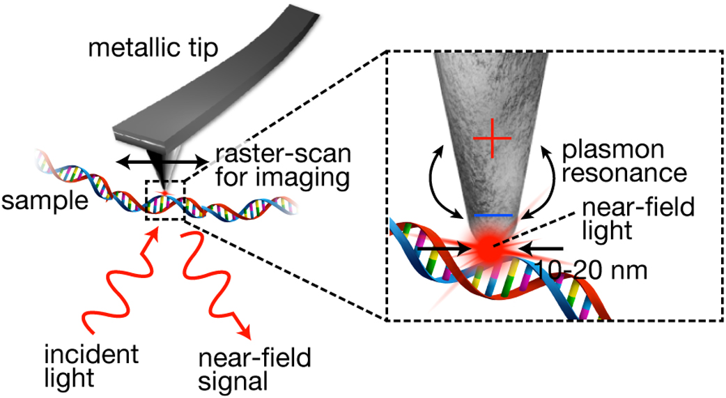
    Near-field scanning optical microscopy (NSOM) is a super-resolution optical microscopy based on nanometrically small near-field light at a metallic tip. It can be combined with various types of optical measurement techniques, including Raman spectroscopy, infrared absorption spectroscopy, and photoluminescence measurements, which provides unique analytical capabilities to a variety of scientific fields. In particular, to understand nanoscale details of advance materials and physical phenomena, NSOM has been often adopted in the fields of material science and physical chemistry. However, owing to the recent critical developments showing the great potential for biological studies, NSOM has also recently gained much attention in the biological field. In this article, we introduce recent developments made in NSOM, aiming at biological applications. The drastic improvement in the imaging speed has shown a promising application of NSOM for super-resolution optical observation of biological dynamics. Furthermore, stable imaging and broadband imaging were made possible owing to the advanced technologies, which provide a unique imaging method to the biological field. As NSOM has not been well exploited in biological studies to date, several rooms need to be explored to determine its distinct advantages. We discuss the possibility and perspective of NSOM for biological applications. This review article is an extended version of the Japanese article, Development of Near-field Scanning Optical Microscopy toward Its Application for Biological Studies, published in SEIBUTSU BUTSURI Vol. 62, p. 128–130 (2022).

  • Tony Z. Jia, Kuhan Chandru
    Article type: Review Article (Invited)
    2023 Volume 20 Issue 1 Article ID: e200012
    Published: 2023
    Released on J-STAGE: March 04, 2023
    Advance online publication: February 22, 2023
    JOURNAL OPEN ACCESS FULL-TEXT HTML

    While it is often believed that the origins of life required participation of early biomolecules, it has been recently proposed that “non-biomolecules”, which would have been just as, if not more, abundant on early Earth, could have played a part. In particular, recent research has highlighted the various ways by which polyesters, which do not participate in modern biology, could have played a major role during the origins of life. Polyesters could have been synthesized readily on early Earth through simple dehydration reactions at mild temperatures involving abundant “non-biological” alpha hydroxy acid (AHA) monomers. This dehydration synthesis process results in a polyester gel, which upon further rehydration, can assemble into membraneless droplets proposed to be protocell models. These proposed protocells can provide functions to a primitive chemical system, such as analyte segregation or protection, which could have further led to chemical evolution from prebiotic chemistry to nascent biochemistry. Here, to further shed light into the importance of “non-biomolecular” polyesters at the origins of life and to highlight future directions of study, we review recent studies which focus on primitive synthesis of polyesters from AHAs and assembly of these polyesters into membraneless droplets. Specifically, most of the recent progress in this field in the last five years has been led by laboratories in Japan, and these will be especially highlighted. This article is based on an invited presentation at the 60th Annual Meeting of the Biophysical Society of Japan held in September, 2022 as an 18th Early Career Awardee.

  • Keiichi Yamaguchi, Kichitaro Nakajima, Yuji Goto
    Article type: Review Article (Invited)
    2023 Volume 20 Issue 1 Article ID: e200013
    Published: 2023
    Released on J-STAGE: March 14, 2023
    Advance online publication: March 02, 2023
    JOURNAL OPEN ACCESS FULL-TEXT HTML

    Much effort has been devoted to elucidate mechanisms of amyloid fibril formation using various kinds of additives, such as salts, metals, detergents, and biopolymers. Here, we review the effects of additives with a focus on polyphosphate (polyP) on amyloid fibril formation of β2-microglobulin (β2m) and α-synuclein (αSyn). PolyP, consisting of up to 1,000 phosphoanhydride bond-linked phosphate monomers, is one of the most ancient, enigmatic, and negatively charged molecules in biology. Amyloid fibril formation of both β2m and αSyn could be accelerated by counter anion-binding and preferential hydration at relatively lower and higher concentrations of polyP, respectively, depending on the chain length of polyP. These bimodal concentration-dependent effects were also observed in salt- and heparin-induced amyloid fibril formation, indicating the generality of bimodal effects. We also address the effects of detergents, alcohols, and isoelectric point precipitation on amyloid fibril formation, in comparison with the effects of salts. Because polyP is present all around us, from cellular components to food additives, clarifying its effects and consequent biological roles will be important to further advance our understanding of amyloid fibrils. This review article is an extended version of the Japanese article, Linking Protein Folding to Amyloid Formation, published in SEIBUTSU BUTSURI Vol. 61, p. 358–365 (2021).

  • Yasuha Watanabe, Kazuharu Arakawa
    Article type: Review Article (Invited)
    2023 Volume 20 Issue 1 Article ID: e200014
    Published: 2023
    Released on J-STAGE: March 21, 2023
    Advance online publication: March 10, 2023
    JOURNAL OPEN ACCESS FULL-TEXT HTML

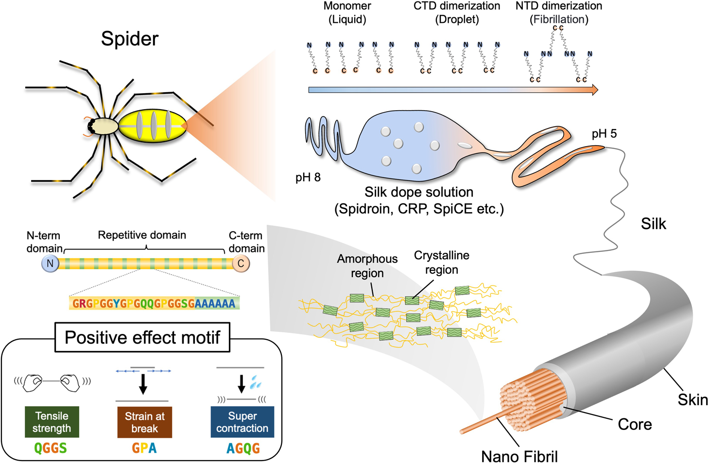
    Spider silk is considered a promising next-generation biomaterial due to its exceptional toughness, coupled with its renewability and biodegradability. Contrary to the conventional view that spider silk is mainly composed of two types of silk proteins (spidroins), MaSp1 and MaSp2, multi-omics strategies are increasingly revealing that the inclusion of complex components confers the higher mechanical properties to the material. In this review, we focus on several recent findings that report essential components and mechanisms that are necessary to reproduce the properties of natural spider silk. First, we discuss the discovery of MaSp3, a newly identified spidroin that is a major component in the composition of spider silk, in addition to the previously understood MaSp1 and MaSp2. Moreover, the role of the Spider-silk Constituting Element (SpiCE), which is present in trace amounts but has been found to significantly increase the tensile strength of artificial spider silk, is explored. We also delve into the process of spidroin fibril formation through liquid-liquid phase separation (LLPS) that forms the hierarchical structure of spider silk. In addition, we review the correlation between amino acid sequences and mechanical properties such as toughness and supercontraction, as revealed by an analysis of 1,000 spiders. In conclusion, these recent findings contribute to the comprehensive understanding of the mechanisms that give spider silk its high mechanical properties and help to improve artificial spider silk production.

  • Takashi Tominaga, Riichi Kajiwara, Yoko Tominaga
    Article type: Review Article (Invited)
    2023 Volume 20 Issue 1 Article ID: e200015
    Published: 2023
    Released on J-STAGE: March 21, 2023
    Advance online publication: March 11, 2023
    JOURNAL OPEN ACCESS FULL-TEXT HTML

    Plasticity is the key feature of our brain function. Specifically, plasticity of hippocampal synapses is critical for learning and memory. The functional properties of the neuronal circuit change as a result of synaptic plasticity. This review summarizes the use of voltage-sensitive dyes (VSDs) to examine neuronal circuit plasticity. We will discuss the significance of plastic changes in circuit function as well as the technical issue of using VSDs. Further, we will discuss the neural circuit level plasticity of the hippocampus caused by long-term potentiation and the entorhinal-perirhinal connection. This review article is an extended version of the Japanese article, Membrane Potential Imaging with Voltage-sensitive Dye (VSD) for Long-term Recording, published in SEIBUTSU BUTSURI Vol. 61, p. 404–408 (2021).

feedback
Top