Biophysics and Physicobiology
Online ISSN : 2189-4779
ISSN-L : 2189-4779
特集号: Biophysics and Physicobiology
20 巻, Supplemental 号
Special Issue: Recent Advances in Retinal Protein Research
選択された号の論文の23件中1~23を表示しています
Special Issue: Recent Advances in Retinal Protein Research
Commentary and Perspective (Memorial Address)
Commentary and Perspective
Review Article (Invited)
  • Mikio Kataoka
    原稿種別: Special Issue: Recent Advances in Retinal Protein Research Review Article (Invited)
    2023 年 20 巻 Supplemental 号 論文ID: e201006
    発行日: 2023年
    公開日: 2023/03/21
    [早期公開] 公開日: 2023/01/19
    ジャーナル オープンアクセス HTML

    It marked half a century since the discovery of bacteriorhodopsin two years ago. On this occasion, I have revisited historically important diffraction studies of this membrane protein, based on my recollections. X-ray diffraction and electron diffraction, and electron microscopy, described the low-resolution structure of bacteriorhodopsin within the purple membrane. Neutron diffraction was effective to assign the helical regions in the primary structure with 7 rods revealed by low-resolution structure as well as to describe the retinal position. Substantial conformational changes upon light illumination were clarified by the structures of various photointermediates. Early trials of time-resolved studies were also introduced. Models for the mechanism of light-driven proton pump based on the low-resolution structural studies are also described. Significantly, they are not far from the today’s understanding. I believe that the spirit of the early research scientists in this field and the essence of their studies, which constitute the foundations of the field, still actively fertilizes current membrane protein research.

  • Elena G. Govorunova, Oleg A. Sineshchekov, John L. Spudich
    原稿種別: Special Issue: Recent Advances in Retinal Protein Research Review Article (Invited)
    2023 年 20 巻 Supplemental 号 論文ID: e201011
    発行日: 2023年
    公開日: 2023/03/21
    [早期公開] 公開日: 2023/02/04
    ジャーナル オープンアクセス HTML

    Since their discovery 21 years ago, channelrhodopsins have come of age and have become indispensable tools for optogenetic control of excitable cells such as neurons and myocytes. Potential therapeutic utility of channelrhodopsins has been proven by partial vision restoration in a human patient. Previously known channelrhodopsins are either proton channels, non-selective cation channels almost equally permeable to Na+ and K+ besides protons, or anion channels. Two years ago, we discovered a group of channelrhodopsins that exhibit over an order of magnitude higher selectivity for K+ than for Na+. These proteins, known as “kalium channelrhodopsins” or KCRs, lack the canonical tetrameric selectivity filter found in voltage- and ligand-gated K+ channels, and use a unique selectivity mechanism intrinsic to their individual protomers. Mutant analysis has revealed that the key residues responsible for K+ selectivity in KCRs are located at both ends of the putative cation conduction pathway, and their role has been confirmed by high-resolution KCR structures. Expression of KCRs in mouse neurons and human cardiomyocytes enabled optical inhibition of these cells’ electrical activity. In this minireview we briefly discuss major results of KCR research obtained during the last two years and suggest some directions of future research.

Regular Article (Invited)
  • Kumari Sushmita, Sunita Sharma, Manish Singh Kaushik, Suneel Kateriya
    原稿種別: Special Issue: Recent Advances in Retinal Protein Research Regular Article (Invited)
    2023 年 20 巻 Supplemental 号 論文ID: e201008
    発行日: 2023年
    公開日: 2023/03/21
    [早期公開] 公開日: 2023/02/03
    ジャーナル オープンアクセス HTML
    J-STAGE Data 電子付録

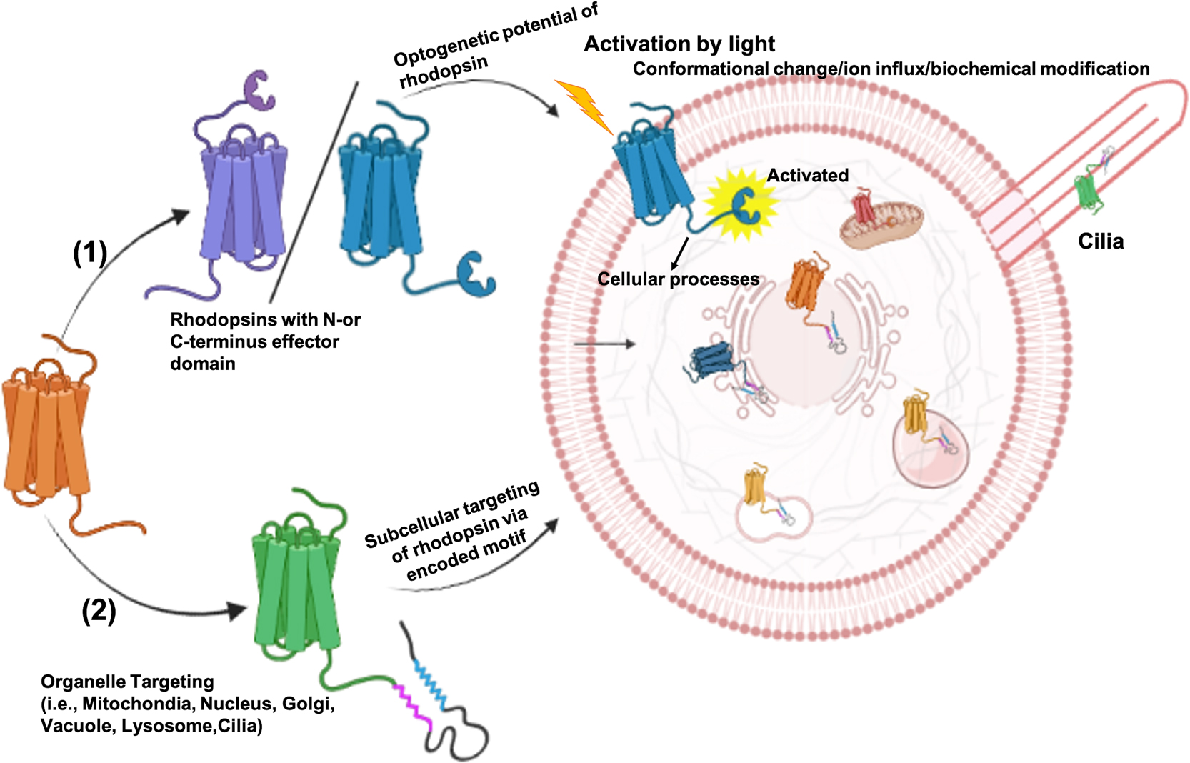
    Rhodopsins have been extensively employed for optogenetic regulation of bioelectrical activity of excitable cells and other cellular processes across biological systems. Various strategies have been adopted to attune the cellular processes at the desired subcellular compartment (plasma membrane, endoplasmic reticulum, Golgi, mitochondria, lysosome) within the cell. These strategies include-adding signal sequences, tethering peptides, specific interaction sites, or mRNA elements at different sites in the optogenetic proteins for plasma membrane integration and subcellular targeting. However, a single approach for organelle optogenetics was not suitable for the relevant optogenetic proteins and often led to the poor expression, mislocalization, or altered physical and functional properties. Therefore, the current study is focused on the native subcellular targeting machinery of algal rhodopsins. The N- and C-terminus signal prediction led to the identification of rhodopsins with diverse organelle targeting signal sequences for the nucleus, mitochondria, lysosome, endosome, vacuole, and cilia. Several identified channelrhodopsins and ion-pumping rhodopsins possess effector domains associated with DNA metabolism (repair, replication, and recombination) and gene regulation. The identified algal rhodopsins with diverse effector domains and encoded native subcellular targeting sequences hold immense potential to establish expanded organelle optogenetic regulation and associated cellular signaling.

  • Jonathan R. Church, Jógvan Magnus Haugaard Olsen, Igor Schapiro
    原稿種別: Special Issue: Recent Advances in Retinal Protein Research Regular Article (Invited)
    2023 年 20 巻 Supplemental 号 論文ID: e201007
    発行日: 2023年
    公開日: 2023/03/21
    [早期公開] 公開日: 2023/01/24
    ジャーナル オープンアクセス HTML
    電子付録

    Multiscale simulations have been established as a powerful tool to calculate and predict excitation energies in complex systems such as photoreceptor proteins. In these simulations the chromophore is typically treated using quantum mechanical (QM) methods while the protein and surrounding environment are described by a classical molecular mechanics (MM) force field. The electrostatic interactions between these regions are often treated using electrostatic embedding where the point charges in the MM region polarize the QM region. A more sophisticated treatment accounts also for the polarization of the MM region. In this work, the effect of such a polarizable embedding on excitation energies was benchmarked and compared to electrostatic embedding. This was done for two different proteins, the lipid membrane-embedded jumping spider rhodopsin and the soluble cyanobacteriochrome Slr1393g3. It was found that the polarizable embedding scheme produces absorption maxima closer to experimental values. The polarizable embedding scheme was also benchmarked against expanded QM regions and found to be in qualitative agreement. Treating individual residues as polarizable recovered between 50% and 71% of the QM improvement in the excitation energies, depending on the system. A detailed analysis of each amino acid residue in the chromophore binding pocket revealed that aromatic residues result in the largest change in excitation energy compared to the electrostatic embedding. Furthermore, the computational efficiency of polarizable embedding allowed it to go beyond the binding pocket and describe a larger portion of the environment, further improving the results.

  • Sui Arikawa, Teppei Sugimoto, Takashi Okitsu, Akimori Wada, Kota Katay ...
    原稿種別: Special Issue: Recent Advances in Retinal Protein Research Regular Article (Invited)
    2023 年 20 巻 Supplemental 号 論文ID: e201017
    発行日: 2023年
    公開日: 2023/03/21
    [早期公開] 公開日: 2023/03/02
    ジャーナル オープンアクセス HTML
    電子付録

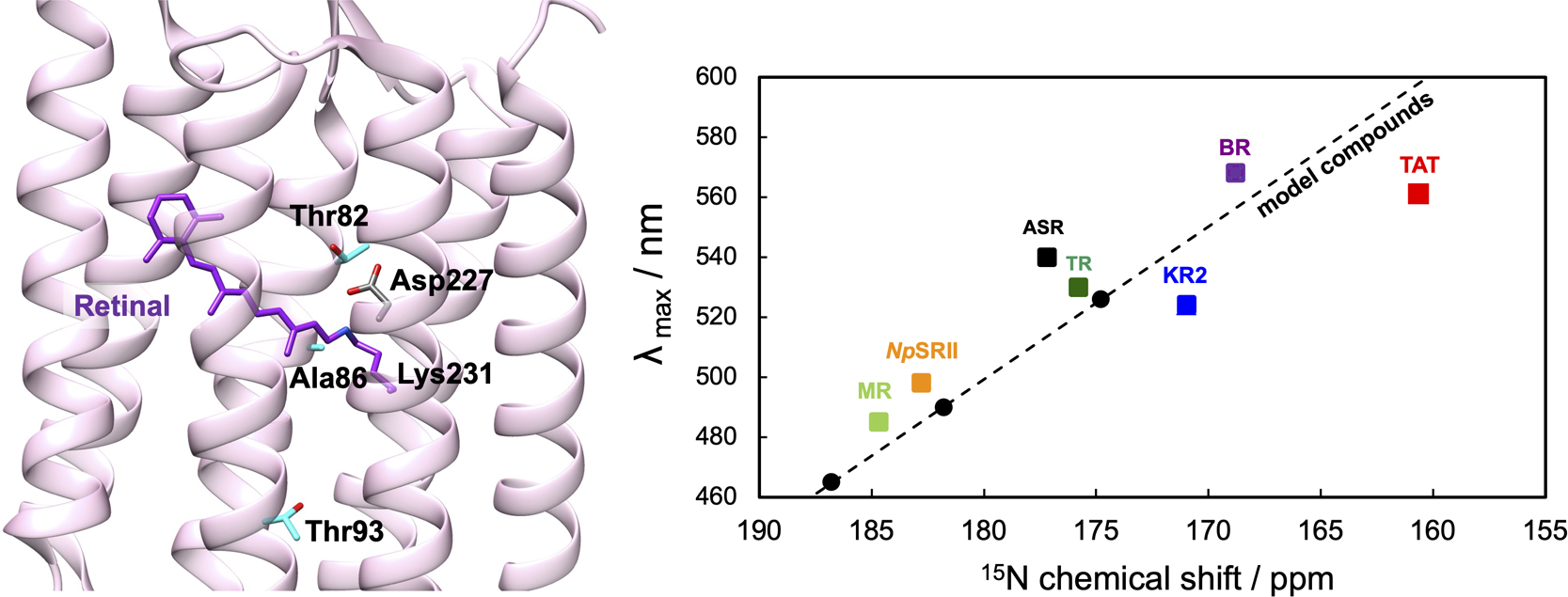
    TAT rhodopsin extracted from the marine bacterium SAR11 HIMB114 has a characteristic Thr-Ala-Thr motif and contains both protonated and deprotonated states of Schiff base at physiological pH conditions due to the low pKa. Here, using solid-state NMR spectroscopy, we investigated the 13C and 15N NMR signals of retinal in only the protonated state of TAT in the 1-palmitoyl-2-oleoyl-sn-glycero-3-phosphoethanolamine/1-palmitoyl-2-oleoyl-sn-glycero-3-phospho (1'-rac-glycerol) (POPE/POPG) membrane at weakly acidic conditions. In the 13C NMR spectrum of 13C retinal-labeled TAT rhodopsin, the isolated 14-13C signals of 13-trans/15-anti and 13-cis/15-syn isomers were observed at a ratio of 7:3. 15N retinal protonated Schiff base (RPSB) had a significantly higher magnetic field resonance at 160 ppm. In 15N RPSB/λmax analysis, the plot of TAT largely deviated from the trend based on the retinylidene-halide model compounds and microbial rhodopsins. Our findings indicate that the RPSB of TAT forms a very weak interaction with the counterion.

  • Akihiro Otomo, Misao Mizuno, Keiichi Inoue, Hideki Kandori, Yasuhisa M ...
    原稿種別: Special Issue: Recent Advances in Retinal Protein Research Regular Article (Invited)
    2023 年 20 巻 Supplemental 号 論文ID: e201016
    発行日: 2023年
    公開日: 2023/03/21
    [早期公開] 公開日: 2023/02/25
    ジャーナル オープンアクセス HTML

    Direct observation of protein structural changes during ion transport in ion pumps provides valuable insights into the mechanism of ion transport. In this study, we examined structural changes in the light-driven sodium ion (Na+) pump rhodopsin KR2 on the sub-millisecond time scale, corresponding with the uptake and release of Na+. We compared the ion-pumping activities and transient absorption spectra of WT and the W215F mutant, in which the Trp215 residue located near the retinal chromophore on the cytoplasmic side was replaced with a Phe residue. Our findings indicated that atomic contacts between the bulky side chain of Trp215 and the C20 methyl group of the retinal chromophore promote relaxation of the retinal chromophore from the 13-cis to the all-trans form. Since Trp215 is conserved in other ion-pumping rhodopsins, the present results suggest that this residue commonly acts as a mechanical transducer. In addition, we measured time-resolved ultraviolet resonance Raman (UVRR) spectra to show that the environment around Trp215 becomes less hydrophobic at 1 ms after photoirradiation and recovers to the unphotolyzed state with a time constant of around 10 ms. These time scales correspond to Na+ uptake and release, suggesting evolution of a transient ion channel at the cytoplasmic side for Na+ uptake, consistent with the alternating-access model of ion pumps. The time-resolved UVRR technique has potential for application to other ion-pumping rhodopsins and could provide further insights into the mechanism of ion transport.

  • María del Carmen Marín, Alexander L. Jaffe, Patrick T. West, Masae Kon ...
    原稿種別: Special Issue: Recent Advances in Retinal Protein Research Regular Article (Invited)
    2023 年 20 巻 Supplemental 号 論文ID: e201023
    発行日: 2023年
    公開日: 2023/03/21
    [早期公開] 公開日: 2023/03/09
    ジャーナル オープンアクセス HTML
    電子付録

    Microbial rhodopsins are photoreceptive transmembrane proteins that transport ions or regulate other intracellular biological processes. Recent genomic and metagenomic analyses found many microbial rhodopsins with unique sequences distinct from known ones. Functional characterization of these new types of microbial rhodopsins is expected to expand our understanding of their physiological roles. Here, we found microbial rhodopsins having a DSE motif in the third transmembrane helix from members of the Actinobacteria. Although the expressed proteins exhibited blue–green light absorption, either no or extremely small outward H+ pump activity was observed. The turnover rate of the photocycle reaction of the purified proteins was extremely slow compared to typical H+ pumps, suggesting these rhodopsins would work as photosensors or H+ pumps whose activities are enhanced by an unknown regulatory system in the hosts. The discovery of this rhodopsin group with the unique motif and functionality expands our understanding of the biological role of microbial rhodopsins.

feedback
Top