Journal of Spine Research
Online ISSN : 2435-1563
Print ISSN : 1884-7137
Volume 11, Issue 10
Displaying 1-21 of 21 articles from this issue
Editorial
Original Article
  • Akihiko Hiyama, Hiroyuki Katoh, Daisuke Sakai, Masahiro Tanaka, Masahi ...
    2020 Volume 11 Issue 10 Pages 1122-1127
    Published: October 20, 2020
    Released on J-STAGE: October 20, 2020
    JOURNAL FREE ACCESS

    Introduction: There has been no report comparing the accuracy of percutaneous pedicle screw (PPS) insertion between fluoroscopy and intraoperative CT navigation.

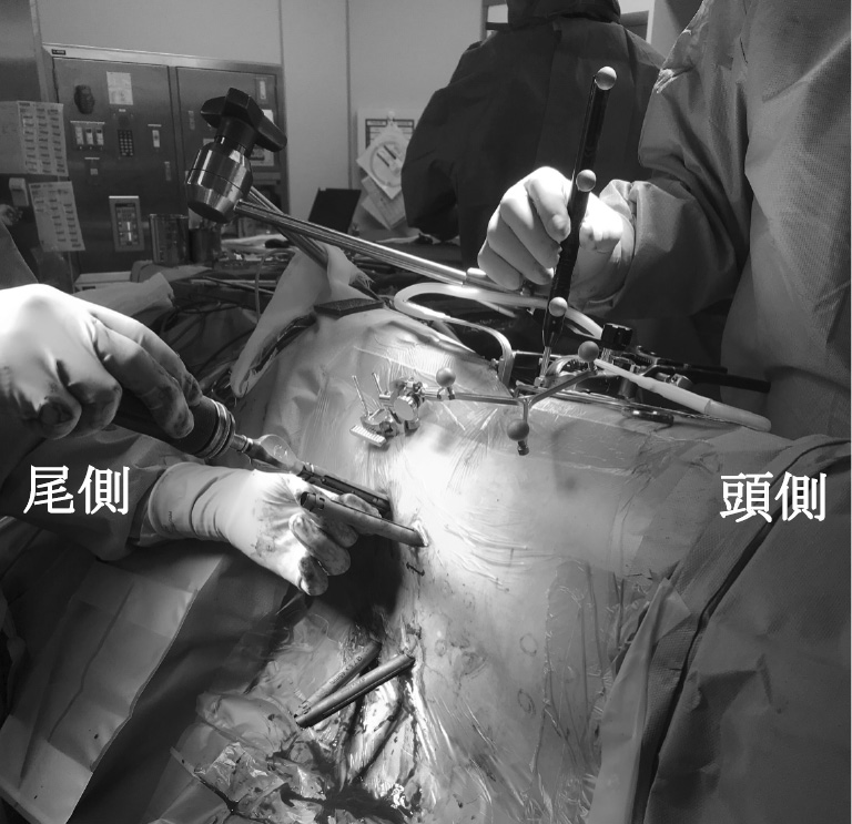
    Methods: Eighty-three patients who underwent indirect decompression with extreme lateral lumbar interbody fusion (XLIF) and PPS were studied. Eighty-three patients were divided into three groups; Dual-position (DP) -PPS group to insert PPS under fluoroscopy in prone position (36 patients); Single position (SP) -PPS group to insert PPS under fluoroscopy in lateral decubitus position (19 patients); SP-PPS+O-arm group in which PPS is inserted in the lateral decubitus position under intraoperative CT navigation (28 patients). We compared the accuracy of PPS insertion among the three groups.

    Results: The results showed that the screw insertion angle of the SP-PPS group was smaller than that of the DP-PPS group, but there was no significant difference in the screw perforation rate among the three groups (DP-PPS: 4.0%, SP-PPS: 5.1%, SP-PPS+O-arm: 3.8%).

    Conclusion: We think that the SP-PPS procedure under fluoroscopy was considered to be a useful method because it does not require an expensive surgical support device and has the same screw accuracy as the DP-PPS procedure.

    Download PDF (1064K)
  • Masaaki Chazono, Shoshi Akiyama
    2020 Volume 11 Issue 10 Pages 1128-1135
    Published: October 20, 2020
    Released on J-STAGE: October 20, 2020
    JOURNAL FREE ACCESS

    Introduction: To avoid surgical morbidity and complications, minimally invasive surgery (MIS) approaches to the treatment of adult spinal deformity (ASD) have been gaining in popularity. In the U. S., some algorithms for the choice of MIS for ASD have been proposed from the Lenke-Silva classification through MisLAT, minimally invasive spinal deformity surgery (MISDEF), and MISDEF-2 algorithms. Whereas, in Japan, three lumbar lordosis (LL) formulae (Fo) such as Niigata Fo (FoN: LL=0.6PI+32.9-0.23age), Dokkyo Fo (FoD: LL=0.59PI+11.1), and Hamamatsu Fo (FoH: LL=0.45PI+31.8) have been developed to target the optimal LL for ASD surgery. In our institute, lateral lumbar interbody fusion (LLIF) followed by minimally invasive posterior surgery (MIS-PSF) has been performed. We sought to verify the most relevant and applicable LL formula for the LLIF+MIS-PSF surgery using two algorithms.

    Material and Methods: Participants included 31 patients (3 male, 28 female) with a Cobb angle > 20° in TL/L scoliotic curves, who underwent the two-stage surgery (LLIF+MIS-PSF). The mean age of the patients at the time of surgery was 71 years. The mean preoperative Cobb angle was 33.2°. The MISDEF algorithm (Mummaneni PV et al, Neurosurg Focus 2014) and MISDEF-J algorithm we have developed were used in this study. First, preoperative spinopelvic parameters in each patient were allocated to the MISDEF algorithm or MISDEF-J algorithm, and then each LL (FoN/FoD/FoH) -LL (MIS-PSF) was calculated, respecively. Participants were divided into two groups: LL (FoN/FoD/FoH) -LL (MIS-PSF) < 5° and with > 5° and differences between the groups were then assessed by constructing 2 × 2 tables using a Cramer V correlation and a Fisher exact test in the MISDEF algorithm and MISDEF-J algorithm.

    Results: There was a distribution with 7 cases of Class 2 and 24 cases of Class 3 in the MISDEF algortihm. The Cramer V correlation and Fisher exact test found V=0.52, p = 0.007 for LL (FoN) -LL (MIS-PSF) and V=0.40, p = 0.033 for LL (FoD) -LL (MIS-PSF), indicating a significant relationship between the MISDEF algorithm and LL (FoN/FoD) -LL (MIS-PSF). By contrast, there was a distribution with 14 cases of MIS-PSF and 17 cases of OPEN in the MISDEF-J algorithm. The test found V=0.48, p = 0.011 for LL (FoN) -LL (MIS-PSF), indicating a significant relationship and the MISDEF-J algorithm and LL (FoN) -LL (MIS-PSF).

    Conclusion: Careful patient selection and evaluation with the algorithm was helpful to select patients with ASD who were appropriate candidates for MIS. Not all cases of deformity can be treated appropriately with MIS, but the algorithm offers a reliable method of patient selection for ASD, especially compared with the FoN.

    Download PDF (1484K)
  • Takumi Hashimura, Shintaro Honda, Eijiro Onishi
    2020 Volume 11 Issue 10 Pages 1136-1143
    Published: October 20, 2020
    Released on J-STAGE: October 20, 2020
    JOURNAL FREE ACCESS

    Purpose: A self-locking stand-alone cage have been used for anterior cervical discectomy and fusion (ACDF). The purpose of this study was to evaluate the clinical and radiographic results in patients who underwent ACDF using this cage and to identify the risk factors for postoperative cage subsidence of this cage.

    Methods: This retrospective study included 15 patients who underwent ACDF (total 22 levels: 9 at 1 level, 5 at 2 levels, and 1 at 3 levels) with a self-locking stand-alone cage from July 2017 to February 2019. In the preoperative radiographical analysis, the C2-7 sagittal vertical axis (SVA), the C2-7 Cobb angle, and the segmental lordotic angle (SLA) were evaluated. Postoperative cage subsidence was considered to be significant when subsidence over 2 mm occurred at the midpoint interbody height (IBH) at each level. The clinical outcomes were evaluated by Japanese Orthopaedics Association (JOA) scoring system.

    Results: Posteoperative JOA score improved significantly after surgery (p< 0.01). Eighteen (82%) of 22 cages achieved bony fusion. Postoperative cage subsidence over 2 mm occurred in 9 (41%) of 22 cages. There was no significant difference in age, sex, BMI, follow-up period, C2-7 SVA, C2-7 Cobb angle, JOA score, and fusion rate between the patients with subsidence under 2 mm (Group I) and the patients with subsidence over 2 mm (Group S). The preoperative SLA was significantly lower in Group S (p=0.04). When differences were compared in SLAs between before and just after the operation, Group S showed significantly higher differences (p=0.03). As a major complication, 1 patient developed aspiration pneumonia due to dysphagia, and 1 patient underwent posterior decompression and fusion due to neurological deterioration caused by cage subsidence.

    Conclusion: ACDF with this cage achieved satisfactory clinical and radiographical outcomes with a low incidence of postoperative complications. However, this study indicated that postoperative subsidence of this cage system might be caused by overcorrection of preoperative segmental kyphosis in patients with lower SLA.

    Download PDF (2018K)
  • Tomoaki Shimizu, Yousuke Takeuchi, Ikuo Aita, Masao Koda, Masashi Yama ...
    2020 Volume 11 Issue 10 Pages 1144-1151
    Published: October 20, 2020
    Released on J-STAGE: October 20, 2020
    JOURNAL FREE ACCESS

    Introduction: We developed a new technique for cervical spine pedicle screw (CPS) insertion to avoid lateral misplacement and to increase accuracy of insertion with C-arm fluoroscopy, 45°drill guide from posterolateral approach and cannulated screw systems (alternative technique). Our objective is to show the technique and to examine the usefulness.

    Method: We placed 115 CPS for 46 patients of traumatic cervical injuries during the period March 2005 to March 2019. We used 58 screws with alternative technique (group A) and 57 screws with conventional technique (group C) via posterior median approach. Ratio of screw misplacement and CPS angle on horizontal plane were evaluated by postoperative CT.

    Result: The mean CPS angle of group A was 41.6° and group C was 37.6°. The angle of group A was statistically larger than that of group C. The misplacement rate of group A was 1.7% and group C was 19%. The misplacement rate was statistically lower in group A.

    Conclusion: We could insert CPS more safety with alternative technique. This technique was useful for traumatic cervical injuries because it can be carried out without restriction of time and institution.

    Download PDF (1435K)
  • Kyohei Sakaki, Kenichiro Sakai, Yoshiyasu Arai, Ichiro Torigoe, Masaki ...
    2020 Volume 11 Issue 10 Pages 1152-1156
    Published: October 20, 2020
    Released on J-STAGE: October 20, 2020
    JOURNAL FREE ACCESS

    Introduction: A prospective study was conducted to evaluate the incidence and to identify risk factors for the postoperative dysphagia after anterior cervical spine surgery.

    material and method: Postoperative dysphasia and several radiographical parameters were prospectively investigated for 87 patients who underwent anterior cervical surgery in our institution. Dysphasia was evaluated by using Bazaz dysphagia score at the postoperative 6 months and 1year. Risk factors of postoperative dysphasia were estimated by multivariate analysis.

    result: Postoperative dysphagia was observed in 25 patients (31.3%) at postoperative 6 months and remained in 22 patients (25.3%) at postoperative one year. Logistic regression analysis using remained dysphagia one year after surgery as an objective variable indicated that multi-level fixation, increased C2-7 lordosis angle, and neck pain were identified as risk factors.

    conclusions: This study identified three risk factors. The only risk factor that could be controlled by the surgeon was the increase in C2-7 lordotic angle.

    Download PDF (774K)
  • Yosuke Ogata, Masaki Tatsumura, Fumihiko Eto, Kohei Murakami, Kai Fuji ...
    2020 Volume 11 Issue 10 Pages 1157-1162
    Published: October 20, 2020
    Released on J-STAGE: October 20, 2020
    JOURNAL FREE ACCESS

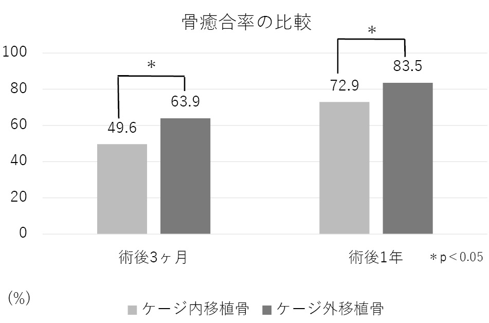
    Introduction: We examined and compared the union rate of autogenous bones implanted inside cages and in the gap between the cages (outside cages) in transforaminal lumbar interbody fusion (TLIF).

    Methods: A total of 133 patients who underwent single-level TLIF for degenerative lumbar disease at our hospital from April 2014 to March 2018 were included. CT imaging was performed 3 months and 1 year after surgery, and bony union was determined. Bony union was defined on CT coronal and sagittal images with bone bridges over the vertebral endplates and without non-union sign (gas phenomenon at the vertebral endplates, loosening of pedicle screw, and cysts of the vertebral endplates).

    Results: The rate of bony union was 49.6% (66 of 133 cases) vs 63.9% (85 of 133 cases) at 3 months after surgery inside cages vs outside cages, which were significant differences (p<0.05). Similarly, the rate of bony union was 72.9% (97 of 133 cases) vs 83.5% (111 of 133 cases) at 1 year after surgery inside cages vs outside cages, which were significant differences (p<0.05).

    Conclusions: In this study, we identified the bony union rate of bone graft outside cages presented higher than that inside cages. We didn't compare the bony union rate between inside only and both sides of cages, so we could not approve that bone grafts outside cages promote bone fusion. However, the estimation of bony union outside cages was appropriate for spinal fusion compared to inside cages.

    Above all, the bony union outside cages may be a good indicator of spinal fusion and the procedure of bone graft outside cages may have a possibility of promoting bony union in TLIF.

    Download PDF (1007K)
  • Takayuki Fujiyoshi, Mitsuhiro Kitamura, Takuma Otagiri, Fumio Hasue, K ...
    2020 Volume 11 Issue 10 Pages 1163-1168
    Published: October 20, 2020
    Released on J-STAGE: October 20, 2020
    JOURNAL FREE ACCESS

    The aim of this study was to investigate efficacy of posterior decompression with segmental fusion at a maximum cord compression level (sPDF) for the K-line (+/-/+) OPLL cases. This study enrolled 29 patients with K-line (+/-/+) OPLL who underwent laminoplasty (LMP) or sPDF. These patients were included in propensity score calculation and 1-to-1 matching resulted in 7 pairs of LMP and sPDF cases. The JOA recovery rate and the acquired score were significantly higher in sPDF group. For the K-line (+/-/+) OPLL case, sPDF seems to be a useful operative method.

    Download PDF (1420K)
  • Takamasa Kudo, Yousuke Takeuchi, Ikuo Aita
    2020 Volume 11 Issue 10 Pages 1169-1176
    Published: October 20, 2020
    Released on J-STAGE: October 20, 2020
    JOURNAL FREE ACCESS

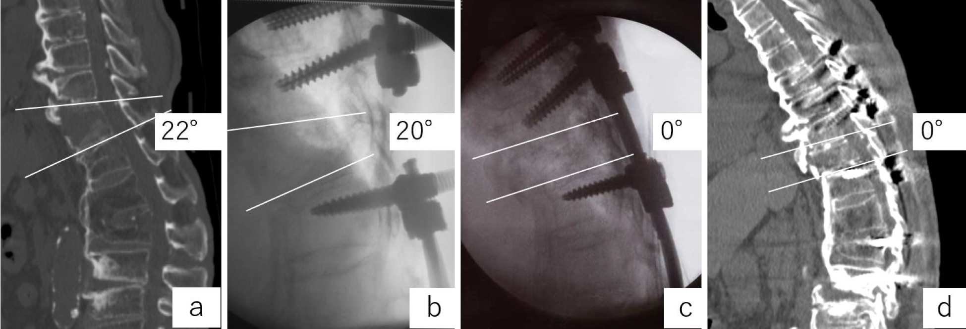
    Introduction: Patients with diffuse idiopathic skeletal hyperostosis (DISH) -related vertebral fracture may suffer from implant failure soon after an operation when they stand up. Insufficient repair in the anterior dilatation after prone fixation has been proposed behind this complication. We developed a new technic to avoid this complication.

    Methods: In three cases of DISH-related vertebral fracture, which is not reduced in the prone position, we inserted the pedicure screw (PS) in the prone position. PS was inserted into each of the three vertebral bodies above and below the fracture, and then, we used four short rods to make each of the fixed vertebral bodies a lump and attached an open domino connector to each rod. After primary closure, put the patient in the forward flexed lateral decubitus position and reopen only between connectors. Finally, we attached other rods to each connector and fixed the fracture.

    Results: No screw back out or neurological deterioration was observed after surgery in all cases.

    Conclusions: This method is considered to be a useful technique for overextended DISH-related vertebral fractures of the thoracolumbar spine which is not reduced in the prone position.

    Download PDF (2118K)
  • Yousuke Takeuchi, Ikuo Aita, Tomoaki Shimizu
    2020 Volume 11 Issue 10 Pages 1177-1183
    Published: October 20, 2020
    Released on J-STAGE: October 20, 2020
    JOURNAL FREE ACCESS

    Introduction: C7 pedicle screw (C7 PS) insertion is usually performed under lateral fluoroscopy, but the shoulder girdles frequently obstruct a clear image of the pedicles. As a solution to this problem, we performed C7 PS insertion using an anteroposterior fluoroscopic image. The aim of this study was to introduce and evaluate the efficacy of the new C7 PS placement technique.

    Methods: From April 2015 to October 2018, 36 PS were inserted using an anteroposterior fluoroscopic image in 21 consecutive cases (A group). When the probe was inserted at a depth of 10 mm from the anatomical entry point, the tip position of the probe was confirmed at the inner edge of the pedicle by anteroposterior fluoroscopy. Before April 2015, 40 PS were inserted under lateral fluoroscopy in 20 cases (L group). We analyzed the rate of screw malposition using postoperative CT, and compared the both groups. Screw deviation of 2 mm or more, grade 2 or higher in the Neo classification, was defined as malposition. Complication associated with the screw malposition was investigated.

    Results: There were no differences in demographic factors (age, gender, disease, BMI) between two groups. The rates of malposition were 20% (8 screws) in L group and 2.7% (1 screw) in A group, respectively. This difference was statistically significant (P=0.04). No complications associated with the screw malposition were found in both groups.

    Conclusions: Safe and accurate C7 pedicle screw placement was performed by confirming the tip position at 10 mm probing by anteroposterior fluoroscopy.

    Download PDF (1568K)
  • Toshikatsu Mamada, Hidemasa Umekoji
    2020 Volume 11 Issue 10 Pages 1184-1192
    Published: October 20, 2020
    Released on J-STAGE: October 20, 2020
    JOURNAL FREE ACCESS

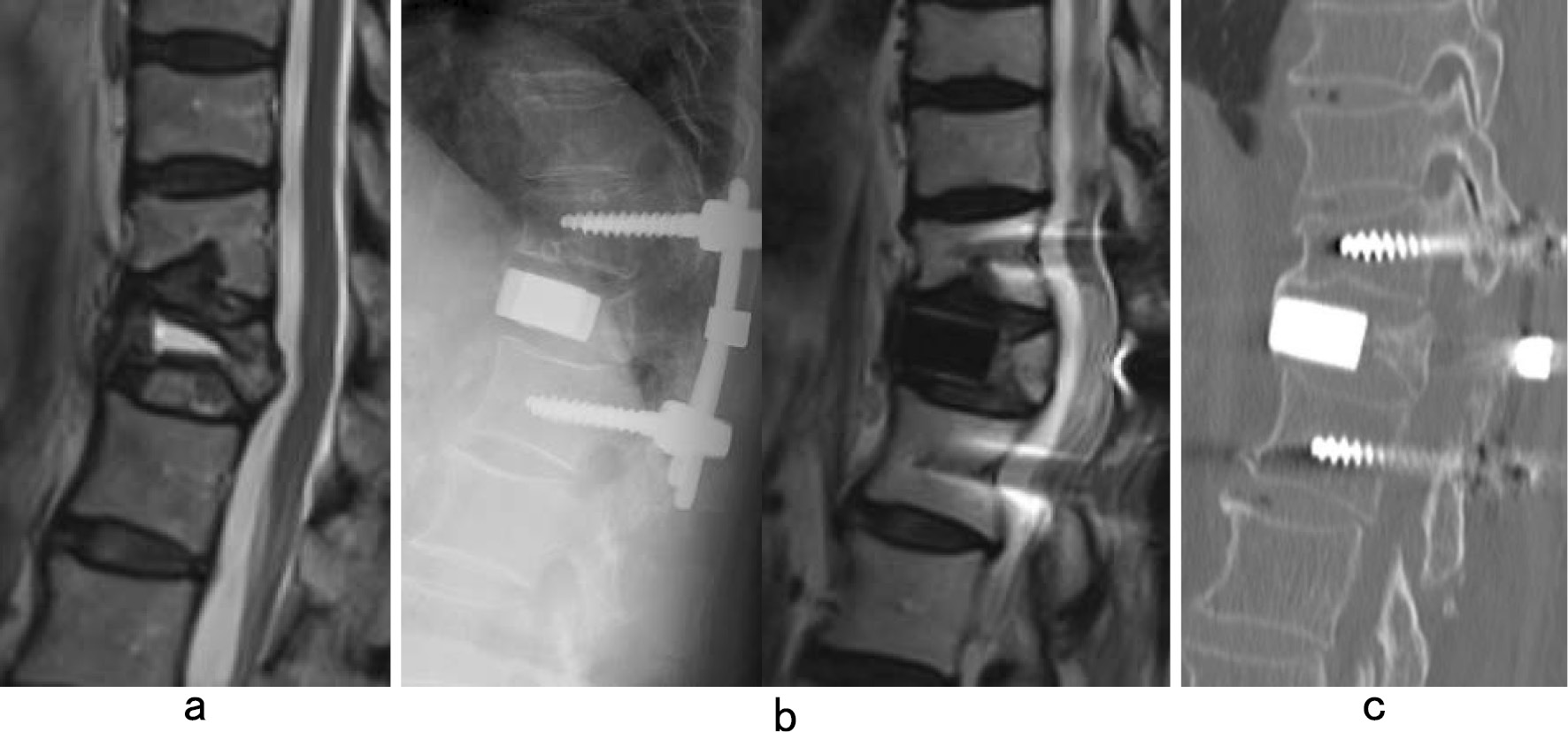
    We report a method we have used to reconstruct a collapsed vertebral body by posterior insertion of an HA (hydroxyapatite) spacer. We performed this procedure in 26 patients diagnosed with delayed neurologic paralysis since 6/2013. The average age at surgery was 75.7 years. The methods are as follows,

    A: Complete resection of the vertebral body and insertion of HA spacer.

    B: Resection of only the upper endplate and intervertebral disk, then fusion with the adjacent vertebral body.

    C: Leave upper and lower endplates intact, and insert HA spacer into the vertebral body for reconstruction (Insertion of HA spacer between both endplates).

    All cases recovered from back pain and weakness of lower extremities. The average correction loss of local kyphosis was 5.0° (range, 1°-15°). Type C allows minimally invasive reconstruction of a collapsed vertebral body without removing vertebral discs. The short-term outcomes indicate that this technique is effective.

    Download PDF (1419K)
  • Yoichi Tani, Takanori Saito, Shinichiro Taniguchi, Masaaki Paku, Masay ...
    2020 Volume 11 Issue 10 Pages 1193-1201
    Published: October 20, 2020
    Released on J-STAGE: October 20, 2020
    JOURNAL FREE ACCESS

    Introduction: The utilization of extreme lateral interbody fusion (XLIF) coupled with percutaneous pedicle screw (PPS) instrumentation has revolutionized the methodology for surgical correction of sagittal plane deformity of the lumbar spine, which used to be treated with open closing osteotomies. Further advancement of the minimally invasive correction technique, proposed in 2012 as the anterior column realignment (ACR) procedure, comprised sectioning the anterior longitudinal ligament (ALL) via the lateral transpsoas approach followed by a hyperlordotic cage placement. At the present, however, few studies have provided confirmatory evidence to support the clinical utility of this technique. Therefore, we report our experience on the ACR applied to adult spinal deformity (ASD) as an additional technique to combined XLIF and PPS procedures, describing its effects on global and regional sagittal spinal balance and indirect neural decompression.

    Methods: The ACR procedure consisted of ALL release and 30-degree hyperlordotic cage placement. Eighteen patients, who had ASD and [pelvic incidence (PI) -lumbar lordosis (LL) ] mismatch (i.e., [PI-LL] ≥ 10 degrees), underwent the ACR procedure at a single level except for one patient who had a two-level ACR. All but one patient had ACR either at L2-3 (6) or L3-4 level (10) or both. The remaining one patient had ACR at L4-5 level. We compared various imaging studies performed preoperatively with those obtained at 6 months postoperatively, and preoperative Oswestry Disability Index (ODI) results with those tested at 12 months postoperatively.

    Results: The full-spine standing X-ray showed significant increases in LL (from 12.1° to 34.6°), segmental lordosis (from 3.8°to 22.5°), and thoracic kyphosis (from 20.7° to 26.7°) with significant decrease in sagittal vertical axis (from 96.1 mm to 42.3 mm), pelvic tilt (from 31.1°to 25.1°) and PI-LL (from 29.9°to 7.2°) postoperatively, resulting in an improvement in the global sagittal balance. The sagittal reformatted CT scans revealed significant increases in disc space height, and bilateral intervertebral foraminal height at the ACR level. Axial T2-weighted MRIs at the disc level that received ACR revealed a significant increase in cross-sectional area of the dural sac (from 107.9 mm2 to 156.3 mm2) postoperatively. The ODI scores significantly decreased from 45.8% preoperatively to 26.2% postoperatively.

    Conclusions: The current results confirmed that the ACR could provide a useful addition to XLIF procedure in achieving not only better sagittal spinal alignment but also indirect neural decompression as a result of restoration of disc height as well as intervertebral foraminal height.

    Download PDF (1880K)
  • Masayuki Nakahara, Yasukazu Hijikata, Jyunji Morii, Naoki Okubo, Nozom ...
    2020 Volume 11 Issue 10 Pages 1202-1206
    Published: October 20, 2020
    Released on J-STAGE: October 20, 2020
    JOURNAL FREE ACCESS

    Introduction: In diffuse idiopathic skeletal hyperostosis (DISH), fracture of the continuous ankylosing vertebral body might cause severe stress on the damaged vertebral body. In addition, screws placed in traditional trajectories might become loose or back out to severe vertebral bone fragility caused by stress shielding. Therefore, introduction of a penetrating endplate screw (PES), which improves the insertion torque by penetrating the cranial vertebral endplate of the screw-inserted vertebra, is increasingly being performed instead of the traditional trajectory screw (TTS) for spinal injury in DISH. The aim of this study was to evaluate surgical outcomes using the PES method for thoracolumbar injury in DISH.

    Method: From April 2014 to March 2018, we treated patients with DISH thoracolumbar injury with either percutaneous pedicle screw insertion using the PES method (PES method group, including PES alone or PES combined with TTS) or the TTS alone method (TTS group). We investigated the characteristics of patients, cases of reoperation due to implant failure and screw loosening in both groups. The results were expressed as mean±standard deviation and were statistically analyzed using Wilcoxon's rank sum test, chi-square test and Fisher's exact test.

    Results: The study included 21 patients, 9 in the PES group (1 male and 8 females) and 12 in the TTS group (3 males and 9 females). Mean patient age in the two groups was 80±7 years and 79±7 years (p = 0.72), respectively, and mean body weight was 51±10 kg and 63±19 kg (p = 0.19), respectively. YAM values in the PES and TTS groups were 72±11% and 71±14% (p = 0.97), respectively. The number of ankylosing vertebral bodies extending cranially and caudally from the fractured vertebrae was 11±3 vertebral bodies and 11±2 vertebral bodies (p = 0.97), respectively. One patient in the TTS group underwent reoperation due to implant failure, with no such cases in the PES group (p = 0.57). Loosening of screws occurred in 22% (2/9 cases) of patients in the PES group, and 50% (6/12 cases) of patients in the TTS group (p = 0.20). The number of loosened screws was 5/85 (6%) in the PES group, 2/13 (15%) of the TTS in PES group patients who also received TTS, and 20/140 (14%) in the TTS group (p = 0.12).

    Conclusion: Compared to the TTS method, the PES method showed less tendency for screw loosening and no reoperations due to implant failure. The PES method with penetration of cortical bone might be useful in the management of thoracolumbar spine injury in elderly DISH patients with severe bone fragility.

    Download PDF (1046K)
  • Tomohiko Hirose, Yoichi Inoue, Tomohiro Inoue, Hisanori Ikuma
    2020 Volume 11 Issue 10 Pages 1207-1213
    Published: October 20, 2020
    Released on J-STAGE: October 20, 2020
    JOURNAL FREE ACCESS

    Introduction: We have performed simultaneous parallel anterior and posterior combined lumbar spine surgery under intraoperative CT navigation (SPAPS) or minimally invasive transforaminal lumbar interbody fusion (MIS-TLIF) for degenerative lumbar disease. The purpose of this study was to compare the clinical outcomes of SPAPS with MIS-TLIF.

    Method: A total of 40 patients who underwent either of the two type of elective surgeries for degenerative lumbar disease at our hospital were retrospectively reviewed. Eighteen patients who underwent SPAPS (group S) and 22 patients who underwent MIS-TLIF (group T) from April, 2016 to December, 2018 were enrolled. All patients were postoperatively followed up for a minimum period of 6 months. The average age of group S patients was 62.3 years and that of group T was 67.0 years. The following items were evaluated preoperatively and at 6 months after surgery: operative time, intraoperative blood loss, perioperative complications, Japanese Orthopaedic Association Back Pain Evaluation Questionnarie (JOABPEQ), visual analog scale (VAS) score, and profit ratio.

    Results: Operative time in groups S and T was 113.8 and 159.5 min, respectively. Intraoperative blood loss volume was 72 ml in group S and 132.8 ml in group T. None of the cases in either group had perioperative complications. The JOABPEQ indicated significant improvements in Low back pain, Lumbar ability, Walking ability, Social life ability and Mental health in group S and significant improvements in Low back pain, Walking ability, Social life ability and Mental health in group T. VAS scores in both groups of group S and T were significantly improved in all evaluation items. Operative time was shorter in group S than in group T. Low back pain according to the JOABPEQ was significantly improved in group S as compared to group T, as was the VAS score of leg numbness. The profit ratio of group S was significantly higher than that of group T, at 53% and 42%, respectively.

    Conclusion: We compared the clinical outcomes of SPAPS and MIS-TLIF. This study showed that group S had good results in terms of operative time, low back pain, leg numbness, and profit ratio.

    Download PDF (1224K)
  • Takashi Kuniya, Kenichi Watanabe, Hiroaki Abe, Takuya Kawai, Akiro Hig ...
    2020 Volume 11 Issue 10 Pages 1214-1219
    Published: October 20, 2020
    Released on J-STAGE: October 20, 2020
    JOURNAL FREE ACCESS

    We compared the use of balloon kyphoplasty (BKP) and posterior fixation with that of posterior vertebral column resection (P-VCR) and posterior fixation to treat osteoporotic vertebral body fractures with posterior wall damage at the thoracolumbar junction. BKP and posterior fixation was a less invasive procedure with a significantly shorter operating time and significantly lower blood loss. Screw loosening and correction loss due to new vertebral body fractures occurred in both groups, but there was no revision surgery due to correction loss, indicating that BKP with posterior fixation is a suitable procedure for older patients.

    Download PDF (1137K)
  • Masayuki Ishihara, Shinichiro Taniguchi, Yoichi Tani, Masaaki Paku, Ta ...
    2020 Volume 11 Issue 10 Pages 1220-1227
    Published: October 20, 2020
    Released on J-STAGE: October 20, 2020
    JOURNAL FREE ACCESS

    Introduction: Recent increases in the prevalence of osteoporotic vertebral fractures are attributable to rapid aging of the population. Conservative treatment usually achieves successful bone fusion; however, some patients show delayed union and vertebral collapse resulting in neurological deficits and kyphotic deformities. These patients are candidates for various surgical procedures; however, surgical invasion and implant-induced complications can occur. The X-core2® system was introduced in Japan in 2015. This device is useful for minimally invasive anterior vertebral corpectomy and vertebral replacement for osteoporotic vertebral collapse. However, the surgical procedure remains controversial. We investigated the surgical procedure and clinical outcomes in 28 consecutive patients with kyphotic deformities secondary to osteoporotic vertebral fractures (KOVF).

    Patients and methods: This study included 28 patients with KOVF who underwent lateral access corpectomy (LAC) performed by the same surgeon at a single hospital between 2017 and 2018. All patients were followed-up for at least 18 months postoperatively. LAC followed by posterior thoracolumbar fixation using a percutaneous pedicle screw (PPS) was performed as corrective surgery for KOVF. The surgical procedure, number of fixed vertebral bodies, intraoperative blood loss, operative time, pre- and postoperative radiological measurements-including various spinopelvic parameters-bone fusion rates, and intra- and postoperative complications were evaluated.

    Results: No patients showed bone fusion of the posterior element. All patients underwent anterior corpectomy followed by posterior fixation (AP). The mean fixed number of vertebral bodies was 5.1±0.57. The mean operation time was 207±92 min, and the mean intraoperative blood loss was 456±156 ml. The following spinopelvic parameters (pre-/postoperative) improved significantly: lumbar lordosis (LL) 28°/45°, pelvic incidence-LL 17°/2°, pelvic tilt 29°/20°, and local kyphosis angle 23.3°/−1.4°. The mean correction loss was 0.7°. The anterior interbody distance improved from 21.8 mm to 35.5 mm, and the posterior interbody distance improved from 25.4 mm to 33.7 mm. Bone union rates were 67% and 85% at 6 and 12 months, respectively. Observed complications included endplate injury (n=2), cage subsidence (n=3), proximal junctional kyphosis (n=2), distal junctional failure (n=1), and parietal pleura injury (n= 8). No patients required reoperation.

    Conclusions: AP achieves good correction. Posterior fixation is possible with PPS in patients without posterior bone fusion.

    Download PDF (2538K)
  • Kazuya Kishima, Keishi Maruo, Fumihiro Arizumi, Kazuki Kusuyama, Toshi ...
    2020 Volume 11 Issue 10 Pages 1228-1233
    Published: October 20, 2020
    Released on J-STAGE: October 20, 2020
    JOURNAL FREE ACCESS

    Introduction: Nonunion is still major complication after transforaminal lumbar interbody fusion (TLIF). Recently, titanium Ti-coated polyetheretherketone (PEEK) has been introduced, which have advantages of PEEK and titanium. We compared the radiological outcomes of TLIF using a titanium Ti-coated PEEK cages and PEEK cages with a minimum of 1-year follow-up.

    Methods: A total of 80 patients who underwent TLIF using Ti-coated cages (2016-2017) and PEEK cage (2014-15) by a single surgeon were retrospectively reviewed. A total of 37 patients with a total of 48 segments used Ti-coated PEEK cages (TP group), and 43 patients with a total of 51 segments used PEEK cages (P group). The mean age was 69 years old with 38 males and 42 females. Endplate cyst, radiolucent zone, screw loosening, cage subsidence (>2 mm), and fusion rate were evaluated by CT at 3- and 12-months. Bony union was assessed by sagittal and coronal CT-MPR to confirm osseous continuity through the vertebral endplate and cage or to confirm a bony bridge around the vertebrae and less than 2 degrees of motion on flexion-extension in the radiograph. Nonunion was assessed by the presence of a visible gap.

    Results: Solid fusion was achieved in 92% of patients in the TP group and 82% in the P group at 12-months after surgery (P=0.24). There was no significant difference in age, gender, number of fused segments, cage height, operation time, and estimated blood loss between the TP group and the P group. The incidence of endplate cyst was 52% in the TP group and 59% in the P group (P = 0.55) at 3 months after surgery, and 50% in the TP group and 47% in the P group (P = 0.84) at 12-months after surgery. The incidence of screw loosening (TP group 27% vs P group 29%, P=0.83), cage subsidence (TP group 29% vs P group 24%, P=0.65), and radiolucent zone around cage (TP group 17% vs P group 20%, P=0.80) were similar in both groups.

    Conclusions: The present study has demonstrated that Ti-coated PEEK cage did not affect on radiographical outcomes. There are various factors affecting bony fusion in lumbar interbody fusion surgery. The incidences of endplate cysts were also similar in the TP group and P group at 3- and 12-months after surgery. Further studies are needed to identify the predictive factors associated with nonunion.

    Download PDF (1305K)
  • Masakazu Toi, Keishi Maruo, Fumihiro Arizumi, Kazuki Kusuyama, Kazuya ...
    2020 Volume 11 Issue 10 Pages 1234-1240
    Published: October 20, 2020
    Released on J-STAGE: October 20, 2020
    JOURNAL FREE ACCESS

    Introduction: Surgical site infection (SSI) is still an important complication after spinal instrumentation surgery. The decision whether to removal of implant, especially management of cage, after transforaminal lumber interbody fusion (TLIF) is difficult to make. The purpose of this study was to investigate the treatment strategy for deep SSI after posterior lumber interbody fusion.

    Method: We performed mini-open TLIF on 504 patients between January 2014 and February 2018, of which 14 patients (2.8%) developed SSI. The retrospective study cohort consisted of the 14 patients (mean age, 71.3 years) who had required surgical intervention for SSI. Patients were treated with aggressive surgical irrigation and debridement (I and D) with retention of implants in cases of early infection. Implant removal, extension of fusion, and re-implantation were considered in cases of screw or cages loosening or persistent infection. Each treatment course was reviewed for the type of surgery, management of implants, additional procedures. Outcomes were categorized into two groups: 1) Treatment success, which was defined as eradication of infection after I and D, 2) treatment failure, which was defined as a recurrent infection, removal of implants to eradicate the infection, or re-implantation due to loosening of screw or cages.

    Result: The median time from index surgery to initial treatment was 18.5 days (range 5-320). SSI occurred within 30 days in 8 cases (57%), within 30-90 days in 5 cases (36%), 1 case (7%) after the index surgery. Treatment success was achieved in 5 (36%) cases. Of the remaining 9 cases (64%) from the treatment failure group, 5 cases were treated with removal of cages and replacement with iliac bone graft, 3 cases were treated with re-implantation without removal of the cage, and only one case required removal of screws and cages. The average degree of correction loss was 5.7±5.1°, and the fusion rate was 71% at final follow-up. Higher rate of treatment success was observed in cases with presence of osteomyelitis (20% vs. 78%, P=0.091). There were no significant differences in terms of duration of diagnosis for SSI, operation time, and estimated blood loss between the two groups. 57% (8/14) out of all cases were treated with successfully without the removal of cages and screws.

    Conclusions: Postoperative spine infections were treated with aggressive surgical irrigation and debridement in cases that showed early infection. Evaluations using CT and MRI are helpful to decide the treatment strategies. Removal of the cage and extension of the fixation or re-implantation may be useful strategies in cases of osteomyelitis.

    Download PDF (1302K)
  • Koju Hayashi, Daishi Nakahara, Hiroshi Sakuramoto
    2020 Volume 11 Issue 10 Pages 1241-1245
    Published: October 20, 2020
    Released on J-STAGE: October 20, 2020
    JOURNAL FREE ACCESS

    Purpose: Osteoporotic vertebral fractures (OVF) with T2 diffuse low in MRI cause early vertebral collapse. Spinal kyphosis caused by early vertebral collapse have major negative impact on both ADL and QOL. We performed BKP soon after patient experienced OVF with T2 diffuse low in MRI. We investigated whether it was possible to stop vertebral collapse by operating BKP.

    Methods: 18 patients were diagnosed with OVF with T2 diffuse low in MRI at our facilities between 2018 and 2019. The BKP was performed to 9 patients soon after the fractures. The other 9 patients received conservative treatment. In this study, we compared the BKP group with conservative treatment group.

    Results: Progress of the kyphosis in BKP group was significantly lower than that of conservative group. In addition, VAS was decreased, and ADL were significantly improved in the group who received BKP. Early BKP could be the effective treatment to prevent deterioration of the kyphosis by the vertebral fracture of T2 diffuse low in MRI.

    Download PDF (842K)
Case Report
  • Hiroaki Kimura, Juichi Miura, Bunichiro Wadayama
    2020 Volume 11 Issue 10 Pages 1246-1251
    Published: October 20, 2020
    Released on J-STAGE: October 20, 2020
    JOURNAL FREE ACCESS

    Introduction: Previous studies reported the usefulness of pedicle subtraction osteotomy (PSO) for kyphotic deformity caused by osteoporotic vertebral fracture. However, several reports have described about correction loss after the surgery due to loosening or migration of pedicle screw. Therefore, we applied staged surgery to prevent the problem as follows; four-segment posterior spinal fusion was performed using PSO at the collapsed vertebra as the first stage. After a week, reconstruction of anterior support was performed with placement of an expandable cylindrical cage through the osteotomy vertebra and placement of interbody cage at the caudal interbody space.

    Case report: Two patients received the surgery for kyphosis associated with L1 vertebral fracture. The local kyphotic angles (T12-L2) were reduced from 41° to 8° in case 1 and 70° to 9° in case 2, respectively. The corrections were maintained during 2-year follow-up in both cases.

    Conclusion: We advocate that reconstruction of anterior support is important to prevent correction loss after PSO for kyphotic deformity caused by osteoporotic vertebral fracture.

    Download PDF (1220K)
  • Toru Funayama, Kentaro Mataki, Kosuke Sato, Kousei Miura, Tetsuya Abe, ...
    2020 Volume 11 Issue 10 Pages 1252-1258
    Published: October 20, 2020
    Released on J-STAGE: October 20, 2020
    JOURNAL FREE ACCESS

    Introduction: Among individuals with Parkinson's disease, outcomes of spinal surgery are generally poor, and the risk of reoperation is high. We report a case of implant failure early after combined poteroanterior fusion for osteoporotic lumbar vertebral collapse in a patient with severe Parkinson's disease.

    Case Report: A 70-year-old man with a 10 year history of Parkinson's disease (Hoehn & Yahl Scale: Stage III) fell in a public bath. Over approximately 6 weeks, the L4 vertebral body collapsed rapidly, and he developed pain in his legs, a drop foot, and incontinence. When he visited our hospital, the L4 vertebral body completely collapsed, and the bone fragment from the posterior wall retropulsed into the spinal canal. To rule out a metastatic spinal tumor or pyogenic spondylitis, we first performed a biopsy of L4 vertebral body and carried out L2-S1 posterior decompression and fusion. After a possibility of a tumor and infection were ruled out, we diagnosed the patient with an osteoporotic vertebral collapse with delayed palsy. Two weeks later he underwent an anterior vertebral body replacement surgery using an expandable cage with a wide plate. However, the back out of the pedicle screws and the cage dislocation occurred soon after surgery. Therefore, fixation was extended up to T6-ilium. The revision surgery effectively stabilized the anterior cage. Due to another fall during rehabilitation, the vertebral body adjacent to the fixation (T5) fractured and caused myelopathy. The fixation was extended again up to C7. At present, 2 years after the last surgery, the L4 bony fusion was completed, and the patient is now able to walk without any assistance.

    Conclusion: There is no consensus regarding the appropriate fusion range for osteoporotic vertebral collapse with Parkinson's disease. However, due to the significant bone fragility, a sufficient posterior fusion range had to be established, even for a combined anterior and posterior fusion.

    Download PDF (2610K)
feedback
Top