Magnetic Resonance in Medical Sciences
Online ISSN : 1880-2206
Print ISSN : 1347-3182
ISSN-L : 1347-3182
19 巻, 4 号
選択された号の論文の14件中1~14を表示しています
Innovative Clinical Images
Review
  • Yuriko Suzuki, Noriyuki Fujima, Matthias J.P. van Osch
    2020 年 19 巻 4 号 p. 294-309
    発行日: 2020年
    公開日: 2020/12/01
    [早期公開] 公開日: 2019/11/22
    ジャーナル オープンアクセス

    In the 1980’s some of the earliest studies of arterial spin labeling (ASL) MRI have demonstrated its ability to generate MR angiography (MRA) images. Thanks to many technical improvements, ASL has been successfully moving its position from the realm of research into the clinical area, albeit more known as perfusion imaging than as MRA. For MRA imaging, other techniques such as time-of-flight, phase contrast MRA and contrast-enhanced (CE) MRA are more popular choices for clinical applications. In the last decade, however, ASL-MRA has been experiencing a remarkable revival, especially because of its non-invasive nature, i.e. the fact that it does not rely on the use of contrast agent. Very importantly, there are additional benefits of using ASL for MRA. For example, its higher flexibility to achieve both high spatial and temporal resolution than CE dynamic MRA, and the capability of vessel specific visualization, in which the vascular tree arising from a selected artery can be exclusively visualized. In this article, the implementation and recent developments of ASL-based MRA are discussed; not only focusing on the basic sequences based upon pulsed ASL or pseudo-continuous ASL, but also including more recent labeling approaches, such as vessel-selective labeling, velocity-selective ASL, vessel-encoded ASL and time-encoded ASL. Although these ASL techniques have been already utilized in perfusion imaging and their usefulness has been suggested by many studies, some additional considerations should be made when employing them for MRA, since there is something more than the difference of the spatial resolution of the readout sequence. Moreover, extensive discussion is included on what readout sequence to use, especially by highlighting how to achieve high spatial resolution while keeping scan-time reasonable such that the ASL-MRA sequence can easily be included into a clinical examination.

MajorPapers
  • Yoshiko Matsubara, Toru Higaki, Chihiro Tani, Shogo Kamioka, Kuniaki H ...
    2020 年 19 巻 4 号 p. 310-317
    発行日: 2020年
    公開日: 2020/12/01
    [早期公開] 公開日: 2019/10/15
    ジャーナル オープンアクセス
    電子付録

    Purpose: CT is a useful modality for the evaluation of fetal skeletal dysplasia but radiation exposure is unavoidable. The purpose of this study is to compare the usefulness of MRI and CT for evaluating the fetal skeletal shape.

    Methods: This study was approved by our Institutional Review Board. Fetal specimens (n = 14) were scanned on a 3T MRI scanner using our newly-developed sequence. It is based on T2*-weighted imaging (TR, 12 ms; TE for opposed-phase imaging, 6.1 ms, for in-phase imaging, 7.3 ms; flip angle, 40°). The specimens were also scanned on a 320 detector-row CT scanner. Four radiologists visually graded and compared the visibility of the bone shape of eight regions on MRI- and CT-scans using a 5-point grading system.

    Results: The diagnostic ability of MRI with respect to the 5th metacarpals, femur, fibula, and pelvis was superior to CT (all, P < 0.050); there was no significant difference in the evaluation results of observers with respect to the cervical and lumbar spine, and the 5th metatarsal (0.058 ≤ P ≤ 1.000). However, the diagnostic ability of MRI was significantly inferior to CT for the assessment of the bone shape of the thoracic spine (observers A and C: P = 0.002, observers B and D: P = 0.001).

    Conclusion: The MRI method we developed represents a potential alternative to CT imaging for the evaluation of the fetal bone structure.

  • Marie-Luise Kromrey, Satoshi Funayama, Daiki Tamada, Shintaro Ichikawa ...
    2020 年 19 巻 4 号 p. 318-323
    発行日: 2020年
    公開日: 2020/12/01
    [早期公開] 公開日: 2019/10/24
    ジャーナル オープンアクセス

    Purpose: To compare the image quality of three-dimensional magnetic resonance cholangiopancreatography (MRCP) acquired with respiratory triggering against breath-hold 3D MRCP with compressed sensing (CS) and parallel imaging (PI) in a clinical setting.

    Methods: This study included 93 patients (45 men, mean age: 69.7 ± 9.3 years), in whom three types of 3D MRCP were performed: 3D breath-hold MRCP with CS and PI reconstruction (BH-CS-MRCP) and PI only reconstruction (BH-PI-MRCP) additionally to 3D respiratory triggered MRCP with navigator echoes (Nav-MRCP). Duct visualization and overall image quality were blindly evaluated on a four-point scale by two independent radiologists. Quantitative analysis was performed by calculating the relative duct-to-periductal contrast (RC) of three main biliary segments. Comparison between the methods was performed using paired t-test.

    Results: Acquisition time was 23 s for both breath-hold MRCP protocols and 1 min 29 s for Nav-MRCP. Mean grading (Nav/CS/PI) for common bile duct (2.74/2.87/2.94), common hepatic duct (2.82/2.92/3.00), central right hepatic duct (2.75/2.85/2.98), central left hepatic duct (2.75/2.85/2.92) and cystic duct (2.22/2.34/2.42) was higher in BH-CS- and BH-PI-MRCP, whereas Nav-MRCP showed higher grading in the peripheral segments (peripheral right hepatic duct: 2.24/2.01/2.12; peripheral left hepatic duct: 2.23/2.02/2.13). Overall image quality of Nav-MRCP (2.91 ± 0.7) was not different from BH-PI-MRCP (2.92 ± 0.6) (P = 0.163), but higher than BH-CS-MRCP (2.80 ± 0.7) (P = 0.031). Quantitative analysis showed lower RC values for CS- and PI-MRCP than Nav-MRCP (P < 0.001).

    Conclusion: Breath-hold 3D MRCP were feasible using PI and CS. Visualization of the greater ductal system was even superior in breath-hold MRCP than in Nav-MRCP by considerably reducing acquisition time. Both breath-hold methods are suitable for revised MRI protocols notably in patients with irregular respiratory cycle.

  • Yasuhiko Tachibana, Akifumi Hagiwara, Masaaki Hori, Jeff Kershaw, Misa ...
    2020 年 19 巻 4 号 p. 324-332
    発行日: 2020年
    公開日: 2020/12/01
    [早期公開] 公開日: 2019/12/27
    ジャーナル オープンアクセス
    電子付録

    Purpose: A current algorithm to obtain a synthetic myelin volume fraction map (SyMVF) from rapid simultaneous relaxometry imaging (RSRI) has a potential problem, that it does not incorporate information from surrounding pixels. The purpose of this study was to develop a method that utilizes a convolutional neural network (CNN) to overcome this problem.

    Methods: RSRI and magnetization transfer images from 20 healthy volunteers were included. A CNN was trained to reconstruct RSRI-related metric maps into a myelin volume-related index (generated myelin volume index: GenMVI) map using the MVI map calculated from magnetization transfer images (MTMVI) as reference. The SyMVF and GenMVI maps were statistically compared by testing how well they correlated with the MTMVI map. The correlations were evaluated based on: (i) averaged values obtained from 164 atlas-based ROIs, and (ii) pixel-based comparison for ROIs defined in four different tissue types (cortical and subcortical gray matter, white matter, and whole brain).

    Results: For atlas-based ROIs, the overall correlation with the MTMVI map was higher for the GenMVI map than for the SyMVF map. In the pixel-based comparison, correlation with the MTMVI map was stronger for the GenMVI map than for the SyMVF map, and the difference in the distribution for the volunteers was significant (Wilcoxon sign-rank test, P < 0.001) in all tissue types.

    Conclusion: The proposed method is useful, as it can incorporate more specific information about local tissue properties than the existing method. However, clinical validation is necessary.

  • Roshani Perera, Haruo Isoda, Kenta Ishiguro, Takashi Mizuno, Yasuo Tak ...
    2020 年 19 巻 4 号 p. 333-344
    発行日: 2020年
    公開日: 2020/12/01
    [早期公開] 公開日: 2020/01/17
    ジャーナル オープンアクセス
    電子付録

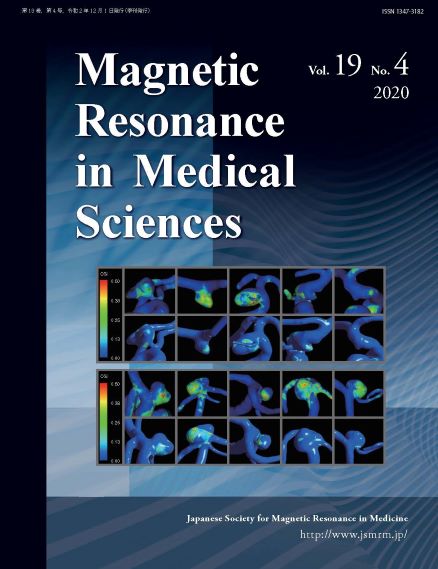
    Purpose: Evaluate in vivo hemodynamic and morphological biomarkers of intracranial aneurysms, using magnetic resonance fluid dynamics (MRFD) and MR-based patient specific computational fluid dynamics (CFD) in order to assess the risk of rupture.

    Methods: Forty-eight intracranial aneurysms (10 ruptured, 38 unruptured) were scrutinized for six morphological and 10 hemodynamic biomarkers. Morphological biomarkers were calculated based on 3D time-of-flight magnetic resonance angiography (3D TOF MRA) in MRFD analysis. Hemodynamic biomarkers were assessed using both MRFD and CFD analyses. MRFD was performed using 3D TOF MRA and 3D cine phase-contrast magnetic resonance imaging (3D cine PC MRI). CFD was performed utilizing patient specific inflow–outflow boundary conditions derived from 3D cine PC MRI. Univariate analysis was carried out to identify statistically significant biomarkers for aneurysm rupture and receiver operating characteristic (ROC) analysis was performed for the significant biomarkers. Binary logistic regression was performed to identify independent predictive biomarkers.

    Results: Morphological biomarker analysis revealed that aneurysm size [P = 0.021], volume [P = 0.035] and size ratio [P = 0.039] were statistically significantly different between the two groups. In hemodynamic biomarker analysis, MRFD results indicated that ruptured aneurysms had higher oscillatory shear index (OSI) [OSI.max, P = 0.037] and higher relative residence time (RRT) [RRT.ave, P = 0.035] compared with unruptured aneurysms. Correspondingly CFD analysis demonstrated significant differences for both average and maximum OSI [OSI.ave, P = 0.008; OSI.max, P = 0.01] and maximum RRT [RRT.max, P = 0.045]. ROC analysis revealed AUC values greater than 0.7 for all significant biomarkers. Aneurysm volume [AUC, 0.718; 95% CI, 0.491–0.946] and average OSI obtained from CFD [AUC, 0.774; 95% CI, 0.586–0.961] were retained in the respective logistic regression models.

    Conclusion: Both morphological and hemodynamic biomarkers have significant influence on intracranial aneurysm rupture. Aneurysm size, volume, size ratio, OSI and RRT could be potential biomarkers to assess aneurysm rupture risk.

  • Yasuo Amano, Yuko Omori, Fumi Yanagisawa, Chisato Ando, Naoki Shinoda, ...
    2020 年 19 巻 4 号 p. 345-350
    発行日: 2020年
    公開日: 2020/12/01
    [早期公開] 公開日: 2020/01/17
    ジャーナル オープンアクセス

    Purpose: Modified Look-Locker inversion recovery (MOLLI) using a 5s(3s)3s scheme is robust to tachycardia, but some errors are occasionally observed in myocardial T1 mapping. We sought to evaluate the relationship between measurement errors in T1 mapping and heart rate (HR) using a confidence map.

    Methods: We enrolled 69 male patients with normal native T1 values of the septal myocardium measured by a 5s(3s)3s MOLLI. The degree of measurement errors in the septal myocardium was assessed by two independent observers on a confidence map using a 4-point scale: 0, no errors; 1, errors located on the myocardial contour; 2, errors extended into the myocardial contour; and 3, errors extended into the midwall. We compared the scores of measurement errors and the average, maximum, minimum or variability of the HR indicated during the MOLLI scan (iHR), image phases of MOLLI or left ventricular ejection fraction (LVEF).

    Results: Patients with score >1 for the septal myocardium had significantly lower minimum iHR than those with a score ≤1 (P < 0.01; 49.8 ± 10.1 vs. 59.6 ± 9.7 beat per min).

    Conclusion: The confidence map shows more measurement errors in patients with lower minimum iHR. The myocardial T1 values should be measured carefully in patients with bradycardia during MOLLI scanning.

  • Ryusuke Irie, Yujiro Otsuka, Akifumi Hagiwara, Koji Kamagata, Kouhei K ...
    2020 年 19 巻 4 号 p. 351-358
    発行日: 2020年
    公開日: 2020/12/01
    [早期公開] 公開日: 2020/01/22
    ジャーナル オープンアクセス

    Purpose: Idiopathic normal pressure hydrocephalus (iNPH) and Alzheimer’s disease (AD) are geriatric diseases and common causes of dementia. Recently, many studies on the segmentation, disease detection, or classification of MRI using deep learning have been conducted. The aim of this study was to differentiate iNPH and AD using a residual extraction approach in the deep learning method.

    Methods: Twenty-three patients with iNPH, 23 patients with AD and 23 healthy controls were included in this study. All patients and volunteers underwent brain MRI with a 3T unit, and we used only whole-brain three-dimensional (3D) T1-weighted images. We designed a fully automated, end-to-end 3D deep learning classifier to differentiate iNPH, AD and control. We evaluated the performance of our model using a leave-one-out cross-validation test. We also evaluated the validity of the result by visualizing important areas in the process of differentiating AD and iNPH on the original input image using the Gradient-weighted Class Activation Mapping (Grad-CAM) technique.

    Results: Twenty-one out of 23 iNPH cases, 19 out of 23 AD cases and 22 out of 23 controls were correctly diagnosed. The accuracy was 0.90. In the Grad-CAM heat map, brain parenchyma surrounding the lateral ventricle was highlighted in about half of the iNPH cases that were successfully diagnosed. The medial temporal lobe or inferior horn of the lateral ventricle was highlighted in many successfully diagnosed cases of AD. About half of the successful cases showed nonspecific heat maps.

    Conclusion: Residual extraction approach in a deep learning method achieved a high accuracy for the differential diagnosis of iNPH, AD, and healthy controls trained with a small number of cases.

  • Akiko Ohki, Shigeyoshi Saito, Eri Hirayama, Yusuke Takahashi, Yuko Oga ...
    2020 年 19 巻 4 号 p. 359-365
    発行日: 2020年
    公開日: 2020/12/01
    [早期公開] 公開日: 2020/01/31
    ジャーナル オープンアクセス

    Purpose: This study aimed to evaluate the effect of chemical exchange saturation transfer (CEST) on the ischemic regions in hypoxic-ischemic encephalopathy (HIE) in comparison with diffusion-weighted imaging (DWI) and magnetic resonance spectroscopy (MRS) using a 7T-MRI.

    Methods: We used neonatal rats (n = 8), aged 8 days, to clarify the progression of HIE. The rat model of HIE was developed by ligating and severing the left common carotid artery, followed by 45 minutes of recovery, and 60 minutes of hypoxia (8% O2/92% N2; 34°C). At 0–2 and 24 hours after the onset of HIE, CEST imaging, DWI, and MRS were performed with a 7T-MRI. The magnetization transfer ratio (MTR) asymmetry curves and four MTR asymmetry maps at 0.5, 1.0, 2.0, and 3.5 ppm were calculated using the CEST images. Fractional anisotropy (FA) and apparent diffusion coefficient (ADC) maps were calculated by DWI, and brain metabolites were assessed by MRS.

    Results: In the ischemic regions of neonatal rats, FA was significantly increased at 0–2 hours and decreased at 24 hours after the onset of HIE. ADC in the ipsilateral side was significantly lower than that of contralateral side. All rats with HIE showed hypointense areas on MTR asymmetry maps (2.0 and 3.5 ppm), that did not correspond with the hyperintense areas on DWI. In addition, a significant increase in lactate levels was observed at 0–2 and 24 hours after the onset of HIE.

    Conclusion: CEST MTR maps did not correspond with the hyperintense areas on DWI at 0–2 and 24 hours after the onset of HIE. The change of multi offset CEST signal may be primarily related to the brain metabolites and pH alterations, such as that caused by lactate, after the onset of HIE.

  • Masataka Sugiyama, Yasuo Takehara, Masanori Kawate, Naoki Ooishi, Masa ...
    2020 年 19 巻 4 号 p. 366-374
    発行日: 2020年
    公開日: 2020/12/01
    [早期公開] 公開日: 2020/01/31
    ジャーナル オープンアクセス

    Purpose: 2D cine phase contrast (PC)-MRI is a standard velocimetry for the superior mesenteric artery (SMA); however, the optimal localization of the measurement plane has never been fully discussed previously. The purpose of this Institutional Review Board approved prospective and single arm study is to test whether flow velocimetry of the SMA with combined use of 2D cine PC-MRI and meal challenge is dependent on the localizations of the measurement planes and to seek optimal section for velocimetry.

    Methods: Seven healthy volunteers underwent cardiac phase resolved ECG gated 2D cine PC-MRI pre- and 30 min post-meal challenge at three measurement planes: proximal, curved mid section and distal straight section of the SMA at 3T. 4D Flow using 3D cine PC-MRI with vastly undersampled isotropic projection imaging (PC VIPR) was also performed right after 2D cine PC-MRI to delineate the flow dynamics within the SMA using streamline analysis. Two radiologists measured flow velocities, and rated the appearances of the abnormal flow in the SMA on streamlines derived from the 4D Flow and the computational fluid dynamics (CFD).

    Results: 2D cine PC-MRI measured increased temporally averaged flow velocity (mm/s) after the meal challenge only in the proximal (129.3 vs. 97.8, P = 0.0313) and distal section (166.9 vs. 96.2, P = 0.0313), not in the curved mid section (113.1 vs. 85.5, P = 0.0625). The average velocities were highest and their standard errors (8.5–26.5) were smallest at the distal straight section both before and after the meal challenge as compared with other sections. The streamline analysis depicted more frequent appearances of vertical or helical flow in the curved mid section both on 4D Flow and CFD (κ: 0.27–0.68).

    Conclusion: SMA velocimetry with 2D cine PC-MRI was dependent on the localization of the measurement planes. Distal straight section, not in the curved mid section is recommended for MR velocimetry.

  • Shinji Naganawa, Rintaro Ito, Hisashi Kawai, Toshiaki Taoka, Tadao Yos ...
    2020 年 19 巻 4 号 p. 375-381
    発行日: 2020年
    公開日: 2020/12/01
    [早期公開] 公開日: 2020/02/06
    ジャーナル オープンアクセス

    Purpose: It has been reported previously that intravenously administered gadolinium-based contrast agent (GBCA) leaks into the subarachnoid space around the cortical veins at 4 h after injection in all old people over 37 years, but not in younger people up to 37 years of age in 3D-real IR images. The purpose of this study was to investigate whether there was a strict threshold of 37 years of age for the leakage of the GBCA into the subarachnoid space.

    Methods: The subjects included 190 patients, that were scanned for 3D-real IR images at 4 hours after intravenous injection of GBCA as a diagnostic test for endolymphatic hydrops. The patient’s age ranged from 14 to 81 years. Two experienced neuroradiologists evaluated the images to determine whether the GBCA leakage around the cortical veins was positive or negative. Any discrepancies between the two observers were discussed and a consensus was obtained.

    A Mann–Whitney U test and receiver operating characteristic (ROC) curve analysis were used to compare the positive and the negative group and to set the age cut-off value for the prediction of GBCA leakage.

    Results: The GBCA leakage around the cortical veins was negative in 35 patients and positive in 155 patients. The average age was 33 ± 11 years in the negative group, and 55 ± 12 years in the positive group (P < 0.01). In the ROC analysis for the age and leakage of the GBCA, an area under the curve was 0.905 and the cut-off age was 37.317 years (sensitivity of 0.942 and specificity of 0.771).

    Conclusion: Intravenously administered GBCA leaks into the subarachnoid space around the cortical veins in most patients over 37 years of age. However, it should be noted that it can be found occasionally in patients under 37 years of age.

Technical Notes
feedback
Top