Progress of Digestive Endoscopy
Online ISSN : 2187-4999
Print ISSN : 1348-9844
ISSN-L : 1348-9844
60 巻, 2 号
選択された号の論文の30件中1~30を表示しています
掲載論文カラー写真集
内視鏡の器械と技術
  • 榎本 祥太郎, 矢作 直久, 藤城 光弘, 岡 政志, 三好 秀征, 森山 義和, 川邊 隆夫, 一瀬 雅夫, 小俣 政男
    2002 年 60 巻 2 号 p. 22-24
    発行日: 2002/06/05
    公開日: 2014/05/22
    ジャーナル フリー
     症例は67歳,男性。健診にて便潜血陽性を指摘され,近医を受診した。注腸検査で直腸に隆起性病変を認めたため当科を紹介され,精査加療目的で入院した。大腸内視鏡検査では直腸(Rs)に径50mm大の腫瘍を認めた。生検では絨毛腺腫で,超音波内視鏡では第3層(SM層)が保たれており,内視鏡的切除の適応と考えた。大型で屈曲部に存在する病変のため,通常の内視鏡的粘膜切除術(EMR)は困難であると考えられ,細径スネア先端を用いて粘膜下層を直接剥離する方法(cutting EMR)にて病変を一括切除した。術後の病理学的検索では50×39mm大の腺腫内癌であり,深達度は一部sm浸潤が疑われるも微小浸潤であり,脈管侵襲像はなく,断端は陰性で完全切除と判断した。腫瘍性病変の内視鏡的切除に際しては,遺残再発を防止し切除標本の評価を厳密に行うために病変を一括切除することが望ましい。本法は大型病変にも対処可能であり極めて有用な手技と考えられる。
  • 大竹 陽介, 阿部 剛, 倉岡 隆, 太田 昭彦, 佐藤 浩一郎, 玉山 隆章, 石塚 俊一郎, 掛村 忠義, 藤沼 澄夫, 酒井 義浩
    2002 年 60 巻 2 号 p. 25-27
    発行日: 2002/06/05
    公開日: 2014/05/22
    ジャーナル フリー
     より広い深達距離を目的に試作されたオリンパス光学社製7.5MHz 3D超音波プローブ(以下7.5MHz 3Dプローブ)の描出能について,12MHz 3D超音波プローブ(以下3D-2R)と比較検討した。基礎的検討で7.5MHz 3Dプローブと3D-2Rとは共に1mmシリコン板を超音波画像上3層構造に描出可能な解像度を有し,描出範囲において7.5MHz 3Dプローブが5mm広かった。臨床的検討では消化管病変における同一部位の走査で,7.5MHz 3Dプローブは平均約6mm描出範囲が広がった。
     7.5MHz 3Dプローブは壁外リンパ節などの構造を描写するための十分な超音波分解能を有すると考えられたが,臨床においては超音波画像の描出範囲と安定性の改善がさらに必要と考えられた。
臨床研究
  • 足立 淳一郎, 普光江 嘉広, 村上 雅彦, 花田 裕之, 李 雨元, 大塚 耕司, 五藤 哲, 草野 満夫
    2002 年 60 巻 2 号 p. 28-32
    発行日: 2002/06/05
    公開日: 2014/05/22
    ジャーナル フリー
     高齢者逆流性食道炎の5例(平均75.5±8.5歳)に対して腹腔鏡下噴門形成術を施行した。3例に混合型食道裂孔ヘルニアを合併した。術前の上部消化管内視鏡所見ではロサンゼルス分類で食道炎はGrade Bが2例,Grade C 1例,grade Dが2例であった。手術は腹腔鏡下噴門形成術(Nissen法3例,Toupet法2例)を施行した。手術時間は平均142.5±40.5分,出血量は全症例少量であった。術後の経口摂取開始は第2病日から可能であり合併症も認められなかった。術後観察期間は平均9.4カ月(5~19カ月)であるが再発など認めていない。以上より高齢者に対しても低侵襲に施行でき有用な治療法と考えられた。
症例
  • 高砂 憲一, 楡井 和重, 加藤 達洋, 中村 学, 岩崎 良和, 菊池 浩史, 鴨志田 佐智子, 蓮沼 理, 栗原 竜一, 加藤 公敏, ...
    2002 年 60 巻 2 号 p. 34-36
    発行日: 2002/06/05
    公開日: 2014/05/22
    ジャーナル フリー
     症例は62歳の男性,主訴は心窩部痛,制酸剤にて経過観察していたが改善しないため,上部内視鏡検査を施行したところ,体中部後壁にⅡa+Ⅱc型の早期胃癌を認めた。これより21日目に上部消化管造影を施行したところ,同部位に,襞集中を伴う隆起性病変を認め,再び26日目に上部内視鏡検査を施行したところ,Ⅰ型と診断した。リンパ節転移を認めるものの,他臓器転移を認めず,幽門側胃切除術が施行され肉眼的診断においてもⅠ型と診断された。約1カ月という短期間に肉眼的変化を来した早期胃癌の症例は少ないため,報告した。
  • 志村 謙次, 中村 朗, 村越 直人, 浅田 学
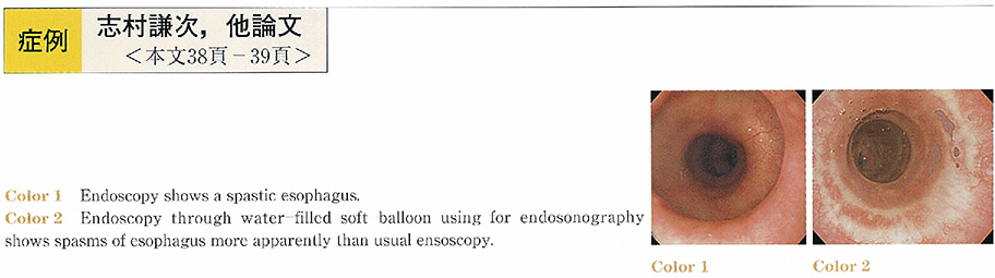
    2002 年 60 巻 2 号 p. 38-39
    発行日: 2002/06/05
    公開日: 2014/05/22
    ジャーナル フリー
    We report a case of diffuse esophageal spasm diagnosed after computed tomography and endosonography. A 46-year-old male was referred to our hospital because of nausea. Recurrent upper abdominal pain lasted for 10 years, and coronary angiography had disclosed a normal finding five years ago. After admission, computed tomography and endoscopic ultasonography revealed the wall thickening of the lower-half esophagus, and endoscope showed spastic contactions of the same part of esophageal wall. X-ray study showed the“rosary bead”appearance, and manometory at 3cm,8cm,and 13cm above the LES revealed the simultaneous contractions. The patient did not wish surgical procedure, and the combination of recurrent hydrostatic balloon-dilation of lower esophagus and medication of nifedipine, lorazepam, and famotidine leaded to symptom improvement.
  • 増尾 貴成, 小野里 康博, 石原 弘, 坂元 一郎, 濱田 哲也, 柳澤 正敏, 新井 弘隆, 阿部 毅彦, 小川 哲史, 棚橋 美文, ...
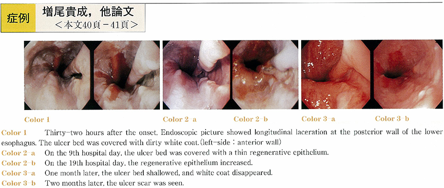
    2002 年 60 巻 2 号 p. 40-41
    発行日: 2002/06/05
    公開日: 2014/05/22
    ジャーナル フリー
    A 56-year-old man was admitted to our hospital because of vomiting after drinking and subsequently a fever and anterior chest pain at swallowing. Endoscopic picture showed longitudinal laceration at the posterior wall of the lower esophagus. Chest CT scan revealed inflammatory esophageal wall thickness and a small amount of pleural effusion. According to the clinical course, endoscopic findings and CT findings, spontaneous esophageal rupture was diagnosed. The patient's physical condition was good even after 24 hours following the onset. Chest pain occurred only during swallowing. In addition, CT scan did not detect mediastinitis or mediastinal emphysema. Thus, we performed conservative treatment. Symptoms and test results showed improvement, he was discharged from the hospital on the 28th day. Surgery is the primary treatment of spontaneous esophageal rupture, however, conservative treatment is applicable in cases that the rupture is limited in the mediastinum and the physical condition of the patient is good.
  • 高橋 裕之, 一原 亮, 池内 哲, 鵜梶 実, 星野 和彦
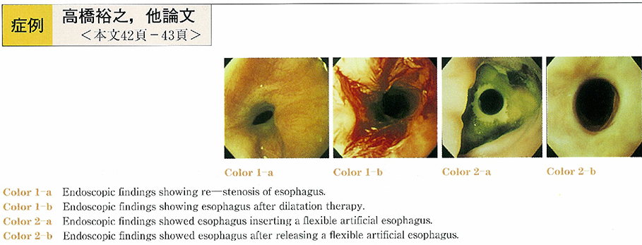
    2002 年 60 巻 2 号 p. 42-43
    発行日: 2002/06/05
    公開日: 2014/05/22
    ジャーナル フリー
    A 16-year-old male injured in his mouth by firework. 2 month later, esophageal stricture developed and esophageal dilatation using a dilater tube was performed every 2 weeks and 48 times in total. But esophageal dilatation could not be preserved. A flexible artificial esophagus is easy of insert and release, so we tried insertion of this artificial esophagus. 2 month later, we released this artificial esophagus from his esophagus. Since this treatment, his symptom has improved. Treatment of esophageal dilatation using a flexible artificial esophagus is useful for benign esophageal stricture because of easy of insert and release.
  • 澤田 幸久, 上平 晶一, 平川 隆一, 吉田 行雄, 井廻 道夫
    2002 年 60 巻 2 号 p. 44-46
    発行日: 2002/06/05
    公開日: 2014/05/22
    ジャーナル フリー
    A 71-year-old woman suffering from liver cirrhosis, underwent gastrointestinal endoscopy. Just after the examination, she had lost the memory of having undergone the examination due to retrograde amnesia, and was disoriented in regard to time and place. She asked a nurse repeatedly in bewilderment,“What am I doing?”“Why am I in this hospital?”. Her situation was explained to her, and was understood, but the answer provided was forgotten within moments due to anterograde amnesia, and once again repeatedly asked the same questions. She could answer her name, birthday, and the name of her physician in charge correctly. But she had lost her memory between the evening before and the moment she finished the examination. Her consciousness was clear, and neurological examinations were normal. Then transient global amnesia (TGA) , presumed to be precipitated by gastrointestinal endoscopy, was diagnosed. The next morning, about 19 hours after the examination, she recovered form TGA. But ultimately, she had lost her memory of about half of the day she underwent the examination, with no neurological deficits. TGA is usually related to physical, emotional stress, and gastrointestinal endoscopy is a stressful examination for examinees. We must therefore be aware that gastrointestinal endoscopy is one of the precipitating events in TGA, and prepare examination circumstances such that nervousness felt by examinees can be significantly reduced.
  • 桑原 公亀, 山田 博文, 石田 秀行, 坂口 大介, 牧田 陽一郎, 小高 明雄, 藤岡 正志, 村田 宣夫, 星野 高伸, 橋本 大定, ...
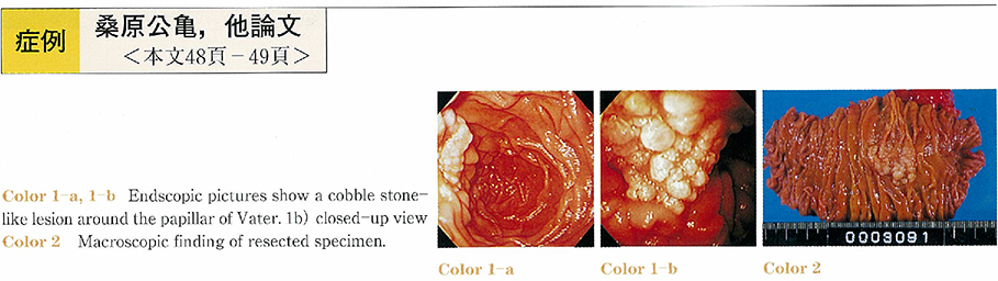
    2002 年 60 巻 2 号 p. 48-49
    発行日: 2002/06/05
    公開日: 2014/05/22
    ジャーナル フリー
    A very rare case of malignant lymphoma of the duodenum is reported. A 67-year-old woman was found to have a cobble stone-like lesion around the papillar of Vater by screening gastroduodenalscope. Histopathology revealed follicular malignant lymphoma. Pylorus preserving pancreatoduodenectomy was successfully performed. The patient has been alive without tumor recurrence since 24 mouths postoperatively. There have been 70 cases of malignant lymphoma of the duodenum in the Japanese reports. And then, follicular lymphoma of the duodenum was detected three cases. Two of them were cobble stone-like appearance endoscopically.
  • 佐々木 克行, 藤木 和彦, 鈴木 俊雄, 渡辺 守
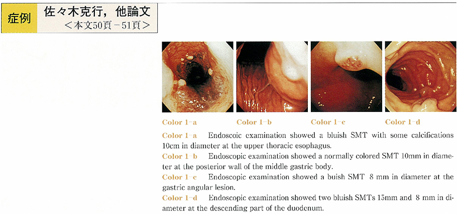
    2002 年 60 巻 2 号 p. 50-51
    発行日: 2002/06/05
    公開日: 2014/05/22
    ジャーナル フリー
    The patient was a 40-year-old woman. She visited our hospital because of hoarseness and difficulty of breathing. A computed tomographic (CT) scan of the chest revealed isodensity mass of esophagus with some calcifications. Endoscopic examination showed a bluish submucosal tumor (SMT) at the upper thoracic esophagus with some calcifications. It also showed two SMTs in stomach and two SMTs at the duodenum. These were similar color to the esophgeal lesion. A magnetic resonance imaging (MRI) of the chest showed the esophageal tumor that was very high intensity on T2-weighted. We suspected these SMTs hemangiomas. In other studys there was one hemangioma of the liver.
    Many cases of bleeding of the gastrointestinal hemangioma are reported, therefore it is necessary for this patient screening of small and large intestin. But it is not done yet because of her rejection.
    Gastrointestinal hemangioma is a benign tumor comparatively rare. We report a very rare case which had been susupected multiple hemangiomas at the upper gastrointestinaltruct.
  • 小澁 敬治, 仁多 寅彦, 吉田 美奈子, 床井 伸介, 藤本 秀明, 小野澤 祐輔, 天川 孝則, 河内 洋, 山田 哲夫, 中村 恭一, ...
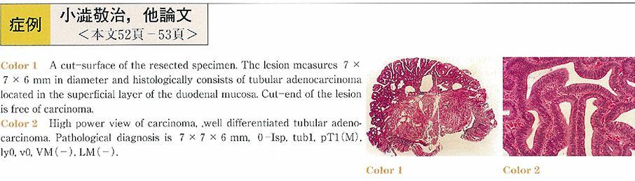
    2002 年 60 巻 2 号 p. 52-53
    発行日: 2002/06/05
    公開日: 2014/05/22
    ジャーナル フリー
    A 73-year-old male visited our hospital with a complaint of appetite loss in 2001. Endoscopic examination showed a semipedunculated polypoid lesion, about 7 mm in size, at the opposite side of the papilla Vater. Endoscopic biopsy revealed adenocarcinoma. Endoscopic mucosal resection was performed after confirmation of negative non-lifting sign, because the lesion was measured less 10 mm in largest diameter. The resected specimen was 7×7×6 mm in dimensions and histologically consisted of tubular adenocarcinoma limited to the superficial layer of the duodenal mucosa (m, ly 0, v 0) . Cut-end of the lesion was free of carcinoma. Duodenal malignant lesion is relatively rare, especially early cancer. Meanwhile, there are many borderline cases of epithelial tumors between benignancy and malignancy in the duodenum. Therefore, complete excisional biopsy such as endoscopic mucosal resection is recommended.
  • 池田 啓浩, 水野 立也, 原田 久, 別府 加寿子, 鈴木 聡明, 越川 均, 岡田 安郎, 丸山 俊秀
    2002 年 60 巻 2 号 p. 54-55
    発行日: 2002/06/05
    公開日: 2014/05/22
    ジャーナル フリー
    A 52-year-old male, who had been on follow-up for his multiple submucosal tumors of the stomach in other clinic since 1988, was referred to our department for further investigation of the tumors showing increase in size and number in August, 2000. The initial x-ray and endoscopic examinations of the gastrointestinal tract detected multiple gently sloping tumors in the stomach, small and large intestine. He, however, was found to have newly developed polypoid lesions in the stomach and cecum at gastroscopy and colonoscopy performed 4 months later than the initial examinations. Biopsy specimens taken from the newly developed lesion were reported as showing carcinoid tumor.
    Soon after the examinations, he suddenly developed unconsciousness due to multiple metastatic tumors of the brain, and he died one month later. Autopsy disclosed a primary lung cancer of small cell type with muitiple secondary deposits in the stomach, small intestine and colon together with other organs. Retrospectively, newly developed polypoid lesions are endoscopically different from carcinoid tumor, which was reported as showing on the biopsied specimen by a pathologist.
    There have been no reports on gastrointestinal lipomatosis co-existent with multiple foci of metastatic lung cancer. Careful analyses of endoscopic findings, however, are considerd to be extremely important to make a diagnosis even in very rare case.
  • 富澤 さやか, 三梨 桂子, 河村 修, 前田 正毅, 下山 康之, 森 昌朋, 草野 元康
    2002 年 60 巻 2 号 p. 56-57
    発行日: 2002/06/05
    公開日: 2014/05/22
    ジャーナル フリー
    Endoscopic removal of sharp and pointed foreign bodies in the upper gastrointestnal tract still poses technical difficulties. We present here our experience with a new endoscopic end lucent skirt protector having the unique characteristic of maintaining its bell portion inverted during the progression of the scope through the gut and flipping back to its shape during withdrawal through the lower esophageal sphincter. This prevents exposure of the esophageal and pharyngeal wall to injuries from the edges of the foreign body. We used this device to remove large sharp and pointed dental prosthesis in the fourth part of the duodenum. We belived that this device enhance the safe removal of a wide variety of potentially dangerous gastroesophageal foreign bodies.
  • 米沢 麻利亜, 井手 徳彦, 宮崎 英史, 土橋 展子, 足立 ヒトミ, 吉田 一成
    2002 年 60 巻 2 号 p. 58-59
    発行日: 2002/06/05
    公開日: 2014/05/22
    ジャーナル フリー
    A 64-year-old male suffering from anemia and tarry stools was comfirmed gastric erosions and duodenitis by endoscopic examination in 1998. Subsequently, he was beewing monitered with prescribed oral H2 blockers.
    However in May 2000, the patient complained of tarry stools just like the previous conditions. No special findings were found to show the source of the bleeding.
    On December 15th 2000, the patient was admitted into our hospital, due to his complaint of tarry stools. The urgent endoscopic examination revealed the bicephalic diverticulum on the adoral papilla. There was palsatile bleeding found on the saddle which placed inbetween the two chambers consisting of the bicephalic diverticulum.
    Even after the treatment of electric-coagulation, the bleeding reoccurred twice. However after using the additional clipping, that condition was completely under control.
    We present this as being extremely interesting case.
  • 秋谷 行宏, 横山 滋彦, 羽尾 邦彦, 小嶋 隆行, 柳 健, 松下 晃, 恩田 昌彦
    2002 年 60 巻 2 号 p. 60-61
    発行日: 2002/06/05
    公開日: 2014/05/22
    ジャーナル フリー
    A 61-year-old man was admitted with the chief complaints of nausea and vomiting. Abdominal CT revealed dilatation of the second and third portions of the duodenum, and a tumor compressing the inferior vena cava. Duodenography revealed obstruction at the third portion of the duodenum. Endoscopy revealed a type 2 tumor causing stenosis of the third portion of the duodenum. The distance between the papilla of Vater and the tumor was 10mm. Biopsy revealed a moderately differentiated adenocarcinoma.
    A stomach tube was inserted after admission. The volume of drainage from the stomach tube was about 5000-8000ml/day, and consequently, the patient developed marked hypochloremia. To correct this electrolyte abnormality, we performed percutaneous endoscopic gastrostomy and enterostomy via a laparotomy, and connected the two so as to allow the digestive juices to flow from the stomach into the intestinal tract. After two weeks, the marked hypochloremia was corrected. Pyloruspreserving pancreaticoduodenectomy was then performed. The resected specimen revealed a type 3 carcinoma in the third portion of the duodenum. Histopathological findings revealed a moderately differentiated adenocarcinoma. No postoperative complications occurred.
    Thus, connecting the gastrostomy with the enterostomy so as to allow the digestive juices to flow from the stomach into the small intestine proved to be a very useful measure to correct the electrolyte abnormality.
  • 名越 淳人, 鄭 義弘, 菱川 健一郎, 田島 博人, 斎藤 哲彦, 多田 有紀, 三輪 剛, 峯 徹哉
    2002 年 60 巻 2 号 p. 62-63
    発行日: 2002/06/05
    公開日: 2014/05/22
    ジャーナル フリー
    A case of male of ischemic colitis followed by mesenteric panniculitis is reported. The patient complained of hematochezia and lower abdominal pain. Laboratory findings on admission were increased WBC and positive CRP. Colonoscopy showed mucosal edema, and multiple erosions and ulcers in sigmoid colon. The patient was diagnosed as ischemic colitis and we treated him conservatively with intravenous drip infusion of antibiotics. His symptoms were promptly improved. After he started oral intakes, he complained of abdominal pain, and laboratory findings revealed further increased WBC and CRP. Abdominal roentgenogram showed a“stepladder”sign as ileus. Colonoscopy showed mucosal edema and narrowing without erosions and ulcerations in the sigmoid colon. Contrast-enhanced CT image demonstrated wall-thickning of the sigmoid colon surrounded by high density which suggests inflammation in soft tissues around sigmoid colon. Under the diagnosis of ischemic colitis followed with mesenteric panniculitis, he was treated with total parenteral nutrition and prednisolone. Clinical symptoms were improved and laboratory findings turned to be normal.
  • 福島 淑隆, 吉川 望海, 熊野 雄一, 青柳 有司, 佐藤 貴也
    2002 年 60 巻 2 号 p. 64-65
    発行日: 2002/06/05
    公開日: 2014/05/22
    ジャーナル フリー
    A 67-year-old woman admitted with a chief complaint of fever and left lower abdominal pain. Abdominal enhanced CT demonstrated an inhomogenous density mass in left iliac lesion. Six days on admission, she suddenly had bloody stool. Colonoscopic findings showed the edematous and easy-bleeding mucosa with multiple erosions and longitudinal ulcers in the sigmoid to descending colon. Histological findings of uterine tube revealed the infiltration of inflammatory cells mainly including neutrophils and the proliferation of fibrosis due to pyosalpinx. Colonoscopic findings after surgical treatment showed deformities of the internal wall and normal mucosa in the sigmoid to descending colon. It is rare that pyosalpinx was complicated with ischemic colitis, which would be precious to discuss the pathogenesis.
  • 三橋 淳, 五十嵐 正広, 安達 滋, 吉澤 繁, 佐田 美和, 小林 清典, 勝又 伴栄, 西元寺 克禮, 三富 弘之, 岡安 勲
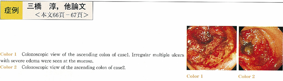
    2002 年 60 巻 2 号 p. 66-67
    発行日: 2002/06/05
    公開日: 2014/05/22
    ジャーナル フリー
    We experienced two cases of hemorrhagic colitis due to Enterohemorrhagic Escherichia coli : O-157 (EHEC : O-157) diagnosed by endoscopy.
    A 45 year old woman was admitted to our hospital because of abdominal pain and bloody watery diarrhea. Abdominal echogram showed edema and swelling of the right-sided colon. Emergent colonoscopy revealed severe hemorrhagic colitis extending from the transverse colon to the ascending colon. Multiple ulcers and erosions, and severe edema were seen at the colon. EHEC : O-157 and verotoxin were confirmed by stool. She was given fosfomycin during 7 days.
    A-26-year old man was admitted to our hospital because of abdominal pain and bloody watery diarrhea. Abdominal echogram showed swelling of the colonic wall of the right-sided colon. Emergent colonoscopy revealed severe hemorrhagic colitis with severe edema and erosion extending from the ascending colon to the cecum. EHEC : O-157 and verotoxin were confirmed by stool. Fosfomycin was effective and he left the hospital after 10 days.
    It is concluded that abdominal echogram, emergent colonoscopy and stool culture are useful for the diagnosis of hemorrhagic colitis due to EHEC : O-157.
  • 大久保 力, 細井 董三, 入口 陽介, 中井 呈子, 中橋 栄太, 中村 尚志, 小田 丈二, 大浦 通久, 水谷 勝, 大久保 明, 小 ...
    2002 年 60 巻 2 号 p. 68-69
    発行日: 2002/06/05
    公開日: 2014/05/22
    ジャーナル フリー
    A 62 years-old male visited us to undergo further detailed examination following the positive fecal occult blood test. Type-Ip lesion revealing a fine villous structure at the top was discerned endoscopically which resulted in the diagnosis of serrated adenoma. EMR (endoscopic mucosal resection) was well indicated. At the second endoscopic examination for EMR, however, in the lower part of the lesion, an irregularly-shaped concavity, endoscopically rigid, revealing the loss of villous structure was present, leading to the suspect diagnosis of sm2 invasive cancer in adenoma. Accordingly, the treatment was switched from EMR to surgical operation. Furthermore, through endoscopy just before the surgical operation, the concavity deepened along with the whitish coating, which was strongly indicative of sm2 cancer.
    Laparoscopic assisted partial resection was carried out in the ascending colon. Pathologically diagnosed as moderately differentiated adenocarcinoma in serrated adenoma, sm2, ly2, v2, n0. The malignant potential of serrated adenoma is considered to be almost equal to that of ordinary adenoma. Taking into consideration that the larger the diameter of a lesion becomes, the greater the malignant incidence grows, and that there actually exists sm invasive cancer originating from serrated adenoma requiring surgical operation, we are expected to scrutinize the lesion of serrated adenoma through endoscopy as to whether it is cancerous or not.
  • 穴澤 康志, 佐田 美和, 奥野 順子, 吉澤 繁, 小林 清典, 五十嵐 正広, 勝又 伴栄, 西元寺 克禮, 三富 弘之
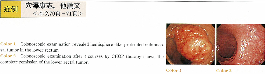
    2002 年 60 巻 2 号 p. 70-71
    発行日: 2002/06/05
    公開日: 2014/05/22
    ジャーナル フリー
    A 68-year-old woman was admitted to our hospital because of bloody stool and submucosal tumor of the rectum. Colonoscopic examination and barium enema revealed hemisphere-like protruded submucosal tumor 5.6 cm in diameter in the lower rectum. Endoscopic ultrasonography shows that the lesion was low echoic mass and extraluminal proturuded, that finding was suggested a1 to a2 invasion. Histological findings of the biopsied specimens showed that it was a non-Hodgkin's, diffuse, small to medium sized cell, B-cell type of malignant lymphoma and that origened to mucosa-associated lymphoid tissue type (MALT) lymphoma.
    After four courses by CHOP therapy, the bloody stool had disappeared, and colonoscopic examination revealed that submucosal tumor comprised of malignant lymphoma had disappeared. The patient has had no recurrence of the disease during 7 months since the last chemotherapy.
  • 船津 康裕, 遠藤 豊, 吉田 生馬, 徳永 晋, 黒木 優一郎, 加藤 健史, 唐沢 隆文, 服部 隆志, 安田 宏, 井上 和明, 与芝 ...
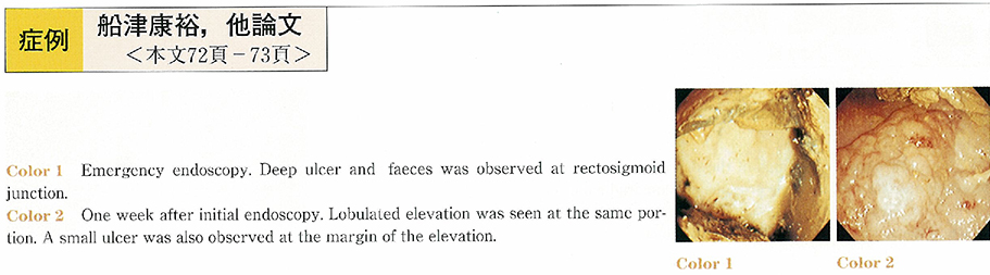
    2002 年 60 巻 2 号 p. 72-73
    発行日: 2002/06/05
    公開日: 2014/05/22
    ジャーナル フリー
    A 74-year-female was admitted to our hospital complaining of lower abdominal pain and hematochezia. A large and deep ulcer was observed at rectosigmoid junction by emergency colonoscopy. After one week TPN therapy, follow up study was performed. The ulcer was altered into elevated lesion with lobulation like laterally spreading tumor granular type (LST-G) . A small ulcer was also detected at the margin of the elevation. The endoscopic diagnosis was confused whether neoplastic tumor or stercoraceous ulcer. The initial endoscopy was performed in poor preparation. Large faeces interrupted observation and only a part of the ulcer was seen. However, there was no pit on the surface of the elevated lesion in magnification. The biopsy revealed the elevation consisting of inflammatory granulation tissue. The elevation was disappeared completely and only ulcer scar was observed after 4 month later. The inflammatory Granulomatous elevation appears in gastric ulcer healing processes, especially treated with PPI. There was no case report of stercoraceous ulcer making inflammatory Granulomatous elevation in Japanese literature. And it is also very rare colonic ulcer making inflammatory Granulomatous elevation. This case is very rare.
  • 森下 慎二, 川畑 正博, 松本 政雄, 中村 昭子, 新村 和平, 佐藤 芳之, 小沢 卓夫, 羽原 隆, 黄田 光博, 井野元 勤, 池 ...
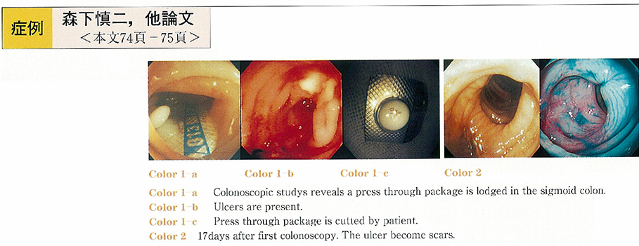
    2002 年 60 巻 2 号 p. 74-75
    発行日: 2002/06/05
    公開日: 2014/05/22
    ジャーナル フリー
    A 80-year-old woman was admitted because of abdominal pain. On the third day of admission, she had bloody stool with increasing abdominal pain. Colonoscopy revealed that a press-through package (PTP) was lodged in the sigmoid colon and ulcers were present adjacent to sharp edge. The package was removed by endoscopic forceps and the patient was discharged in good capsules. PTPs are rapidly becoming popular as packages for tablets and capsules. Reports of accidental swallowing of a PTP have been increasing but progression of PTPs to the colon is rare. We were able to remove it endoscopically. We propose that drugs should not be dispended in PTPs especially to elderly patients. It is also important to inform patients that accidental swallowing of PTPs is disastrus and warn them not to swallow PTPs. Collaboration of co-medical staff such as a pharmacist is necessary.
  • 角嶋 直美, 藤城 光弘, 矢作 直久, 岡本 真, 榎本 祥太郎, 三好 秀征, 森山 義和, 山道 信毅, 建石 綾子, 岡 政志, 辻 ...
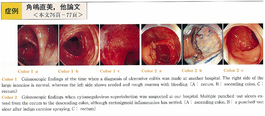
    2002 年 60 巻 2 号 p. 76-77
    発行日: 2002/06/05
    公開日: 2014/05/22
    ジャーナル フリー
    A 56-year-old Japanese man with ulcerative colitis was admitted because of being judged as resistance to treatment with intravenous prednisolone (60-100 mg/day) and oral sulfasalazine (3 g/day) over 1.5 months at another hospital. Colonoscopy at our hospital showed multiple punched-out ulcers from the cecum to the descending colon without active inflammation in the rectum. Although cytomegalovirus superinfection after long-term steroid administration was highly suspected and the treatment for it was planned, he developed massive bleeding and progressive clinical deterioration that necessitated emergency total colectomy in several days after the colonoscopic examination. Histopathologically, cytomegalic inclusion bodies were identified on the ulcer rim and base, while the activity of ulcerative colitis was kept under control with steroid. Because the antigenemia of cytomegalovirus continued positive even after colectomy, ganciclovir was administered until the antigenemia was turned into negative. The present case showed that cytomegalovirus superinfection should be considered in patients with inflammatory bowel disease having long-term steroid administration and immediate serological and histological investigation should be performed when the clinical course is unusual.
  • 山根 建樹, 内山 幹, 中村 眞, 石井 隆幸, 佐藤 泰弘, 藤瀬 清隆, 加藤 弘之, 小林 正之, 戸田 剛太郎
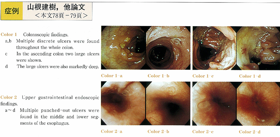
    2002 年 60 巻 2 号 p. 78-79
    発行日: 2002/06/05
    公開日: 2014/05/22
    ジャーナル フリー
    A 21-year-old female entered our hospital complaining of diarrhea, abdominal pain and body weight loss. Colonoscopic examination revealed multiple small and middle sized discrete ulcers throughout the whole colon and two large and deep ulcers in the ascending colon. Biopsy specimens taken from the small ulcers of the left sided colon showed non-caseating epithelioid cell granuloma histologically. And upper gastrointesinal endoscopic examination revealed multiple punched-out ulcers in the middle and lower segments of the esophagus. Granuloma was not evident in biopsy specimens taken from the esophageal ulcers. Though the large colonic ulcers and punched-out esophageal ulcers suggested Behçet's disease or simple ulcer, we suspected her illness as Crohn's disease due to detection of granuloma and prescribed her Prednisolone. Then, she recovered and follow-up endoscopic examination showed improvement of the ulcers. This case was difficult to make definite diagnosis.
  • 濵野 耕靖, 泉 並木, 川村 央信, 鈴木 比有万, 土谷 薫, 板倉 潤, 朝比奈 靖浩, 野口 修, 内原 正勝, 三宅 祥三, 堺 ...
    2002 年 60 巻 2 号 p. 80-81
    発行日: 2002/06/05
    公開日: 2014/05/22
    ジャーナル フリー
    Laparoscopic radiofrequency ablation is considered first-line therapy especially for liver cancer on the liver surface and located in the subphrenic area. We have recently used a sector ultrasonic probe for laparoscopic treatement with a guiding tract to obtain accurate ablation of targeting HCC nodule.
  • 朴 勝春, 鈴木 秀明, 菅原 崇, 斎藤 聖磨, 里田 誠, 香山 秀之, 林 弘美, 高谷 育男, 松岡 幹雄, 深澤 信悟, 細井 英 ...
    2002 年 60 巻 2 号 p. 82-83
    発行日: 2002/06/05
    公開日: 2014/05/22
    ジャーナル フリー
    A 74-year-old man, who had a history of choledocho-lelithiasis with obstructive jaundice treated by endoscopic papillary balloon-dilation method, admitted to our hospital, because of treatment of diabetes mellitus.
    Abdominal CT showed cholelithiasis and wall thickening of the gallbladder. We suspected gallbladder cancer, and performed endoscopic retrograde cholangiography (ERC) . When we inserted endoscope, cholelith was found in the antrum of the stomach, and ERC visualized common bile duct (CBD) , atrophic gallbladder and also the stomach. We found fistula on the greater curvature of the antrum, and injected contrast agents under balloon-occlusion, therefore atrophic gallbladder and CBD was seen, and made confirmation as cholecystogastric fistula. Then we collected cholelith endoscopically. Surgical operation was performed with a diagnosis of chronic cholecystitis.
    Cholecystogastric fistula is very rare among the spontaneous internal biliary fistulae. It is difficult to make a preoperative diagnosis of cholecystogastric fistula, but in our case, we timely performed an examination of ERC, therefore we found cholelith and fistula in the stomach, and were able to confirm as cholecystogastric fistula.
  • 神澤 輝実, 藤原 崇, 鈴木 瑞佳, 雨宮 こずえ, 江川 直人, 門馬 久美子, 中嶋 均, 榊 信廣
    2002 年 60 巻 2 号 p. 84-85
    発行日: 2002/06/05
    公開日: 2014/05/22
    ジャーナル フリー
    A 72-year-old man had been followed up for branch-type mucin producing pancreatic tumor for three years. Endoscopic retrograde cholangiopancreatography (January, 1998) showed a 1.5 cm-sized irregular cystic lesion in the neck of the pancreas, and amorphous filling defects of mucin in the main pancreatic duct. Though the patient had been asymtomatic, endoscopic retrograde cholangiopancreatography (February, 2001) showed the main pancreatic duct obstructed near the origin of the inferior branch. Magnetic resonance cholangiopancreatography showed cluster of cyst near the obstructed main pancreatic duct, and irregular dilatation of upstream main pancreatic duct. Pylorus preserving pancreaticoduodenectomy was performed. Histologically, 1.1-sized invasive carcinoma derived from intraductal papillary carcinoma was detected around the main pancreatic duct near the cyst. In follow-up of the branch-type mucin producing pancreatic tumor, it is necessary to note the possibility of the tumor in the main pancreatic duct near the cyst.
  • 中川 望, 山田 拓司, 杉山 和子, 奥田 浩史, 粕谷 健司, 間下 信昭, 中 佳一, 堀 亮太, 吉岡 伊作, 山下 巌, 野村 直 ...
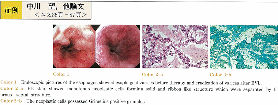
    2002 年 60 巻 2 号 p. 86-87
    発行日: 2002/06/05
    公開日: 2014/05/22
    ジャーナル フリー
    A 62 year-old male was admitted to our hospital for detailed examinations of a low density mass in the spleen by abdominal CT scan at a physical checkup. Abdominal CT scan also demonstrated portal obstruction and collateral veins around pancreatic head. Furthermore, abdominal angiograhy showed a hypovascular lesion in the spleen. Upper gastroendoscopy demonstrated esophageal varices, which were eradicated after EVL. The above examinations, however, could not make the definitive diagnosis, therefore splenectomy was carried out on 5 July 2000.
    The tumor size of the cut surface was 4×5cm in diameter. Histopathological diagnosis was non-functioning pancreatic endocrine tumor. The patient remains well with no evidence of recurrence one year and seven months after the operation.
  • 今村 綱男, 坂本 仁, 池上 覚俊, 江口 潤一, 柳川 達郎, 塙 勝博, 三代川 章雄, 吉田 仁, 野津 史彦, 草間 一成, 片寄 ...
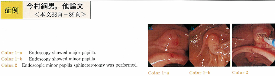
    2002 年 60 巻 2 号 p. 88-89
    発行日: 2002/06/05
    公開日: 2014/05/22
    ジャーナル フリー
    A 65-year-old male was admitted to our hospital because of repeated abdominal pain. The patient had been previously diagnosed as having chronic pancreatitis with pancreatic stones on the basis of finding of abdominal computed tomography (CT) . Endoscopic retrograde pancreatography revealed pancreas divisum and chronic pancreatitis with pancreatic stones. We judged that chronic dorsal pancreatitis was caused by insufficiency of pancreatic juice drainage from minor papilla, so we performed endoscopic minor papilla sphincterotomy for the improvement of pancreatic juice drainage. After the treatment, the patient has been free of episode of abdominal pain. Our experience suggests that endoscopic minor papilla sphincterotomy is one of the effective and noninvasive treatments of chronic dorsal pancreatitis with pancreas divisum.
feedback
Top