Progress of Digestive Endoscopy
Online ISSN : 2187-4999
Print ISSN : 1348-9844
ISSN-L : 1348-9844
83 巻, 1 号
選択された号の論文の79件中1~50を表示しています
掲載論文カラー写真集
内視鏡の器械と技術
  • 廣岡 映治, 松山 秀樹, 伊藤 眞史, 佐藤 純, 福田 千文, 岡本 史樹
    2013 年 83 巻 1 号 p. 43-46
    発行日: 2013/12/14
    公開日: 2013/12/21
    ジャーナル フリー
    大腸悪性狭窄によるイレウスは,日常よく遭遇するoncological emergencyである。従来はHartmann手術や人工肛門造設術による二期的手術が行われていたが,近年経肛門的イレウス管留置や大腸ステントが導入され緊急手術が回避されるようになった。大腸ステントは非手術症例に対する姑息的治療や手術までの待機的治療(bridge to surgery : BTS)として安全に行われるようになったが穿孔や逸脱などの偶発症を起こす可能性もあり,留置する際には細心の注意が必要である。今回我々は緩和医療として3例,BTSとして3例に対する大腸ステント治療を経験した。全例でステント留置は成功したが1例は留置後早期に敗血症を来しステントによる影響が示唆された。しかし5例は穿孔や逸脱などの偶発症を認めずイレウス管による苦痛と人工肛門を回避でき,患者QOLの向上に大きな役割を果たした。
  • 柿本 年春, 辻 忠男, 篠崎 博志, 桂 英之, 金田 浩幸, 水谷 友美, 三浦 邦治, 宮永 亮一, 松波 幸寿, 山藤 和夫, 竹島 ...
    2013 年 83 巻 1 号 p. 47-50
    発行日: 2013/12/14
    公開日: 2013/12/21
    ジャーナル フリー
    急性膵炎の合併症の1つであるwalled-off necrosis(WON)は,感染を合併すると治療に難渋する。我々は2009年5月〜2013年5月までに,急性膵炎によるWON 7例(男性6例/女性1例,年齢45〜77歳)に経皮的ドレナージとその後,必要に応じて経皮的内視鏡的necrosectomyを行う治療(step-up approach)(経皮的ドレナージのみ4例,内視鏡的necrosectomy3例)を施行した。Necrosectomyは平均3.6回施行し,重篤な合併症は認めなかった。急性膵炎に伴うWONのstep-up approachは,保存的治療のみでは治療困難な症例に試みるべき治療の1つと考える。なお,経皮的アプローチで危惧される膵液漏については,経乳頭的なルート等で内瘻化することで予防可能である。
臨床研究
  • 清水 智樹, 山本 頼正, 富田 英臣, 岡本 恒平, 堀内 裕介, 石川 寛高, 松尾 康正, 吉澤 奈津子, 大前 雅実, 石山 晃世志 ...
    2013 年 83 巻 1 号 p. 51-55
    発行日: 2013/12/14
    公開日: 2013/12/21
    ジャーナル フリー
    上部消化管内視鏡治療時の麻酔として,一般的に鎮静剤〔benzodiazepine(BZ)系など〕と鎮痛剤が併用されるが,時に効果不十分な症例を経験する。その際,haloperidol(HLP)やpropofolが使用されるが,どの症例で効果不十分となるかは明確になっていない。鎮静不十分となる症例の予測因子を明らかにすべく,HLP追加投与を要した症例の解析を行った。2011年11月〜2012年6月に当院で専門医が内視鏡的粘膜下層剥離術を施行した,単発の食道・胃の上皮性腫瘍性病変169例を対象とした。Midazolam(MZ)+pethidine hydrochloride(PH)で鎮静した群152例(90%)とMZ+PHにHLPを併用した群17例(10%)の2群でそれぞれの臨床像を比較検討した。HLP併用群では,有意に平均年齢が低く,男性が多く,体表面積が大きく,1年以内の飲酒・喫煙歴を有し,食道病変が多く,病変径も大きかった。また,術前内視鏡時からMZ,PHを多く要し,治療時もPH投与量が多く麻酔時間が長かった。有意差を示した項目に対してさらに多変量解析を行った結果,有意な独立因子は,①60歳未満,②病変径30mm以上,③術前検査時のMZ投与量≧0.06mg/kgの3点であった。これらはMZ+PHによる鎮静が不十分となることを予測する因子として有用である。
  • 山本 泰漢, 金子 和弘, 小西 一男, 倉橋 利徳, 伊藤 紘朗, 片桐 敦, 粂川 陽祐, 桑原 芽衣子, 久保田 祐太郎, 村元 喬, ...
    2013 年 83 巻 1 号 p. 56-59
    発行日: 2013/12/14
    公開日: 2013/12/21
    ジャーナル フリー
    近年,社会の高齢化に伴い,脳・心血管障害や整形外科疾患を持つ患者は増加傾向である。それに伴ってaspirinやNSAIDs服用者による消化管障害の有害事象も出現し,胃・十二指腸潰瘍出血を来すことも多い。潰瘍出血に内視鏡的止血術は有用であるが,再出血例も散見される。そして出血性潰瘍患者の中には,aspirinやNSAIDs服用者も少なくない。またaspirinには抗血小板作用があり,一般的に出血要因と考えられている。本検討では,当院で胃・十二指腸潰瘍出血と診断した188例(男性136例,女性52例)をNSAIDs群(aspirin潰瘍群,その他NSAIDs潰瘍群)と消化性潰瘍群に分類して,再出血率について各群で比較検討した。再出血率はNSAIDs全体では10.8%(8/74例)であった。Aspirin潰瘍群14.0%(6/43例),その他NSAIDs潰瘍群6.5%(2/31例),消化性潰瘍群11.4%(13/114例)であり,aspirin潰瘍群と消化性潰瘍群でやや高い傾向であったが,再出血率で有意差を認めなかった。以上より胃・十二指腸潰瘍出血において,aspirinを含むNSAIDs服用歴は再出血のリスク因子の可能性が低いことが示唆された。
  • 橋爪 真之, 佐川 俊彦, 佐藤 賢, 大塚 修, 田中 秀典, 飯塚 賢一, 土岐 譲, 井上 照基, 田中 寛人, 草野 元康, 森 昌 ...
    2013 年 83 巻 1 号 p. 60-64
    発行日: 2013/12/14
    公開日: 2013/12/21
    ジャーナル フリー
    小腸疾患診療は,カプセル内視鏡(CE)やバルーン内視鏡(DBE)の登場とともに,飛躍的な向上を遂げた。ただしこれらCE,DBEの病院普及の程度や実臨床での使用経験など不明な点は多い。2012年4月時点での群馬県内(以下,県内)すべての病院120施設におけるCE,DBEの所有状況を検討した。また,普及の進んでいるDBEおよびCEによる診療現況と群馬大学大学院医学系研究科病態制御内科学(以下,当科)が構築したカプセル内視鏡ネットワーク(CEN)の現況については,当科とその関連19施設を対象として検討を行った。CEやDBEの所有状況は県内で偏在化がみられる。しかし,機器レンタルも含め,導入医療機関は増えており,今後の拡充が期待される。DBEでの施行目的は出血精査が3分の2を占め,全小腸観察率は64.0%,偶発症では膵炎が1件(0.5%)と低率で安全に施行することができた。また,CEでの施行目的は出血精査が約4分の3を占め,全小腸観察率は79.4%であった。カプセル滞留3件(1.5%)を認めたが,2件はDBEで,1件は小腸狭窄のため手術で回収することができた。診療現況はDBE,CEいずれも既報とほぼ匹敵するものと考えられた。CENは,構築時の業務の他,当科が中心となって小腸疾患患者の診療アドバイスや当科への紹介受診,また,医師に対してCEの読影方法の教育なども行うシステムへと発展している。したがって,地域医療の均霑化において向上の余地はあるものの,県内の病院においてDBEおよびCEの所有状況の拡充傾向ならびに当科および関連施設において診断治療技術の向上が認められる。
  • 松下 典正, 須藤 泰裕, 芹澤 朗子, 新井 俊文, 窪田 猛, 井上 達夫
    2013 年 83 巻 1 号 p. 65-68
    発行日: 2013/12/14
    公開日: 2013/12/21
    ジャーナル フリー
    当院では2011年1月より急性虫垂炎に対して腹腔鏡手術(laparoscopic appendectomy : LA)を導入している。導入期における従来の小開腹手術(open appendectomy : OA)とLAに関して,術後経過や合併症などについて評価を行った。2009年4月〜2012年12月までの約3年9カ月をOAからLAへの移行期間として観察し,この間の小開腹ならびに腹腔鏡下での虫垂炎手術症例を集計した。発症急性期に手術を行ったOA群とLA群の患者に採血上の炎症反応の差は認めなかった。手術時間はOA群が49.0±22.1分,LA群が71.0±26.6分とLA群が有意に長かった。手術後合併症の主なものは,創部感染症であり,CDC(Centers for Disease Control and Prevention)ガイドライン1)が示す比較的軽度の表層手術部位感染(SSI)を含めれば,OA群では45.4%に合併症の発症を認めたが,LA群では18.5%とLA群で合併症の低下を認めた。LAは術後合併症低下など利点も多く,有用な治療法と考えられ,当院での成績を報告する。
  • 宮澤 志朗, 木田 光広, 山内 浩史, 奥脇 興介, 徳永 周子, 岩井 知久, 菊地 秀彦, 竹澤 三代子, 渡辺 摩也, 今泉 弘, ...
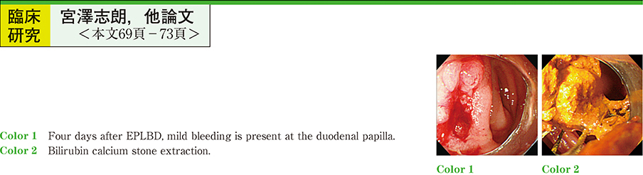
    2013 年 83 巻 1 号 p. 69-73
    発行日: 2013/12/14
    公開日: 2013/12/21
    ジャーナル フリー
     近年,術後再建腸管に伴う総胆管結石症に対し,小腸鏡下胆管結石治療が行われるようになってきた。術後再建腸管症例では,endoscopic sphincterotomy(EST)を安全に行える処置具はなく,endoscopic papillary balloon dilation(EPBD)が行われることが多い。しかし巨大結石や積み上げ結石に対するEPBDでは,完全截石に時間を要し,複数回の処置が必要となる。そこで当院では2011年2月以降,術後再建腸管総胆管結石症に対し小腸鏡下endoscopic papillary large balloon dilation(EPLBD)を16例に施行しており,その有効性と安全性について検討した。対象は男女比15 : 1で平均年齢は76.8歳であった。術後再建方法はB-Ⅱ再建法5例,R-Y再建法 11例であった。十二指腸乳頭部は無処置12例,EST後3例,EPBD後1例であった。結石径中央値は12.0mm,平均最大胆管径は14.8mm,結石数中央値は1個であった。乳頭処置は全例ESTを行わずに一期的にEPLBDを行った。バルーン径中央値は12.8mmで下部胆管径を超えないものとした。全例に完全截石は可能であり,13例(81%)は1回の治療で完全截石が可能であった。平均治療時間は44.2分,EPLBD後の治療時間中央値は15.0分であった。偶発症は1例(6%)で止血術を必要としない軽症出血を認めたが,急性膵炎は認めなかった。
     術後再建腸管に伴う総胆管結石症に対する小腸鏡下EPLBDは,重篤な偶発症もなく有効な乳頭処置と考えられたが,出血傾向のある症例には慎重に適応を選択する必要がある。
経験
  • 細江 直樹, 長沼 誠, 柏木 和弘, 今枝 博之, 金井 隆典, 緒方 晴彦
    2013 年 83 巻 1 号 p. 74-76
    発行日: 2013/12/14
    公開日: 2013/12/21
    ジャーナル フリー
     大腸内視鏡検査専用検査食を使用することにより,腸管洗浄液の減量と患者受容性のさらなる向上が可能かどうか検討した。過去に大腸内視鏡検査を,検査前日の就寝前にピコスルファートナトリウム水和物内用液0.75%,20mlを内服,検査当日にポリエチレングリコール含有電解質溶液(以下,PEG) 2l服用する前処置(PEG2l法)を行った患者30例を対象とし,検査前日の昼食以降(昼食・間食・夕食)の食事に,大腸内視鏡検査専用検査食を摂取してもらい,PEG2l法のPEG服用量を1lに減量して前処置(検査食法)を行った。前処置の完了率,腸管洗浄度を評価した。また患者受容性,有害事象(嘔気,腹満),次回希望する前処置法(検査食法もしくはPEG2l法)についてアンケート調査を行った。1例で同意撤回があり,29例に検査食法による前処置を行った。2例で洗浄不良がありPEG1lを追加内服した。PEG1l内服のみの27例のうち,26例においては,腸管洗浄度は良好であったが,1例で洗浄度が不十分であった。検査食群では全例で前処置を完了できた。検査食法を行った全例で,次回も検査食法を希望した。今後,対照群を設定したランダム化比較試験が必要と考えられるが,大腸内視鏡検査専用検査食を使用することにより,腸管洗浄液減量の可能性が示唆された。
内視鏡の器械と技術
  • 山田 千寿, 新田 宙, 石川 文彦, 藤田 昌久, 釜田 茂幸, 相田 俊明, 高橋 誠, 葛西 豊高, 福田 裕昭, 宮原 庸介, 川辺 ...
    2013 年 83 巻 1 号 p. 78-79
    発行日: 2013/12/14
    公開日: 2013/12/21
    ジャーナル フリー
    These cases included one patient with bile duct cancer, two with pancreatic cancer and one with gallbladder cancer. Duodenal SEMS were deployed prior to biliary SEMS in three patients, and the remaining patient was treated in the reverse order. Duodenal SEMS were placed at the oral side of the papilla via the through-the-scope method because the strictures were proximal to the papilla in all patients. Combined endoscopic stenting was successful and no procedure-related complications occurred in any of the patients. One patient developed recurrent GOO due to tumor ingrowth as a late complication, and was treated by re-insertion of another SEMS using the stent-in-stent method 161days after the first SEMS. The QOL of all patients was well-maintained until the time of death. Combined endoscopic biliary and duodenal stenting is a feasible and effective technique for the palliation of malignant GOO and biliary obstruction.
症例
  • 藤村 匠, 大森 泰, 平岩 訓彦, 川久保 博文, 入江 理恵, 杉浦 仁
    2013 年 83 巻 1 号 p. 80-81
    発行日: 2013/12/14
    公開日: 2013/12/21
    ジャーナル フリー
    The patient was a 69-year-old man diagnosed with type 0-IIa+IIc superficial carcinoma in the right epiglottis and base of the tongue, and type 0-Is+IIb superficial carcinoma in the right pyriform sinus. Lymph node and organ metastases were not detected. Endoscopic laryngo-pharyngeal surgery (ELPS) was performed on these lesions. Pathological findings revealed that the type 0-IIa+IIc lesion was a well differentiated squamous cell carcinoma (SEP, ly0, v0, HM0, VM0) and the type 0-Is+IIc lesion was a moderately differentiated squamous cell carcinoma (SEP, ly1, v1, HM0, VM0) .
    ELPS is video-assisted surgery which is performed under general anesthesia. The concept and technique of ELPS are based on the principles of laparoscopic surgery and endoscopic submucosal resection (ESD) . Using ELPS it is possible to perform large en bloc resection in the oral, supraglottic and pharyngeal areas. We perform ELPS with two doctors of the operator and the endoscopic operator. The endoscopic operator inserts a digestive endoscope through the mouth. The surgeon inserts surgical instruments via the mouth, and removes the lesion while watching the image on a monitor. Our experience indicates that it is possible to safely remove a carcinoma from the base of the tongue using ELPS, and also that the procedure is useful and effective for treatment of superficial carcinoma.
  • 小宮 靖彦, 稲生 優海, 河島 圭吾, 内藤 舞, 飯沼 瑞恵, 藤田 祐司, 江塚 明子, 内山 詩織, 金沢 憲由, 谷 理恵, 川名 ...
    2013 年 83 巻 1 号 p. 82-83
    発行日: 2013/12/14
    公開日: 2013/12/21
    ジャーナル フリー
    Recently, endoscopic submucosal dissection (ESD) has been widely used as a therapeutic procedure for mucosal cancer of the digestive tract, including esophageal disease. However, ESD may result in bleeding, perforation and post-treatment stenosis of the esophagus. In high-risk patients with underlying disease, ESD should be avoided for treatment of superficial esophageal carcinoma. Argon plasma coagulation (APC) has been reported to be a safe and useful procedure for the management of gastric mucosal cancer. We report here two cases of superficial esophageal carcinoma and high-grade dysplasia of the esophagus which were treated using APC.
    Case 1 : An 84-year-old man─with malignant lymphoma─was diagnosed with superficial squamous cell carcinoma of the esophagus. Since he had been receiving steroid therapy and had renal dysfunction, he underwent APC in preference to endoscopic resection to treat the esophageal carcinoma. After ablation of the lesion by APC, the ablated epithelial layer was removed by endoscopic distal attachment (soft hood) and additional APC was performed on the lesion. The esophageal carcinoma disappeared.
    Case 2 : A 55-year-old man─who had lower pharyngeal cancer─was diagnosed with early esophageal cancer. Since he had multiple tiny lesions unstained by iodine, he underwent chemoradiotherapy for the esophageal cancer. High-grade dysplasia of the esophagus developed 20 months after CRT. After repeated APC therapy, the esophageal dysplasia disappeared.
    No complications were observed in either case. This suggests that APC is a safe and effective treatment for superficial esophageal carcinoma that cannot be resected endoscopically because of underlying severe disease, as well as treatment of local recurrence after chemoradiotherapy.
  • 森 麻紀子, 伊藤 紗代, 鈴木 武志, 佐藤 浩一郎, 富永 健司, 加藤 充, 北川 智之, 竹中 由希夫, 平山 圭穂, 吉田 有輝, ...
    2013 年 83 巻 1 号 p. 84-85
    発行日: 2013/12/14
    公開日: 2013/12/21
    ジャーナル フリー
    A 64-year-old man was admitted to our hospital with tarry stool. Esophageal carcinoma was diagnosed based on gastrointestinal endoscopy showing a type 2 advanced cancer of the middle thoracic esophagus. After three courses of chemotherapy and radiation therapy (56 Gy) , he developed a fever and began strong coughing. On chest CT, a passage between the mediastinum and esophagus was identified. The fistula was completely sealed by a covered esophageal stent. Oral intake became possible on day seven after the procedure. Nevertheless, the patient again developed pneumonia one month later. His general condition slowly worsened, and he died on day 96 after stenting.
    Pathological autopsy demonstrated the fistula between esophagus and right bronchi, still sealed by the stent. Esophageal stenting was thus deemed an effective method to improve the patient’s quality of life.
  • 加藤 文彦, 大森 泰, 中村 理恵子, 高橋 常浩, 和田 則仁, 川久保 博文, 竹内 裕也, 才川 義朗, 北川 雄光
    2013 年 83 巻 1 号 p. 86-87
    発行日: 2013/12/14
    公開日: 2013/12/21
    ジャーナル フリー
    A male in his 60’s had undergone hepatectomy twice to treat hepatocellular carcinoma and intrahepatic bile duct cancer, as well as proximal gastrectomy for gastric cancer. The patient had been diagnosed with esophageal varices (EV) caused by alcohol-related cirrhosis (Child Pugh A) and was treated by EVL/EIS in 2010. EV recurred (Lm, F2, Cb, RC+) and were treated by EVL again in February 2012. Additional EVL was conducted seven days after the first EVL. Three days after the second treatment, the patient suddenly vomited blood. Emergency endoscopy was performed, and confirmed spurting hemorrhage from the F0 varix close to the esophago-gastric anastomosis, with EV in the scar site. It was not possible to ligate the bleeding point using EVL due to the presence of scars. EIS was not effective as the mucosa was too hard to penetrate using the injection needle. Finally, the bleeding was successfully controlled using an endoscopic clipping device. Fortunately, it was facile as the bleeding point was clearly visible. In general, EVL or EIS are chosen to stop bleeding of EV and endoscopic clipping is rarely used as a hemostatic device. However, when the bleeding cannot be controlled using standard devices─such as in this case─we should consider endoscopic clipping as an option.
  • 今村 潤, 木村 公則, 佐伯 俊一, 林 星舟
    2013 年 83 巻 1 号 p. 88-89
    発行日: 2013/12/14
    公開日: 2013/12/21
    ジャーナル フリー
    A 60-year-old woman with hepatitis C virus-related liver cirrhosis was admitted to our institute for treatment of recurrent esophageal varices. After endoscopic injection sclerotherapy (EIS) using 1% polidocanol, the patient complained of chest pain, aphagia and dyspnea. At POD 3, upper gastrointestinal endoscopy and computed tomography showed a large submucosal hematoma of the esophagus. The lumen of the esophagus was occluded by the hematoma, which extended proximally to the larynx. Laryngeal endoscopy revealed that the hematoma also resulted in marked swelling of the epiglottis. Tracheotomy was performed as emergency airway management. At POD 6, the hematoma extended to the pharynx and a subcutaneous hematoma was observed in the cervical region. From POD 9 onward, the hematoma reduced in size, and the patient recovered gradually with no aftereffects. Submucosal hematoma of the esophagus should be considered as a potential major complication of EIS.
  • 田崎 修平, 河崎 恒久, 林 一彦
    2013 年 83 巻 1 号 p. 90-91
    発行日: 2013/12/14
    公開日: 2013/12/21
    ジャーナル フリー
    A 47-year-old male was referred to our clinic because GERD was suspected based on the frequency scale for the symptoms of GERD (FFSG) . Upper gastrointestinal endoscopy showed rough whitish mucosa, loss of vascularity, and the presence of linear furrows and mucosal rings throughout the esophagus. Histological examination of biopsy specimens revealed marked infiltration of more than 50 eosinophils per high power field (HPF) in the esophageal mucosa and slight infiltration of about 2 eosinophils/HPF in the gastric mucosa. The patient was diagnosed with eosinophilic esophagitis (EE) based on diagnostic guidelines for the disease. He was prescribed an oral proton pump inhibitor (PPI) and although the patient’s symptoms improved, endoscopic and histologic findings did not change remarkably. Therefore, an oral administration of fluticasone inhalation was initiated, which resulted in improvement of endoscopic and histologic findings. Reports of EE are rare in Japan. Therefore, this case is considered to have some clinical significance in establishing this disease concept.
  • 松野 高久, 中野 茂, 落 裕太, 吉本 憲介, 團 宣博, 土方 一範, 乾山 光子, 熊倉 有里, 小野 真史, 木村 隆輔, 藤本 ...
    2013 年 83 巻 1 号 p. 92-93
    発行日: 2013/12/14
    公開日: 2013/12/21
    ジャーナル フリー
    An 83-year-old woman presented complaining of malaise, poor appetite and vomiting. After upper gastrointestinal endoscopy revealed severe stenosis in the lower esophagus, she was admitted for further investigation and treatment. Whole-body examination detected no malignant findings. A diagnosis of benign esophageal stenosis due to repeated reflux esophagitis was made. A total of 14 regularly scheduled endoscopic balloon dilations were performed at the stenotic site, but no improvement of the stenosis was seen. Therefore ablation was performed on three areas of the stenotic lesion, using microwave coagulation. This resulted in improvement of the stenosis and accompanying symptoms.
  • 伊藤 周二, 伊藤 公博, 高見 慎一郎, 横須 賀淳, 伊芸 秀一, 佐々木 知也, 増渕 正隆
    2013 年 83 巻 1 号 p. 94-95
    発行日: 2013/12/14
    公開日: 2013/12/21
    ジャーナル フリー
    A 54-year-old man suffered tonsillar pain, extending through the chest, resulting in an inability to eat or drink. Initial endoscopic examination showed a diffuse edematous swelling of the esophagus, with no abnormality in the mucosal surface. Inflammation or tumor of the esophagus was suspected, and conservative therapy was commenced. On the fourth day of hospitalization, the patient spat up some coagulated blood, after which his symptoms greatly improved. Esophageal endoscopy on the eighth day of hospitalization showed submucosal dissection in the upper part of the esophagus, with edema and bleeding in the false part of the esophagus. After endoscopic study, an upper GI series and CT were performed. These showed the false diverticulum in the esophagus, with no evidence of extravasation. Submucosal dissection of the esophagus was diagnosed, and conservative therapy continued. Endoscopic examination on the 16th day of hospitalization showed reduction of the edema and bleeding, with mucosal regeneration. Through such a clinical course, organic disease of the esophagus may occur before submucosal dissection.
  • 楠原 光謹, 土岐 真朗, 落合 一成, 太田 博崇, 神保 陽子, 大野 亜希子, 倉田 勇, 田部井 弘一, 畑 英行, 蓮江 智彦, ...
    2013 年 83 巻 1 号 p. 96-97
    発行日: 2013/12/14
    公開日: 2013/12/21
    ジャーナル フリー
    A woman in her seventies underwent surgery for the removal of varicose veins from a lower extremity under general anesthesia. After extubation, the patient developed vomiting and severe precordial chest pain. On the following day, tarry stool was noted. However, as no blood clots were observed in a gastric lavage and tarry stool was seen only once, esophagogastroduodenoscopy (EGD) was not performed. As the postoperative clinical course was uneventful and obvious recovery was observed, the patient was discharged. Subsequently, the woman was referred to our hospital for management of persistent precordial chest pain. An EGD performed as a preoperative procedure about two weeks prior to the surgery had not revealed any abnormality of the esophagus. However, a repeat EGD performed about one month post-surgery showed submucosal dissection and mucosal bridge from the middle to lower esophagus. Judging by the clinical course, the cause of the symptoms was considered to be spontaneous intramural hematoma of the esophagus associated with the vomiting reflex after extubation. EGD was performed again about six months later, which revealed no change in the state of the submucosal dissection. Lugol solution spraying revealed deep staining of the dissection plane, and regeneration of the mucosal surface layer was confirmed by biopsy. We report here this rare case of esophageal submucosal dissection caused by spontaneous intramural hematoma of the esophagus along with a literature review.
  • 金城 譲, 鈴木 晴久, 小田 一郎, 岡本 朋子, 佐藤 知子, 関口 正宇, 眞一 まこも, 谷内田 達夫, 山田 真善, 曽 絵里子, ...
    2013 年 83 巻 1 号 p. 98-99
    発行日: 2013/12/14
    公開日: 2013/12/21
    ジャーナル フリー
    An 88-year-old man was referred to our hospital for treatment of gastric cancer. Endoscopy showed a flat elevation with central protrusion (lesion 1) and a shallow depression (lesion 2) in the antrum. Biopsy specimens revealed a moderately differentiated adenocarcinoma and a moderately to poorly differentiated adenocarcinoma, respectively. Although there was a slight possibility of SM invasion in lesion 1, endoscopic submucosal dissection (ESD) of both lesions was performed as a diagnostic procedure. En-bloc resections with negative margins were achieved, but pathological findings revealed the resections to be non-curative due to the following : well to moderately differentiated and papillary adenocarcinoma invaded SM2 layer with lymphovascular invasion (lesion 1) , and moderately to poorly differentiated adenocarcinoma limited to mucosa with lymphatic invasion (lesion 2) . However, additional gastrectomy was not performed at this time due to the patient’s older age. Endoscopy one year later showed local recurrence. After further evaluation of the patient, he was regarded as a suitable candidate for surgery and gastrectomy was performed. The resected specimen revealed mucinous and poorly to moderately differentiated adenocarcinoma with subserosal invasion and regional lymph node metastasis. After seven months, the patient was in good condition with no evidence of recurrence. Based on the outcome of this case, it is advisable to monitor for local recurrences after diagnosis of non-curative ESD with lymphovascular invasion even when en-bloc resections show negative margins.
  • 堤 菜津子, 宇野 昭毅, 大内 琴世, 増田 あい, 高安 賢太郎, 稲見 真木子, 小松 まゆみ, 藤川 博敏, 木田 和利, 吹野 信 ...
    2013 年 83 巻 1 号 p. 100-101
    発行日: 2013/12/14
    公開日: 2013/12/21
    ジャーナル フリー
    A 62-year-old man presented with aggravation of limb edema. Abdominal enhanced computed tomography revealed an increase in the wall thickness of the gastric corpus with a gastrocolic fistula. Endoscopic examination showed a type 3 tumor on the greater curvature of the middle body of the stomach, shown by tumor biopsy to be poorly differentiated adenocarcinoma. Gastrografin enema demonstrated complete obstruction of the transverse colon and gastrocolic fistula. Total gastrectomy, partial resection of transverse colon and splenectomy were performed. The tumor penetrated the transverse colon at two sites. Final pathological examination revealed poorly differentiated adenocarcinoma. Beyond the serosa, tumor had invaded the mucosa of the transverse colon. The patient died nine months postoperatively of peritonitis carcinomatosa. Gastric cancer with gastrocolic fistula is particularly rare. When a tumor is detected on the greater curvature of the stomach, it is advisable to perform barium enema in order to establish an early prognosis.
  • 大久保 恒希, 矢田 智之, 斉藤 大三, 茶谷 成, 青木 洋一郎, 小飯塚 仁彦, 石田 剛, 上村 直実
    2013 年 83 巻 1 号 p. 102-103
    発行日: 2013/12/14
    公開日: 2013/12/21
    ジャーナル フリー
    A 50-year-old man underwent esophagogastroduodenoscopy as part of a routine health screen. Endoscopic examination showed a small depressed lesion approximately 10 mm in size on the lesser curvature of the lower gastric body. Pathological examination of biopsy specimens revealed signet-ring cell carcinoma. Atrophic findings were not recognized in the background gastric mucosa, and rapid urease and serological tests were negative for Helicobacter pylori infection. Endoscopic submucosal dissection was performed. Pathological examination of the resected specimen revealed signet-ring cell carcinoma, 10×9 mm in diameter. The cancer extended to the level of the glandular neck within the lamina propia and was covered with normal crypt epithelium. It has been reported that most gastric cancers develop in patients with chronic active gastritis or chronic atrophic gastritis caused by H. pylori infection. H. pylori-negative gastric cancer is a rare disease in Japan, hence detection of H. pylori-associated gastritis has been determined a significant indicator for early detection of gastric cancer. However, H. pylori-negative gastric cancer has become more common recently because of a decrease in the incidence of H. pylori infection. Studies on the characteristic endoscopic findings for H. pylori-negative gastric cancer are required to assist diagnosis in the future.
  • 和気 泰次郎, 山本 健一郎, 田中 啓, 倉田 仁, 檀 直彰
    2013 年 83 巻 1 号 p. 104-105
    発行日: 2013/12/14
    公開日: 2013/12/21
    ジャーナル フリー
    A 64-year-old female was diagnosed with advanced type 3 gastric cancer with para-aortic lymph node metastases. Since a curative operation was deemed impossible, S-1/DOC combination therapy was administered. After five courses of chemotherapy, both the primary tumor and metastatic lesions reduced in size but a CT scan showed metastasis of Virchow’s lymph nodes. The patient therefore received 2nd line chemotherapy with CPT-11/CDDP. Repeated CT showed that Virchow’s lymph nodes further diminished in size. After 18 courses of treatment, however, the treatment had an adverse effect (renal dysfunction) . As 3rd line chemotherapy, CPT-11 alone was administered. After 13 courses of treatment, the primary lesion was remarkably improved and para-aortic lymph node metastases had disappeared. We discontinued chemotherapy after an additional 26 courses of treatment, followed by observation. The patient has now been in good health without a recurrence.
  • 落 裕太, 小林 康次郎, 團 宣博, 松野 高久, 吉本 憲介, 土方 一範, 乾山 光子, 木村 隆輔, 小野 真史, 藤本 愛, 平野 ...
    2013 年 83 巻 1 号 p. 106-107
    発行日: 2013/12/14
    公開日: 2013/12/21
    ジャーナル フリー
    An 80-year-old man was diagnosed with adenoma of the main papilla in 1999 and underwent endoscopic papillectomy in January 2003 due to disease progression. Histological findings showed a tubulovillous adenoma. At follow-up examinations, the patient underwent routine upper gastrointestinal endoscopy (GE) , MRCP and CT, with no recurrence noted. However in July 2011, GE revealed a 5 mm 0-IIc and a 12 mm 0-IIc tumor in the posterior wall of the middle and upper stomach, respectively, with histological findings of well-differentiated adenocarcinoma. Endoscopic submucosal dissection was performed for en bloc resection in October 2011. Histological findings were type 0-IIc, pT1a (M) , ly (−) , v (−) , UL (−) , pHM0, pVM0, StageI. This is a rare case of adenoma of the main papilla complicated by gastric cancer.
  • 中沢 哲也, 芝田 敏勝, 高屋敷 典夫
    2013 年 83 巻 1 号 p. 108-109
    発行日: 2013/12/14
    公開日: 2013/12/21
    ジャーナル フリー
    A 51-year-old woman was admitted to our hospital because of tarry stools and dizziness. She had undergone investigation of anemia at another hospital previously, and endoscopy and biopsy had been performed a few days earlier. Endoscopy showed an elevated lesion 6 mm in size at the greater curvature of the middle body of the stomach. The surface of the lesion was covered with normal gastric mucosa and blood was leaking from the lesion center. Forty days later, the lesion had enlarged to 12 mm in size, now had redness at the surface and formed a polypoid lesion with a depression at the center. Magnifying endoscopy findings showed an unclear surface structure of the depression. To reduce the risk of re-bleeding and establish a diagnosis by total biopsy, endoscopic submucosal resection was subsequently performed. Histological examination revealed lobular growth of capillary vessels with hyperplasia of endothelial cells, and a diagnosis of pyogenic granuloma was made. Pyogenic granuloma is rare in the gastrointestinal tract and only seven cases have been reported in the gastric mucosa. Here, we report a case of pyogenic granuloma of the stomach with rapid change of form over a short period of observation.
  • 池宮城 秀和, 片岡 幹統, 鈴木 伸治, 安斎 翔, 白崎 友彬, 間渕 一壽, 藤井 崇, 田中 啓, 齋藤 慶幸, 黒田 純子, 久保 ...
    2013 年 83 巻 1 号 p. 110-111
    発行日: 2013/12/14
    公開日: 2013/12/21
    ジャーナル フリー
    This case report describes a 67-year-old female who underwent upper gastrointestinal endoscopy in 2005, demonstrating a 10-mm sized tumor at the greater curvature of the antrum. In 2010, a second endoscopy showed that the tumor had increased slightly in size. In October 2012, the tumor size had increased to 20 mm and its shape had changed. At this point, the tumor was further investigated. EUS showed a homogeneous low-echoic mass located in the second and third layers (mucosa and submucosa) . In February 2013, endoscopic submucosal dissection (ESD) was performed in order to resect the tumor. The pathological diagnosis was inflammatory fibroid polyp (IFP) . Recently, the standard method of diagnosis of IFP has been endoscopic mucosal resection (EMR) . However, ESD is useful to diagnose IFP effectively in cases where it is difficult to perform en-bloc resection using conventional EMR. We report a case in which ESD was used to diagnose IFP of the stomach.
  • 伊東 傑, 山下 允孝, 井上 悌仁, 尾崎 隼人, 古橋 廣崇, 高城 健, 清水 基規, 安武 優一, 佐藤 宏和, 成松 和幸, 渡辺 ...
    2013 年 83 巻 1 号 p. 112-113
    発行日: 2013/12/14
    公開日: 2013/12/21
    ジャーナル フリー
    The patient was a 74-year-old man who was admitted with a drop in blood pressure and anemia during dialysis. He was referred to our hospital because upper gastrointestinal bleeding was suspected. Upper gastrointestinal endoscopy did not detect any active bleeding, but showed multiple hyperplastic polyps in the stomach. All polyps were less than 20 mm in size, but had increased since an examination performed two years prior. Other endoscopic findings suggestive of cancerous polyps included adhesion of white mucus and increased mucosal irregularity. The patient was also undergoing hemodialysis and oral anti-platelet drugs may promote bleeding. Additionally, the patient wished to receive treatment to reduce the risk of future bleeding. For diagnostic and therapeutic purposes, endoscopic mucosal resection was performed, and diagnosed foveolar hyperplastic polyps containing a focus of intramucosal carcinoma.
  • 吉野 廉子, 今井 幸紀, 岡 政志, 内田 義人, 藤井 庸平, 繁田 貴博, 打矢 紘, 平原 和紀, 中澤 学, 近山 琢, 安藤 さ ...
    2013 年 83 巻 1 号 p. 114-115
    発行日: 2013/12/14
    公開日: 2013/12/21
    ジャーナル フリー
    A 65-year-old man with liver cirrhosis due to HCV infection and receiving hemodialysis for chronic renal failure was admitted to our hospital due to sudden onset hematemesis. Endoscopic examination revealed a large submucosal hematoma on the antrum of the stomach. A repeat examination performed 24 hours later showed formation of an ulcerative lesion due to spontaneous rupture of the hematoma. Two months later, the patient showed melena and anemia associated with hemorrhage from the gastric ulcer due to rupture of the hematoma; similar events repeatedly occurred six times during the subsequent two years. Total gastrectomy was performed, and histological examination revealed amyloid protein deposition─identified by immunostaining as being derived from β2-microgloblin─in the muscularis mucosae and muscularis propria of the stomach. Gastric amyloidosis should be considered a candidate of cause of gastric ulcer in patients without H. pylori infection or NSAIDs intake.
  • 芦谷 啓吾, 大庫 秀樹, 山岡 稔, 筋野 智久, 米野 和明, 菅野 龍, 野口 哲, 小林 威仁, 有馬 博, 木下 俊介, 飯田 慎 ...
    2013 年 83 巻 1 号 p. 116-117
    発行日: 2013/12/14
    公開日: 2013/12/21
    ジャーナル フリー
    A 70-year-old man was admitted to our hospital with weight loss, appetite loss and anemia (Hb 6.7 g/dl) . He had been prescribed diclofenac sodium and methylprednisolone by another clinic to treat rheumatoid arthritis for the previous eight years. Esophagogastroduodenoscopy (EGD) revealed multiple gastric ulcers in the midbody and antrum. Colonoscopy revealed an ulcer in the transverse colon. Both gastric and colonic ulcers were suspected to be induced by the nonsteroidal anti-inflammatory drug (NSAID) . Capsule endoscopy revealed there were several red areas in the small intestine. Diclofenac sodium administration was ceased; the dose of methylprednisolone reduced and a proton pump inhibitor administered. EGD repeated one month later demonstrated healing of the gastric ulcers. The anemia and appetite improved and the patient was discharged. Four months later, a second colonoscopy showed scarring of the colonic ulcer. We report here a case showing simultaneous gastric and colonic ulcers induced by NSAID. Only one previous case with concurrent gastric and colonic ulcers induced by NSAIDs has been reported in Japan. In anemic patients receiving NSAIDs, both upper and lower gastrointestinal tracts should be examined.
  • 綱島 弘道, 梶山 祐介, 小林 猛, 菊池 健太郎, 山川 達郎
    2013 年 83 巻 1 号 p. 118-119
    発行日: 2013/12/14
    公開日: 2013/12/21
    ジャーナル フリー
    A 69-year-old woman presented to our hospital with sudden nausea and epigastric pain. Her abdomen was tympanic, and upper abdominal tenderness without muscular defense was detected. Non-contrast CT scan revealed upside-down stomach, diagnosed as mesentero-axial gastric volvulus. A nasogastric tube was inserted after admission. On the following day, emergency laparoscopic gastropexy was performed because endoscopic repositioning was unsuccessful. The patient’s postoperative course was uneventful and she was discharged from hospital. Although acute gastric volvulus is relatively rare in adults, early diagnosis is critical because this condition has a risk of gastric perforation. In our case, abdominal CT was helpful to diagnose gastric volvulus.
  • 森下 慶一, 小池 伸定, 林 恒男
    2013 年 83 巻 1 号 p. 120-121
    発行日: 2013/12/14
    公開日: 2013/12/21
    ジャーナル フリー
    Rendu-Osler-Weber disease is a rare condition─found only in one or two people in every 100,000─which causes anemia by systemic telangiectasis and repeated hemorrhage. This disease currently has no curative treatment, but it is said that the prognosis is quite good with symptomatic treatment. However, in many cases a blood transfusion is necessary to resolve anemia caused by hemorrhage. We report here a case of Rendu-Osler-Weber disease in a 78-year-old Asian female who underwent effective treatment of bleeding gastric angioectasia with argon plasma coagulation.
  • 森重 健二郎, 山本 頼正, 堀内 裕介, 石川 寛高, 吉澤 奈津子, 大前 雅実, 石山 晃世志, 由雄 敏之, 平澤 俊明, 土田 知 ...
    2013 年 83 巻 1 号 p. 122-123
    発行日: 2013/12/14
    公開日: 2013/12/21
    ジャーナル フリー
    A 59-year-old man was referred in 2007 to our hospital with duodenal adenoma. Esophagogastroduodenoscopy (EGD) showed a slightly elevated whitish lesion at the 2nd portion of the duodenum. The lesion was 15 mm in size and appeared to be accompanied by a scar. Biopsy showed low grade adenoma and hence the lesion was monitored. On endoscopic examination, it gradually changed to a reddish color and became more depressed. In 2012, the lesion was examined using a magnifying endoscope with narrow band imaging (ME-NBI) . This revealed an irregular microvascular pattern of the lesion. At this stage the lesion was diagnosed as a cancer, and endoscopic biopsy of this identified well differentiated adenocarcinoma (tub1) . ESD was performed and the resected specimen showed D, 15 × 10 mm, Type 0-IIa, tub1, pT1a, ly (−) , v (−) , pHM0, pVM0. In this case, ME-NBI was useful to diagnose the cancer. The endoscopy was performed every one year. Repeated biopsy procedures result in fibrosis of the submucosal layer of the duodenum and may make subsequent ESD difficult. Consequently, we recommend the use of ME-NBI as optical biopsy for duodenal adenoma.
  • 小泉 理美, 神澤 輝実, 田畑 拓久, 來間 佐和子, 千葉 和朗, 岩崎 将, 桑田 剛, 江頭 秀人, 藤原 崇, 小泉 浩一, 遠藤 ...
    2013 年 83 巻 1 号 p. 124-125
    発行日: 2013/12/14
    公開日: 2013/12/21
    ジャーナル フリー
    An asymptomatic 57-year-old woman was admitted to our hospital for further investigation of a duodenal tumor detected by screening abdominal CT. Duodenoscopy revealed an ulcerative tumor at the major duodenal papilla fistula on the longitudinal fold. A biopsy specimen from the ampullary tumor showed tubular adenocarcinoma. Endoscopic retrograde cholangiopancreatography through the fistula revealed a communication with the common bile duct. After diagnosis of ampullary carcinoma, pylorus-preserving pancreatoduodenectomy was performed. Microscopically, infiltration of cancer cells was detected beneath the normal duodenal mucosa around the fistula. According to histopathological data, the mechanism of the fistula formation was speculated to have been created by cancer invasion, explaining why this patient was not jaundiced. To our knowledge, choledochoduodenal fistulae formed by cancer invasion are rare.
  • 山本 慶郎, 土方 一範, 新井 典岳, 乾山 光子, 小野 真史, 木村 隆輔, 藤本 愛, 平野 直樹, 中野 茂, 五十嵐 良典
    2013 年 83 巻 1 号 p. 126-127
    発行日: 2013/12/14
    公開日: 2013/12/21
    ジャーナル フリー
    A 59-year-old man with chronic glomerulonephritis─who had undergone kidney transplantation─was incidentally found on routine upper gastrointestinal (GI) endoscopy to have- a 5-mm 0-IIa tumor in the descending portion of the duodenum. Histological findings of a biopsy specimen showed atypical cells. Upper GI endoscopy was therefore repeated, and adenoma was suspected based on these latest biopsy findings. One month later, the structure of the tumor was noted on endoscopy to have changed and elevating the tumor by physiological saline injection was not possible. The decision was made to perform endoscopic mucosal resection (EMR) as the tumor was small. However, perforation occurred immediately upon starting the procedure. The defect was closed by endoscopic clipping, and it healed with conservative treatment. Histological assessment of the EMR specimen showed Brunner’s glands in the submucosal layer beneath the tumor. The second biopsy specimen was reviewed and similarly found to contain Brunner’s glands in the submucosal layer. Perforation likely occurred due to failure of the physiological saline injection for two reasons-firstly because there were Brunner’s glands beneath the tumor, and secondly because the submucosal layer had become thin due to removal of Brunner’s glands by biopsy. Physicians should recognize the presence of Brunner’s glands during EMR for duodenal neoplasms.
  • 福井 崇大, 中野 雅, 加藤 裕佳子, 小林 拓, 芹澤 宏, 渡辺 憲明, 清水 清香, 常松 令, 土本 寛二, 日比 紀文, 細江 ...
    2013 年 83 巻 1 号 p. 128-129
    発行日: 2013/12/14
    公開日: 2013/12/21
    ジャーナル フリー
    An asymptomatic 80-year-old man─who had been taking low-dose aspirin for more than ten years─was recruited for surveillance endoscopy of small intestinal lesions caused by aspirin. Capsule endoscopy was performed, identifying a duodenal adenoma. Laboratory data at this point revealed no abnormal findings. The patient then initially underwent upper gastrointestinal endoscopy and colonoscopy but the lesion could not be observed.
    Single-balloon endoscopy revealed an elevated flattened lesion─10 mm in size with a central ulcer─in the 4th portion of the duodenum. Endoscopic mucosal resection (EMR) was performed. Histopathological diagnosis was tubular adenoma with moderate dysplasia.
    This case suggests that capsule endoscopy may be a useful diagnostic tool in the screening of small intestinal lesions.
  • 柏木 宏幸, 岸野 真衣子, 奥野 奈央, 高橋 麻依, 児玉 和久, 山本 果奈, 石川 一郎, 中村 真一, 白鳥 敬子
    2013 年 83 巻 1 号 p. 130-131
    発行日: 2013/12/14
    公開日: 2013/12/21
    ジャーナル フリー
    A 53-year-old man with alcoholic liver disease was admitted to our hospital with symptoms of gastrointestinal hemorrhage. Emergency upper GI endoscopy was performed, detecting bleeding varices in the second part of the duodenum. Endoscopic obliteration using N-butyl-2-cyanoacrylate (Histoacryl, 0.5 ml) and lipiodol (0.3 ml) was performed. The varices were successfully obliterated, and no recurrent bleeding or complications were encountered. Treatment of bleeding duodenal varices has not been firmly established. Endoscopic procedures such as sclerotherapy or ligation, interventional radiology (IVR) , and surgical procedures have been reported. Endoscopic obliteration using Histoacryl was effective, and could become the first standard technique for bleeding duodenal varices.
  • 高橋 利実, 中河原 浩史, 宮田 隆, 西尾 みどり, 松岡 俊一, 森山 光彦, 窪田 信行, 小橋 恵津
    2013 年 83 巻 1 号 p. 132-133
    発行日: 2013/12/14
    公開日: 2013/12/21
    ジャーナル フリー
    A 76-year-old man developed progressive anemia during treatment for diabetes and was hospitalized for further investigation. No melena was observed, and upper gastrointestinal endoscopy and colonoscopy revealed no findings to indicate the cause of anemia. Abdominal contrast-enhanced computed tomography (CT) identified intussusception in the small bowel, but intestinal obstruction was not observed. A small bowel series─performed on suspicion of intussusception due to a small bowel tumor─revealed a protruded lesion in the jejunum. Enteroscopy confirmed the presence of the protruded lesion and biopsy results found it to be a papillary adenocarcinoma. Laparoscopic resection of a portion of the small bowel was subsequently performed to treat for small bowel cancer. The tumor was in a state of intussusception at the time of surgery and was resected after removing th-e invagination. Pathologically, the tumor was a papillary adenocarcinoma with a postoperative diagnosis of T3N1M0 in accordance with the general classification of stomach cancer. Adult intussusception is a rare pathology that is commonly caused by malignant diseases. The present case was diagnosed with the aid of CT, which identified the intussusception. Since small bowel cancer presents with few symptoms, investigation of anemia of unknown cause may lead to suspicion of small bowel diseases-demonstrating the importance of performing detailed examinations.
  • 遠藤 佑香, 田畑 拓久, 千葉 和朗, 來間 佐和子, 桑田 剛, 藤原 崇, 江頭 秀人, 藤原 純子, 荒川 丈夫, 門馬 久美子, ...
    2013 年 83 巻 1 号 p. 134-135
    発行日: 2013/12/14
    公開日: 2013/12/21
    ジャーナル フリー
    Two cases of small intestinal metastasis from pulmonary cancer are reported.
    Case 1 : A 64-year-old man was diagnosed with non-small-cell lung cancer in November 2011. First-line chemotherapy was ineffective, and in July 2012 the patient was admitted to the hospital with abdominal pain, hematemesis, hematochezia and anemia. Abdominal CT and capsule endoscopy were suggestive of an ulcerative tumor with bleeding in the jejunum. Partial jejunectomy was performed. Pathological diagnosis was small intestinal metastasis from the pulmonary cancer. The patient could eat and his anemia was controlled until he died seven months post-surgery.
    Case 2 : A 46-year-old man received surgery for treatment of a large cell carcinoma of the right lung in February 2012. He was admitted to the hospital in July 2012 with anorexia, abdominal pain and dyschezia. Abdominal CT showed an enhanced tumor in the ileum. Single-balloon assisted endoscopy revealed an ulcerative tumor. Pathological findings based on the biopsy specimen were compatible with small intestinal metastasis from the pulmonary cancer. After palliative ilectomy, the patient’s symptoms were controlled. He received second-line and third-line chemotherapies, until peritoneal metastasis progressed and he died four months after ilectomy.
  • 眞田 和賢, 十倉 淳紀, 岡本 法奈, 堀野 誠, 高畑 彩子, 藤原 裕之, 大前 芳男
    2013 年 83 巻 1 号 p. 136-137
    発行日: 2013/12/14
    公開日: 2013/12/21
    ジャーナル フリー
    A 79-year-old woman was admitted to our hospital with melena. On abdominal computed tomography (CT) , an enhanced mass was shown in the small bowel. Capsule endoscopy (CE) was performed. A submucosal tumor found in the jejunum was suspected to be the source of bleeding. After hospitalization, melena persisted and surgical resection of the tumor was performed to achieve hemostasis. Histopathological findings confirmed the diagnosis as gastrointestinal stromal tumor (GIST) .
  • 村上 昌, 岩本 淳一, 門馬 匡邦, 小西 直樹, 屋良 昭一郎, 伊藤 真典, 平山 剛, 齋藤 吉史, 池上 正, 本多 彰, 小西 ...
    2013 年 83 巻 1 号 p. 138-139
    発行日: 2013/12/14
    公開日: 2013/12/21
    ジャーナル フリー
    An 84-year-old woman was admitted to our hospital complaining of tarry stool and anemia. She also had tarry stool three years previously. Gastrointestinal endoscopy and colonoscopy were performed, but the source of the hemorrhage was not detected. Videocapsule endoscopy was performed and located an actively bleeding lesion. The small intestine was examined using double-balloon endoscopy, and a submucosal tumor with ulceration was found. Laparoscopy-assisted partial resection of small intestine was performed. Immunopathological study of the tumor was positive for c-kit, CD34 and α-SMA but negative for S-100 protein, indicating gastrointestinal stromal tumor. Findings with this case suggest that patients with obscure gastrointestinal bleeding should be examined both by videocapsule endoscopy and double-balloon endoscopy.
  • 永尾 清香, 田畑 拓久, 桑田 剛, 千葉 和朗, 岩崎 将, 來間 佐和子, 藤原 崇, 江頭 秀人, 藤原 純子, 荒川 丈夫, 門馬 ...
    2013 年 83 巻 1 号 p. 140-141
    発行日: 2013/12/14
    公開日: 2013/12/21
    ジャーナル フリー
    A 50-year-old man was admitted to our hospital for investigation of anemia and a positive fecal occult blood test. No obvious pathology was seen on esophago-gastro-duodenoscopy and colonoscopy, and the patient was diagnosed with obscure gastrointestinal bleeding (OGIB) .
    Capsule endoscopy showed an ulcerative lesion in the upper jejunum. Biopsies from the ulcer were taken by single-balloon assisted enteroscopy. Pathological findings were compatible with small intestinal gastrointestinal stromal tumor (GIST) , and partial jejunectomy was performed laparoscopically. Macroscopic examination of the resected specimen showed a tumor measuring 60 mm in diameter. Histopathologically, the tumor cells were spindle-shaped (HE staining) and KIT positive, and the final pathological diagnosis was high risk GIST.
    Although the percentage of small intestinal tumors as the cause of OGIB is only 14%, the ratio of GIST is high in small intestinal tumors. It has previously been reported that 63% of small intestinal GIST diagnosed with balloon assisted enteroscopy are detected from OGIB. Examination of the small intestine should be performed in patients with OGIB while considering the potential presence of small intestinal tumors including GIST.
  • 葛西 豊高, 川辺 晃一, 村松 誠司, 岩前 成紀, 山田 拓郎, 宮原 庸介, 福田 裕昭, 長野 央希, 石川 文彦, 新田 宙, 釜 ...
    2013 年 83 巻 1 号 p. 142-143
    発行日: 2013/12/14
    公開日: 2013/12/21
    ジャーナル フリー
    We report two cases of aspirin-induced multiple small bowel ulceration treated with antiplatelet agent change. An 83-year-old man and a 75-year-old man were referred to our hospital for evaluation of chronic anemia. Both patients had been diagnosed with acute myocardial infarction and treated with percutaneous coronary intervention a few years previously, and were prescribed aspirin (100 mg/day) . Multiple small bowel ulcers were diagnosed using small bowel capsule endoscopy. After treatment involving a change of aspirin to clopidogrel (75 mg/day) , the multiple small bowel ulcers and chronic anemia improved. Changing aspirin to clopidogrel is thought to be an effective treatment for aspirin-induced multiple small bowel ulceration.
  • 西井 慎, 清水 基規, 高城 健, 山下 允孝, 安武 優一, 佐藤 宏和, 成松 和幸, 渡辺 知佳子, 高本 俊介, 穂苅 量太, 緒 ...
    2013 年 83 巻 1 号 p. 144-145
    発行日: 2013/12/14
    公開日: 2013/12/21
    ジャーナル フリー
    A man in his sixties presented at our hospital with a chief complaint of diarrhea accompanied by nail deformation, skin pigmentation, dysgeusia, anorexia, weight loss and edema of the lower leg. Laboratory data showed malnutrition, and Technetium-99m-labeled human serum albumin scan showed positive accumulation in the intestine. On endoscopy, many more polypoid lesions were found in the stomach and colon than in the esophagus, duodenum, and terminal ileum. Background mucosa between polypoid lesions was reddish and edematous. This case was diagnosed as Cronkhite-Canada syndrome, and treatment initiated with intravenous hyperalimentation, prednisolone, sulfasalazopyridine and tranexamic acid. Gastrointestinal endoscopy performed after three months of treatment showed a decrease in the number of polypoid lesions and reduction in mucosal redness and edema. Gastrointestinal endoscopy after nine months’ treatment showed a disappearance of almost all the polypoid lesions.
  • 草野 昌男, 駒沢 大輔, 渡部 敬之, 伊藤 広道, 土佐 正規, 大楽 尚弘, 池田 智之, 上野 孝治, 池谷 伸一, 中山 晴夫, ...
    2013 年 83 巻 1 号 p. 146-147
    発行日: 2013/12/14
    公開日: 2013/12/21
    ジャーナル フリー
    A 66-year-old woman presented at our hospital after a positive result in her annual fecal occult blood test. The patient showed no abdominal symptoms. Colonoscopy revealed a sessile submucosal tumor─with a pellucid and smooth surface─on the ileocecal valve and type 1 advanced cancer in the ascending colon. Biopsy results showed lymphangioma and well differentiated adenocarcinoma (tub1) .
    A fluoroscopic image identified a submucosal tumor-like smooth surface on the ileocecal valve and an elevated lesion in the ascending colon. Right colectomy was performed.
    Macroscopic findings of the resected specimen were submucosal tumor on the ileocecal valve (15×10 mm) and type 1 tumor (35×35 mm) in the ascending colon. Pathological examination revealed lymphatic spaces separated by a thin septum, lined with flattened endothelium on the ileocecal valve of submucosal tumor, and a mainly well differentiated adenocarcinoma with partially mucinous adenocarcinoma in the ascending colon cancer.
    We report here a relatively rare case of colonic lymphangioma associated with ascending colon cancer.
  • 阿部 道子, 渡辺 一宏, 渡邉 俊介, 西畠 瑞希, 武田 剛志, 高原 楠昊, 後藤 絵理子, 瀬戸 元子, 小池 幸宏, 川瀬 建夫, ...
    2013 年 83 巻 1 号 p. 148-149
    発行日: 2013/12/14
    公開日: 2013/12/21
    ジャーナル フリー
    A 68-year-old woman was admitted to our hospital with severe back pain. Laboratory data revealed high serum levels of LDH, CRP, and NSE (101 ng/ml) . The diagnosis of PDNEC was confirmed with a biopsy specimen from the lesion. Conventional cisplatin plus irinotecan chemotherapy was administered. Furthermore, radiation therapy was performed at the primary site and bone metastases, and radiofrequency ablation (RFA) was administered to hepatic metastases for down staging. Nevertheless, chemotherapy could only be continued for two courses because of the patient’s depression, and overall survival was ten months. This is a report describing a case of advanced PDNEC in which multidisciplinary treatment was performed.
  • 菅沼 孝紀, 千野 晶子, 谷口 智香, 鈴木 翔, 森重 健二郎, 檀 清寿, 加藤 薫, 岸原 輝仁, 浦上 尚之, 為我井 芳郎, 五 ...
    2013 年 83 巻 1 号 p. 150-151
    発行日: 2013/12/14
    公開日: 2013/12/21
    ジャーナル フリー
    This report describes three cases where colonic submucosal tumors (SMT) were definitively diagnosed using endoscopic submucosal dissection (ESD) as excisional biopsy. [Case 1] An approximately 8 mm hemispherical SMT with a smooth surface was present in the rectosigmoid colon of a 44-year-old woman. Endoscopic ultrasonography (EUS) showed hypoechogenicity in the third layer from the epithelial lining. ESD was performed, and histopathological examination revealed a leiomyoma. [Case 2] A <10 mm yellowish hemispherical SMT was present in the ascending colon of a 43-year-old man. An SMT with hyperechogenicity was suspected in a region of calcification, with the main locus assumed to be in the tertiary layer based on EUS findings. A granular cell tumor was diagnosed, and ESD was performed. The histopathological diagnosis was leiomyoma with calcification. [Case 3] A 7 mm yellowish SMT with a mildly irregular surface was present in the transverse colon of a 63-year-old man. EUS revealed a hypoechoic spherical mass, assumed to be located in the tertiary layer. The resected specimen showed necrotic nodules in the submucosa. Colonic SMTs are difficult to diagnose in many cases and are simply followed up ; however, ESD as excisional biopsy may aid in diagnosis in such cases.
  • 鈴木 翔, 千野 晶子, 清水 智樹, 谷口 智香, 宮本 勇治, 岡本 恒平, 富田 英臣, 菅沼 孝紀, 岸原 輝仁, 浦上 尚之, 藤 ...
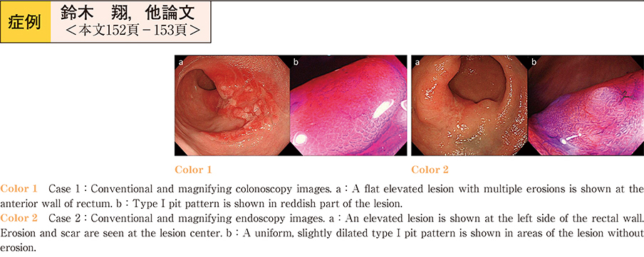
    2013 年 83 巻 1 号 p. 152-153
    発行日: 2013/12/14
    公開日: 2013/12/21
    ジャーナル フリー
    Rectal mucosal prolapse syndrome is often difficult to differentiate from neoplastic lesions─such as advanced rectal carcinoma or rectal malignant lymphoma─using conventional colonoscopy. We report here two cases of rectal mucosal prolapse syndrome diagnosed using endoscopic ultrasonography.
    Case 1 : A 35-year-old man. Colonoscopy showed a flat elevated lesion about 5 cm diameter in the rectum. Multiple erosions were present on the surface of the lesion.
    Case 2 : A 51-year-old woman. Colonoscopy showed an elevated lesion about 4 cm diameter in the rectum. Erosion and scarring were seen at the center of the lesion.
    Endoscopic ultrasonography of both lesions showed smooth, diffuse thickening of the second and third layers of the rectal wall. Neither a solid hypoechoic mass nor a transmural infiltrating lesion was visible, and the five-layer structure of the rectal wall was preserved except for the scarred region. Histological examination of several biopsy specimens obtained from each lesion revealed fibromuscular obliteration. Based on these findings, the lesions were diagnosed as rectal mucosal prolapse syndrome. These findings suggest endoscopic ultrasonography is useful in diagnosis of rectal mucosal prolapse syndrome.
  • 武田 剛志, 渡辺 一宏, 阿部 道子, 西畠 瑞希, 渡邉 俊介, 高原 楠昊, 後藤 絵理子, 瀬戸 元子, 小池 幸宏, 川瀬 建夫, ...
    2013 年 83 巻 1 号 p. 154-155
    発行日: 2013/12/14
    公開日: 2013/12/21
    ジャーナル フリー
    A 78-year-old man underwent total colonoscopy to investigate a positive fecal occult blood test, and was diagnosed with three submucosal tumors (SMTs) of the rectum. He was admitted to our department and endoscopic submucosal dissection (ESD) was performed on the largest lesion. Histological findings confirmed the diagnosis of mucosa-associated lymphoid tissue (MALT) lymphoma. Since MALT lymphoma is generally a low grade lymphoma, additional chemotherapy or surgery was not performed. Subsequently the other two lesions were treated by endoscopic mucosal resection (EMR) . Since the patient was also infected with H. pylori, eradication therapy was administered (and confirmed to be successful) . There has been no evidence of recurrence during the four years following treatment. Colorectal MALT lymphoma is a rare disease. Although H. pylori eradication is the treatment of choice for gastric MALT lymphoma, no standard therapy has been established for colorectal MALT lymphoma. Previously, surgical resection was most common performed, but recent reports even show efficacy of H. pylori eradication against colorectal MALT lymphoma. In this case, H. pylori eradication after ESD was effective and there have been no signs of recurrence in four years after treatment.
  • 渡邉 俊介, 渡辺 一宏, 武田 剛志, 西畠 瑞希, 阿部 道子, 高原 楠昊, 後藤 絵里子, 瀬戸 元子, 小池 幸宏, 岡 輝明, ...
    2013 年 83 巻 1 号 p. 156-157
    発行日: 2013/12/14
    公開日: 2013/12/21
    ジャーナル フリー
    A 75-year-old man underwent total colonoscopy (TCS) after a positive fecal occult blood test, and was diagnosed with submucosal tumor (SMT) in the Bauhin valve. In 2009, the patient had a hot reddish face and easily became angry. In 2010, TCS re-examination showed the enlarged SMT lesion to be approximately 12 mm in size. Laboratory data showed normal serum gastrin and elevated serum serotonin (299 ng/ml) . Abdominal CT scan identified a 12 mm sized enhanced mass on the Bauhin valve. Subsequently, endoscopic submucosal dissection (ESD) of the SMT was performed. Pathological examination was positive for synaptophysin, CD56 and chromogranin, and the results confirmed the diagnosis of neuroendocrine tumor (NET-G1, i.e. carcinoid) . Following ESD, serum serotonin decreased to 133 ng/ml, and the patient’s symptoms of carcinoid syndrome─such as readiness to anger─were gone. In this case we performed ESD to treat colonic carcinoid with carcinoid syndrome. Follow-up shows there have been no signs of recurrence for three years.
  • 牧野 有里香, 青木 順, 岡澤 裕, 高橋 里奈, 水越 幸輔, 河合 雅也, 嵩原 一裕, 高橋 玄, 柳沼 行宏, 小島 豊, 五藤 ...
    2013 年 83 巻 1 号 p. 158-159
    発行日: 2013/12/14
    公開日: 2013/12/21
    ジャーナル フリー
    Colonoscopy in an 81-year-old man undergoing dialysis treatment identified a 20 mm laterally spreading tumor (LST) in the transverse colon, and endoscopic submucosal dissection (ESD) was performed. The tumor was pathologically diagnosed as a tubular adenoma. Parasite eggs were present in the submucosal layer and the case was diagnosed as schistosomiasis japonica. Several theories exist regarding the association between schistosomiasis japonica and colorectal cancer, but no consensus has been reached. Schistosoma japonica ova were present beneath the colon adenoma. This case report is presented with a literature review, with regard to future follow-up and the possibility of carcinogenesis.
feedback
Top