
91 巻, 1 号
選択された号の論文の60件中1~50を表示しています
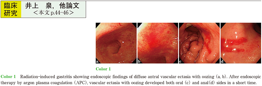
臨床研究
-
2017 年 91 巻 1 号 p. 44-46
発行日: 2017/12/08
公開日: 2017/12/21
 PDF形式でダウンロード (708K)
 -
2017 年 91 巻 1 号 p. 47-51
発行日: 2017/12/08
公開日: 2017/12/21
 PDF形式でダウンロード (1305K)
 -
2017 年 91 巻 1 号 p. 52-56
発行日: 2017/12/08
公開日: 2017/12/21
PDF形式でダウンロード (1062K) -
2017 年 91 巻 1 号 p. 57-61
発行日: 2017/12/08
公開日: 2017/12/21
 PDF形式でダウンロード (836K)
 -
2017 年 91 巻 1 号 p. 62-66
発行日: 2017/12/08
公開日: 2017/12/21
 PDF形式でダウンロード (874K)
 -
2017 年 91 巻 1 号 p. 67-71
発行日: 2017/12/08
公開日: 2017/12/21
 PDF形式でダウンロード (1161K)
 -
2017 年 91 巻 1 号 p. 72-75
発行日: 2017/12/08
公開日: 2017/12/21
PDF形式でダウンロード (724K) -
2017 年 91 巻 1 号 p. 76-80
発行日: 2017/12/08
公開日: 2017/12/21
 PDF形式でダウンロード (847K)
 -
2017 年 91 巻 1 号 p. 81-84
発行日: 2017/12/08
公開日: 2017/12/21
PDF形式でダウンロード (781K) -
2017 年 91 巻 1 号 p. 85-89
発行日: 2017/12/08
公開日: 2017/12/21
 PDF形式でダウンロード (1036K)
 -
2017 年 91 巻 1 号 p. 90-93
発行日: 2017/12/08
公開日: 2017/12/21
 PDF形式でダウンロード (981K)
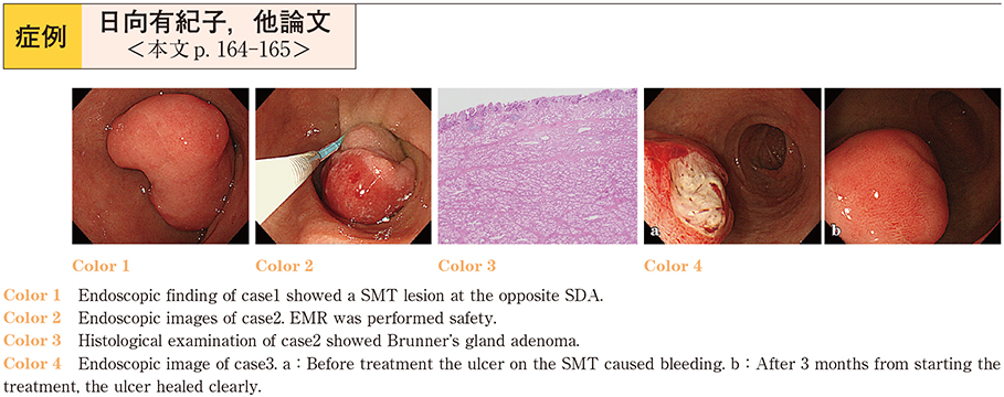
症例
-
2017 年 91 巻 1 号 p. 94-97
発行日: 2017/12/08
公開日: 2017/12/21
 PDF形式でダウンロード (818K)
 -
2017 年 91 巻 1 号 p. 98-101
発行日: 2017/12/08
公開日: 2017/12/21
 PDF形式でダウンロード (1051K)
 -
2017 年 91 巻 1 号 p. 102-105
発行日: 2017/12/08
公開日: 2017/12/21
 PDF形式でダウンロード (783K)
 -
2017 年 91 巻 1 号 p. 106-108
発行日: 2017/12/08
公開日: 2017/12/21
PDF形式でダウンロード (849K) -
2017 年 91 巻 1 号 p. 109-113
発行日: 2017/12/08
公開日: 2017/12/21
 PDF形式でダウンロード (943K)
 -
2017 年 91 巻 1 号 p. 114-117
発行日: 2017/12/08
公開日: 2017/12/21
 PDF形式でダウンロード (1115K)
 -
2017 年 91 巻 1 号 p. 118-119
発行日: 2017/12/08
公開日: 2017/12/21
 PDF形式でダウンロード (724K)
 -
2017 年 91 巻 1 号 p. 120-121
発行日: 2017/12/08
公開日: 2017/12/21
 PDF形式でダウンロード (695K)
 -
2017 年 91 巻 1 号 p. 122-123
発行日: 2017/12/08
公開日: 2017/12/21
 PDF形式でダウンロード (846K)
 -
2017 年 91 巻 1 号 p. 124-125
発行日: 2017/12/08
公開日: 2017/12/21
 PDF形式でダウンロード (629K)
 -
2017 年 91 巻 1 号 p. 126-127
発行日: 2017/12/08
公開日: 2017/12/21
 PDF形式でダウンロード (694K)
 -
2017 年 91 巻 1 号 p. 128-129
発行日: 2017/12/08
公開日: 2017/12/21
 PDF形式でダウンロード (753K)
 -
2017 年 91 巻 1 号 p. 130-131
発行日: 2017/12/08
公開日: 2017/12/21
 PDF形式でダウンロード (868K)
 -
2017 年 91 巻 1 号 p. 132-133
発行日: 2017/12/08
公開日: 2017/12/21
 PDF形式でダウンロード (672K)
 -
2017 年 91 巻 1 号 p. 134-135
発行日: 2017/12/08
公開日: 2017/12/21
 PDF形式でダウンロード (655K)
 -
2017 年 91 巻 1 号 p. 136-137
発行日: 2017/12/08
公開日: 2017/12/21
 PDF形式でダウンロード (705K)
 -
2017 年 91 巻 1 号 p. 138-139
発行日: 2017/12/08
公開日: 2017/12/21
 PDF形式でダウンロード (668K)
 -
2017 年 91 巻 1 号 p. 140-141
発行日: 2017/12/08
公開日: 2017/12/21
 PDF形式でダウンロード (936K)
 -
2017 年 91 巻 1 号 p. 142-143
発行日: 2017/12/08
公開日: 2017/12/21
 PDF形式でダウンロード (907K)
 -
2017 年 91 巻 1 号 p. 144-145
発行日: 2017/12/08
公開日: 2017/12/21
 PDF形式でダウンロード (866K)
 -
2017 年 91 巻 1 号 p. 146-147
発行日: 2017/12/08
公開日: 2017/12/21
 PDF形式でダウンロード (687K)
 -
2017 年 91 巻 1 号 p. 148-149
発行日: 2017/12/08
公開日: 2017/12/21
 PDF形式でダウンロード (833K)
 -
2017 年 91 巻 1 号 p. 150-151
発行日: 2017/12/08
公開日: 2017/12/21
 PDF形式でダウンロード (696K)
 -
2017 年 91 巻 1 号 p. 152-153
発行日: 2017/12/08
公開日: 2017/12/21
 PDF形式でダウンロード (756K)
 -
2017 年 91 巻 1 号 p. 154-155
発行日: 2017/12/08
公開日: 2017/12/21
 PDF形式でダウンロード (592K)
 -
2017 年 91 巻 1 号 p. 156-157
発行日: 2017/12/08
公開日: 2017/12/21
 PDF形式でダウンロード (667K)
 -
2017 年 91 巻 1 号 p. 158-159
発行日: 2017/12/08
公開日: 2017/12/21
 PDF形式でダウンロード (668K)
 -
2017 年 91 巻 1 号 p. 160-161
発行日: 2017/12/08
公開日: 2017/12/21
 PDF形式でダウンロード (784K)
 -
2017 年 91 巻 1 号 p. 162-163
発行日: 2017/12/08
公開日: 2017/12/21
 PDF形式でダウンロード (1022K)
 -
2017 年 91 巻 1 号 p. 164-165
発行日: 2017/12/08
公開日: 2017/12/21
 PDF形式でダウンロード (913K)
 -
2017 年 91 巻 1 号 p. 166-167
発行日: 2017/12/08
公開日: 2017/12/21
 PDF形式でダウンロード (972K)
 -
2017 年 91 巻 1 号 p. 168-169
発行日: 2017/12/08
公開日: 2017/12/21
 PDF形式でダウンロード (737K)
 -
2017 年 91 巻 1 号 p. 170-171
発行日: 2017/12/08
公開日: 2017/12/21
 PDF形式でダウンロード (1387K)
 -
2017 年 91 巻 1 号 p. 172-173
発行日: 2017/12/08
公開日: 2017/12/21
 PDF形式でダウンロード (787K)
 -
2017 年 91 巻 1 号 p. 174-175
発行日: 2017/12/08
公開日: 2017/12/21
 PDF形式でダウンロード (905K)
 -
2017 年 91 巻 1 号 p. 176-177
発行日: 2017/12/08
公開日: 2017/12/21
 PDF形式でダウンロード (837K)
 -
2017 年 91 巻 1 号 p. 178-179
発行日: 2017/12/08
公開日: 2017/12/21
 PDF形式でダウンロード (645K)
 -
2017 年 91 巻 1 号 p. 180-181
発行日: 2017/12/08
公開日: 2017/12/21
 PDF形式でダウンロード (646K)
 -
2017 年 91 巻 1 号 p. 182-183
発行日: 2017/12/08
公開日: 2017/12/21
 PDF形式でダウンロード (865K)