消化器内視鏡の進歩:Progress of Digestive Endoscopy
Online ISSN : 2189-0021
Print ISSN : 0389-9403
最新号
選択された号の論文の37件中1~37を表示しています
掲載論文カラー写真集
内視鏡の器械と技術
  • 木田 芳樹, 木田 光広, 竹澤 三代子, 白須 恭子, 山崎 好喜, 鹿間 伸明, 渡辺 摩也, 西元寺 克禮
    2000 年 57 巻 2 号 p. 20-23
    発行日: 2000/11/15
    公開日: 2014/11/04
    ジャーナル フリー
     胆道系内視鏡診断・治療の成否は,術者の技量はもちろんのこと目的に適した処置具の選択やその使用に際しての容易性,迅速性,確実性に大きく左右される。
     今回われわれは,当施設で開発し臨床試用している①細胞診および遺伝子診断用検体の胆汁採取を目的とした先端側孔付ERCPカテーテル,②挿入が困難な標的胆管分枝へのアプローチのためのガイドワイヤー式ブラッシング・カテーテル,③挿入が困難な胆管分枝や十二指腸乳頭へのアプローチのためのガイドワイヤー式バスケット・カテーテル,④総胆管結石の容易な把持を目的とした回転式バスケット・カテーテル,⑤内視鏡的内瘻術用ステント長の客観的選択を目的としたメジャー付ERCPカテーテルの5つの診断・処置具を報告する。
臨床研究
  • 中村 真一, 光永 篤, 星野 容子, 岸野 真衣子, 小西 洋之, 村田 洋子, 鈴木 茂, 林 直諒
    2000 年 57 巻 2 号 p. 24-28
    発行日: 2000/11/15
    公開日: 2014/11/04
    ジャーナル フリー
     1995年1月から1999年12月の5年間に内視鏡的止血治療を施行した食道静脈瘤出血120例(Lg-cを含む)の治療状況と累積非再出血率,生存率をretrospectiveに検討した。男性87例,女性33例,平均年齢は59.5歳で,出血部位は食道96例,Lg-c24例であった。初期止血法は内視鏡的静脈瘤結紮術(EVL)70例(58.3%),内視鏡的硬化療法(EIS)32例(26.7%),Histoacrylによる硬化療法18例(15.0%)であった。食道出血の67.7%にEVLが,Lg-c出血の54.1%にHistoacrylによる硬化療法が施行された。2例(1.7%)の止血困難例を認めた。3年累積非再出血率は内視鏡的治療法,Child-Pugh分類による肝機能,静脈瘤形態や出血部位では有意差は認めなかった。肝癌合併では肝癌有は45.5%で有意に低かった(P<0.05)。3年生存率は内視鏡的治療法ではEISは65.6%で有意に高かった(P<0.05)。肝機能ではChild-Pugh分類Cは13.7%で,肝癌合併では肝癌有は25.3%で有意に低く(P<0.05),Child-Pugh分類C肝癌有は極めて低かった(P<0.05)。出血例に対する治療法として,約60%の症例にEVLが施行され,第1選択となっている。Lg-cや止血困難例に対しHistoacrylによる硬化療法が有用である。再出血や予後に与える背景に関しては基礎肝機能および肝癌合併の要因が重要である。
  • 藤井 真理, 伊藤 都, 片桐 正人, 中野 茂, 松崎 浩司, 蜂矢 朗彦, 三木 一正
    2000 年 57 巻 2 号 p. 30-33
    発行日: 2000/11/15
    公開日: 2014/11/04
    ジャーナル フリー
     我々の施設では,平均40個のO-ringを用いる撲滅結紮法(eEVL)で,食道静脈瘤を治療している。今回初回治療の食道静脈瘤10症例に対し,カラードプラー超音波内視鏡(CDEUS)を施行し,eEVL前後で静脈瘤供血路である左胃静脈(LGV)の変化を検討した。LGVは6例で血流の検出が可能でありすべて遠肝性であった。eEVL後,この6例のうち3例は遠肝性のまま,1例は求肝性に転じ,残りの6例は描出できなかった。LGV前枝はeEVL前に,遠肝性3例,to and fro 1例の計4例が描出されたが,eEVL後にはこの3例のうち2例が遠肝性に描出されただけで8例は描出不能であった。LGV後枝はeEVL前,1例のみが遠肝性に描出されたが,eEVL後には,2例が遠肝性に描出された。eEVL後,LGVとLGV前枝の描出が不良となり,血管径,血流速度および血流量は減少する傾向を認めたが,LGV後枝にこの傾向はなかった。以上より撲滅結紮法には食道静脈瘤への供血路を遮断する効果がある可能性が示唆された。
  • 松久 威史, 羽山 享宏, 芳村 昇治, 山田 宣孝
    2000 年 57 巻 2 号 p. 34-39
    発行日: 2000/11/15
    公開日: 2014/11/04
    ジャーナル フリー
     Helicobacter pyloriHp)陰性例の萎縮性胃炎,腸上皮化生を観察し,Hp陽性例のそれらと比較した。年齢,性別をマッチさせて得られたHp陽性例,陰性例を比較すると,陽性例では内視鏡的萎縮が36.5%にみられたのに対し,陰性例では17.6%であった。これは,われわれが予測した頻度よりも高い値であった。Hp陰性例の年齢と内視鏡的胃粘膜スコアの間には,陽性例と同様に正の相関がみられ,陰性例も加齢に伴い段階的に萎縮が進行することが明らかとなった。Hp陰性例の3点生検切片3番(胃体下部小彎側)の腸上皮化生スコアは,陽性例と同様に年齢との間に正の相関を示し,軽度から高度の腸上皮化生が各年齢層に分布していた。Hp陽性例との違いは腸上皮化生の出現し始める時期が陽性例よりも約10年遅い点である。また,何れの生検部位においても,Hp陽性例に比し陰性例で腺萎縮のみられる症例の頻度が有意に低かった。腸上皮化生例の頻度も切片2番(胃体上部大彎側)を除き陽性例で高かった。Hp陽性例では3点生検切片の腺萎縮,腸上皮化生率は近似しているが,陰性例では腺萎縮率よりも腸上皮化生率が高く,腸上皮化生が萎縮に先行する症例のある可能性も否定出来ない。Hp陽性例ではHp持続感染により萎縮が進行することをよく反映し,PGⅠ/Ⅱ比と年齢,PGⅠ/Ⅱ比と背景胃粘膜の間に負の相関を認めたが,陰性例ではこのような傾向はみられなかった。
  • 吉村 昇, 松崎 宏治, 永尾 重昭, 川口 淳, 宮崎 純一, 田中 宏史, 中島 弘幸, 又木 紀和, 吉田 由紀子, 加藤 真吾, 青 ...
    2000 年 57 巻 2 号 p. 40-44
    発行日: 2000/11/15
    公開日: 2014/11/04
    ジャーナル フリー
     [背景と目的]井上らの開発したキャップ吸引粘膜切除法であるEMRC法は,優れた粘膜切除法であるが,フルサクションで行う場合,切除深度のコントロールは困難である。今回,我々はEMRCにより得られた病理組織標本を計測することにより,EMRCの安全性,有用性について検討を行った。[対象と方法]当科においてEMRC法で施行された粘膜切除症例42病変を対象とした。切除検体のHE染色標本を用いて切除最深部粘膜表層及び粘膜筋板との距離を計測し,隆起性病変と平坦・陥凹性病変に分け,比較検討した。また,37病変については切除切片の最大径についても計測を行い,切除深度との関係を検討した。[結果]1)切除最深部と粘膜表層及び粘膜筋板との距離はそれぞれ平均2.19±0.83mm,1.11±0.67mmであった。2)平坦・陥凹性病変の方が隆起性病変より深い位置まで採取される傾向にあった。3)切片最大径と切除深度では明らかな相関は認められなかった。[結論]EMRCでは,特に平坦・陥凹性病変の場合,穿孔に対する注意が必要ではあるが,切除前に十分な局注をすることにより安全な切除が可能と考えられた。また,安定して粘膜下層が採取されており,sm浅層胃癌に対する粘膜切除にも対応可能であると考えられた。病変の部位や粘膜の萎縮度も切除深達に影響を与える可能性があり,今後の検討が必要である。
  • 神澤 輝実, 江川 直人, 榊 信廣, 石渡 淳一, 山口 敏和
    2000 年 57 巻 2 号 p. 45-49
    発行日: 2000/11/15
    公開日: 2014/11/04
    ジャーナル フリー
     膵管癌31例,粘液産生膵腫瘍26例,慢性膵炎19例の53例において,ERCP施行時にセクレチンを静注して膵液ないし十二指腸液を採取し,enriched-PCR法とELMA(enzyme linked mini-sequence assay法)を用いて,K-ras癌遺伝子Codon12点突然変異(K-ras遺伝子変異)を検索し,半定量を行った。膵液中のK-ras遺伝子変異(1+以上)は,膵管癌で22例中16例73%,粘液産生膵腫瘍で23例中16例70%,慢性膵炎で15例中6例40%で認められ,膵液中のK-ras遺伝子変異の検索は,膵管癌のhigh risk groupの設定に有用であると考えられる。一方,K-ras変異細胞の半定量値を2+以上とすると,膵管癌では55%,粘液産生膵腫瘍では39%と陽性率は低下するが,慢性膵炎では13%のみであり,膵管癌と慢性膵炎との鑑別に有用であると考えられた。セクレチン静注後に採取した十二指腸液中のK-ras遺伝子変異は,膵管癌で32%,粘液産生膵腫瘍で33%に認められ,膵癌の拾い上げ診断に応用できる可能性が考えられた。
  • 羽山 享宏, 井上 泰夫, 松久 威史
    2000 年 57 巻 2 号 p. 50-51
    発行日: 2000/11/15
    公開日: 2014/11/04
    ジャーナル フリー
    Psychological test was performed in cases of peptic ulcer. We used Fujinami's psychlogical test, which is composed by NI score on fifty-items neurotic index and DI score on four-factors depression index. NI, DI score were the highest in gastroduodenal ulcer and they were higher in positive group according to abdominal symptom. NI, DI score of gastric ulcer were high below 39 and over 70 years old. They were high below 39 years old in duodenal ulcer. NI, DI score of Helicobacter pyloriHp)negative group were higher than that of Hp positive group.
  • 大塚 耕司, 村上 雅彦, 加藤 貴史, 藤岡 稔洋, 普光江 嘉広, 亀山 眞一郎, 町田 健, 草野 満夫
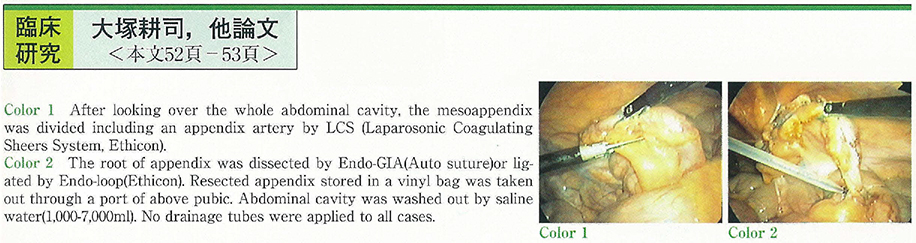
    2000 年 57 巻 2 号 p. 52-53
    発行日: 2000/11/15
    公開日: 2014/11/04
    ジャーナル フリー
    54 cases of acute appendicitis were operated laparoscopically for past 3 years (Three cases were converted to open exploration.) . Patients were divided into three groups according to postoperative pathological findings of appendix. (Group A : catarrhal, Group B : phlegmonous, Group C : gangrenous) . In each groups, operation time, postoperative period of bed rest and NPO (nothing per os) , pyretic term after operation, length of hospital stay and postoperative complications were analyzed to estimate the quality of laparoscopic appendectomy. Three ports were inserted on the abdomen ; under umblication, right upper abdomen (3 or 5 mm) and above left pubic (12 mm in a diameter) . After looking over the whole abdominal cavity, the mesoappendix was divided including an appendix artery by LCS (Laparosonic Coagulating Sheers System, Ethicon) and then, the root of appendix was dissected by Endo-GIA (Auto suture) or ligated by Endo-loop (Ethicon) . Resected appendix stored in a vinyl bag was taken out through a port of above pubic. Abdominal cavity was washed out by saline water (1,000-7,000 ml) . No drainage tubes were applied to all cases.
    In Group C, period of bed rest was longer than Group A. No significant difference was detected in another classification. There were no remarkable complications as wound infection or abdominal abscess after surgery except paralytic ileus in two patients. These results demonstrate laparoscopic surgery is appropriate, and should be standard method for acute appendicitis.
  • 長浜 雄志, 丸山 道生, 吉田 達也
    2000 年 57 巻 2 号 p. 54-55
    発行日: 2000/11/15
    公開日: 2014/11/04
    ジャーナル フリー
    Endoscopic treatment and observation of colon for poorly prepared patients is difficult from poor visibility during procedures. We have experienced transparent cap fitted colonoscopy for five patient who could not tolerate mechanical bowel preparation. Insertion of colonoscopy to the level of cecum was easily attained for colon bleeding and colon volvulus under good visualization. Insertion to the level of stricture was also easily attained for patients with sigmoid colon cancer. With this transparent cap fitted procedure it was easy to maintain a visual field between colonic mucosa and the tip of colonoscope. Therefore adhesion of stool and blood to the surface of the tip could be easily avoided. Maneuverbility of scope during insertion was relatively poor than that without transparent cap as elongation of bending section of endoscopy. However attaining fair visibility is more beneficial for endoscopic observation and treatment.
    We can conclude that transparent cap is a useful device for colonoscope with poor bowel preparation.
  • 藤井 陽一朗, 室久 俊光, 岡本 裕, 須藤 康司, 渡辺 秀考, 玉野 正也, 小熊 資男, 飯島 誠, 柳沢 伸嘉, 前原 操, 菅谷 ...
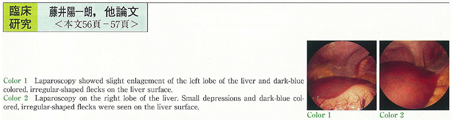
    2000 年 57 巻 2 号 p. 56-57
    発行日: 2000/11/15
    公開日: 2014/11/04
    ジャーナル フリー
     症例は48歳,男性。日本酒3合/日,28年間の飲酒歴あり。1997年健診にて初めて肝機能障害を指摘され,1999年8月近医を受診,HCV抗体陽性,AST 59IU/l,ALT 156IU/lと肝機能障害あり。同年11月C型肝炎に対するインターフェロン治療を希望し当科紹介受診,12月精査目的に入院。入院時,皮膚に異常所見なし。確定診断の目的に腹腔鏡,肝生検を施行した。
     腹腔鏡では肝は腫大し,表面には小陥凹を認め,慢性肝炎に合致する所見を,また肝表面には地図状に暗青色部を認め,腹腔鏡上ポルフィリン症の存在を疑った。その後ポルフィリン代謝産物の測定にてポルフィリン代謝異常と診断した。
     C型肝炎に晩発性皮膚ポルフィリン症の合併は知られているが,本例では皮膚所見がなく腹腔鏡検査にて初めてポルフィリン代謝異常が疑われ,診断の契機となった。
症例
  • 林 祐二, 関田 吉久, 谷 雅夫, 斎藤 直也, 河野 辰幸, 岩井 武尚, 竹下 公矢, 中村 隆
    2000 年 57 巻 2 号 p. 58-60
    発行日: 2000/11/15
    公開日: 2014/11/04
    ジャーナル フリー
     症例は17歳女性で,1999年8月24日に上腹部痛および吐血を認めて近医を受診,上部消化管内視鏡検査にて胃体中部大彎側に直径3cm強の山田Ⅳ型の隆起性病変を認め,中央部の潰瘍からの出血を認めた。同9月1日に精査加療目的にて当料へ紹介入院となった。入院後の上部消化管内視鏡検査で,有茎性で潰瘍を合併した粘膜下腫瘍を認めた。超音波内視鏡検査では筋層は保たれていたことから,留置スネアを併用し,内視鏡的腫瘍摘出術が可能であった。病理組織学的にはS-100蛋白のみが陽性であり,胃神経鞘腫と診断された。
  • 小野寺 大吾, 永尾 重昭, 川口 淳, 吉田 由紀子, 又木 紀和, 中島 弘幸, 北川 敬丈, 田中 宏史, 宮崎 純一, 松崎 宏治, ...
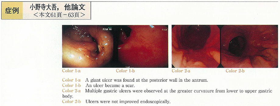
    2000 年 57 巻 2 号 p. 61-63
    発行日: 2000/11/15
    公開日: 2014/11/04
    ジャーナル フリー
     症例1は87歳,女性。食欲不振を主訴に来院。胃内視鏡にて前庭部にA1 stageの巨大潰瘍を認めた。Helicobacter pyloriH.P)陽性,low-grade胃MALTリンパ腫と診断し,除菌療法を施行した。除菌後巨大潰瘍は瘢痕化し,病変部は褪色調の胃粘膜となり,完全寛解となった。症例2は83歳,男性。心窩部痛を主訴に来院。胃内視鏡にて胃体上部~下部の大弯側に多発性潰瘍を認め,H.Pは病理,培養,呼気テスト全てが陰性だったが,生検の結果,low-grade MALTリンパ腫と診断し,除菌療法を施行した。投与後も潰瘍は残存し,完全寛解には至らなかったが内視鏡的には増悪・進展はみられていない。2例とも外来にて経過観察中である。高齢者における胃MALTリンパ腫の治療は,全身状態および内視鏡像を考慮し,除菌療法を試みてよいと考えられた。
  • 川島 ルミ子, 今井 幸紀, 新井 晋, 栫 弘太郎, 真下 由美, 朝倉 泰, 柿沼 徹, 石川 恵子, 木下 学, 下地 克典, 太田 ...
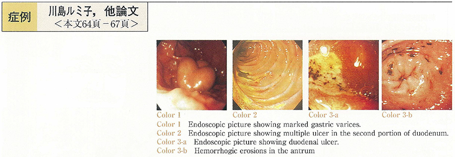
    2000 年 57 巻 2 号 p. 64-67
    発行日: 2000/11/15
    公開日: 2014/11/04
    ジャーナル フリー
     81歳,男性。平成7年9月C型肝硬変,肝細胞癌と診断された。平成11年11月29日胃静脈瘤に対して予防的にバルーン下逆行性経静脈的塞栓術(B-RTO)施行。その翌日より腹水貯留,5日後心窩部痛,黒色便出現。内視鏡検査にて食道炎,多発性出血性胃びらん,多発性十二指腸潰瘍を認めた。絶食とし,PPI投与10日後の内視鏡検査では十二指腸潰瘍はH1 stageまで改善し胃びらん,食道炎は消失した。利尿剤増量にて腹水は減少し退院となった。退院後もPPI服用は継続していた。平成12年1月5日ごろより腹水増量し1月22日入院。その2日後に吐血あり,内視鏡検査にて前回と同様の広範な消化管病変を認めた。本症例における上部消化管粘膜障害は腹水の増加に一致して出現しており,その発症にはB-RTOによる門脈圧の上昇が関与している可能性が考えられた。
  • 杉田 光司, 北川 敬丈, 吉田 由紀子, 中島 弘幸, 叉木 紀和, 吉村 昇, 池松 弘朗, 竹林 晃一, 小野寺 大吾, 松崎 宏治, ...
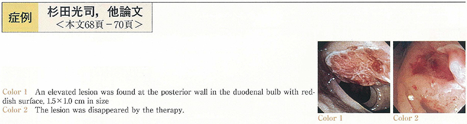
    2000 年 57 巻 2 号 p. 68-70
    発行日: 2000/11/15
    公開日: 2014/11/04
    ジャーナル フリー
     腎細胞癌の十二指腸転移は比較的稀であり,またインター
    フェロン治療により十二指腸転移巣の消失を認めた報告はほとんどない。我々は腎細胞癌の十二指腸への転移再発に対してインターフェロンが有効であった一例を経験したので報告する。症例は55歳の男性で,昭和61年に当院にて右腎癌に対して根治的右腎摘出術が施行された(RCC : tubular type,mixed subtype,G2,pT2,pN0,pM0,pV0)。
     以後再発を認めなかったが,術後13年目の検診にて貧血ならびに便潜血陽性を指摘され,当科を紹介され受診した。平成11年3月の上部消化管内視鏡検査にて十二指腸球後部に約1.5cm大の発赤した粘膜下腫瘍様病変を認め,生検にて腎癌の十二指腸への転移と診断された。入院後,他に膵・肝・左肩甲骨・左肺の転移巣にも再発を認め,泌尿器科にてインターフェロン治療を施行した。十二指腸腫瘍の縮小を認め,平成12年2月には肉眼的に腫瘍は消失し,同部位よりの生検においても腫瘍像は認められなかった。
  • 長場 静香, 小林 清典, 佐田 美和, 吉澤 繁, 五十嵐 正広, 勝又 伴栄, 西元寺 克禮, 大谷 剛正, 三富 弘之
    2000 年 57 巻 2 号 p. 71-74
    発行日: 2000/11/15
    公開日: 2014/11/04
    ジャーナル フリー
     患者は21歳,女性。15歳時に下痢,貧血を認め当院小児科を受診。全大腸炎型潰瘍性大腸炎(UC)と診断され,副腎皮質ステロイド剤を中心とした薬物療法で加療するも,ステロイド依存性で再燃を繰り返していた。1999年8月に下血と下腹部痛を認め近医に入院。腸閉塞を伴う直腸癌と診断され当科に転院となった。大腸内視鏡所見で,直腸に全周性狭窄を伴う腫瘍を認め,生検で低分化腺癌と診断された。回腸への瘻孔形成がみられたが,遠隔転移は認めなかった。UCに合併した進行直腸癌と診断し,大腸亜全摘,腹会陰式直腸切断術を行った。病理組織学的には,直腸歯状線から口側22cmまで結節状隆起が連続し,同部に低分化腺癌が認められた。癌は直腸壁を越えて回腸に直接浸潤し,リンパ節転移が陽性で,一部にhigh grade dyplasiaの合併も認めた。左側~全大腸炎型で活動性が高いUCは,若年者でも短期間で癌化する場合があり注意が必要である。
  • 村井 信二, 元神 賢太, 藤田 晃司, 半田 真一, 中村 明彦, 山内 秀夫, 松井 淳一, 雨宮 哲
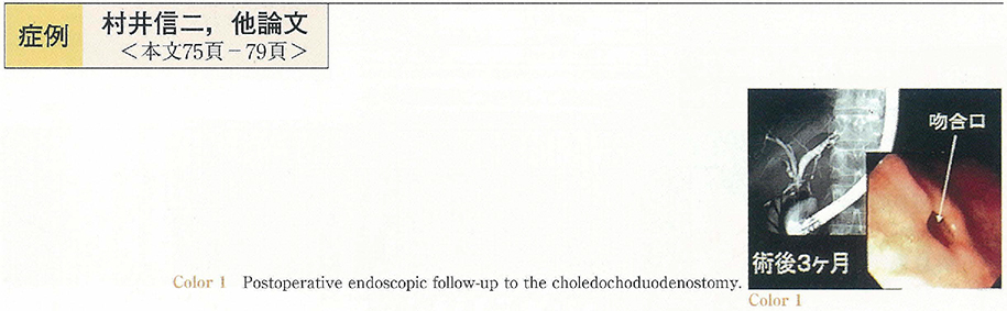
    2000 年 57 巻 2 号 p. 75-79
    発行日: 2000/11/15
    公開日: 2014/11/04
    ジャーナル フリー
     症例1は30歳女性,右季肋部痛を主訴に来院し,精査にて総胆管の囊腫状拡張を伴う膵管胆道合流異常と診断し,腹腔鏡下に総胆管拡張部切除および総肝管十二指腸吻合を施行した。症例2は53歳女性,上腹部痛を主訴に近医受診し,USにて胆囊ポリープを指摘され当院に紹介された。USおよびEUSにて,胆囊に複数の隆起性病変を認め,最大径は25mmであった。ERCPにて,総胆管非拡張型の膵管胆道合流異常の合併を認めた。腹腔鏡下胆囊摘出術を施行し,術中迅速にて良性腫瘍であることを確認した後,腹腔鏡下に総胆管切除および総肝管十二指腸吻合術を施行した。症例1,2ともに手術は完全腹腔鏡下に行い,術後5日目より食事摂取を開始し10日目に退院した。平均手術時間は7時間31分であった。本法は,術後の内視鏡的経過観察も可能であり,適応を選択することにより,膵管胆道合流異常に対する侵襲の少ない優れた手術法の1つになり得ると考え報告する。
  • 星野 光典, 石井 博, 中村 明央, 高村 光一, 大山 祥, 新谷 隆, 太田 篤, 御子神 哲也, 榎澤 哲司, 草野 満夫, 青柳 ...
    2000 年 57 巻 2 号 p. 80-83
    発行日: 2000/11/15
    公開日: 2014/11/04
    ジャーナル フリー
     症例は69歳女性。主訴は発熱。1997年5月より膵管狭細型慢性膵炎の診断で当院内科にて経過観察中,1998年8月より胆管狭窄に伴う黄疸が出現し,経乳頭的ステンテイングとステロイド治療を施行するも反応せず,頻回の胆管炎を繰り返すため,1999年12月当科に転科した。血液所見では糖尿病を呈するも免疫学的検査に異常なく,CTでは膵はびまん性に腫大し,ERCPでは主膵管のほぼ全域に至るびまん性狭細像と膵内胆管の狭窄が認められた。右PTBDルートを設置し拡張後,経皮経肝胆道鏡下に胆管狭窄部を確認し,マイクロ波凝固療法用ボール型電極(径1.8mm)を用いて50Wで10秒間の接触凝固を数回施行した。その後狭窄は改善し内瘻化チューブを皮下埋込み留置し外来通院中である。胆道鏡下マイクロ波凝固療法は低侵襲かつ安全な局所療法であり,切除不能膵胆道癌やpoor riskの良性胆道狭窄などの症例に対して応用可能と思われ症例を蓄積し長期経過を観察する必要はあるものの有用な方法と思われ報告した。
  • 神澤 輝実, 佐仲 雅樹, 屠 聿揚, 江川 直人, 榊 信廣, 石渡 淳一
    2000 年 57 巻 2 号 p. 84-87
    発行日: 2000/11/15
    公開日: 2014/11/04
    ジャーナル フリー
     症例は81歳,女性。中部胆管癌症例で,高齢のため金属ステントを挿入して退院した。腫瘍のステント内への発育による黄疸で3ケ月後再入院した。温熱・化学・放射線の三者併用療法を施行し,胆管の再疎通を認めた。しかし,胆道出血及び消化管出血を起こし,ショック状態に陥った。緊急血管造影にて下膵十二指腸動脈と右肝動脈における仮性動脈瘤からの出血と診断し,マイクロコイルを用いて塞栓止血した。仮性動脈瘤の形成機序として,動脈瘤が金属ステントを挿入し局所温熱療法を施行した部位に存在することより,三者併用療法により脆弱した胆管壁に金属ステントがくい込み,動脈壁を損傷した可能性が考えられた。金属ステントを挿入した胆道癌症例の消化管出血の原因のひとつとして,肝動脈の仮性動脈瘤からの胆道出血を考慮する必要がある。
  • 倉持 英和, 林 和彦, 井手 博子, 村田 洋子, 鈴木 茂, 高崎 健
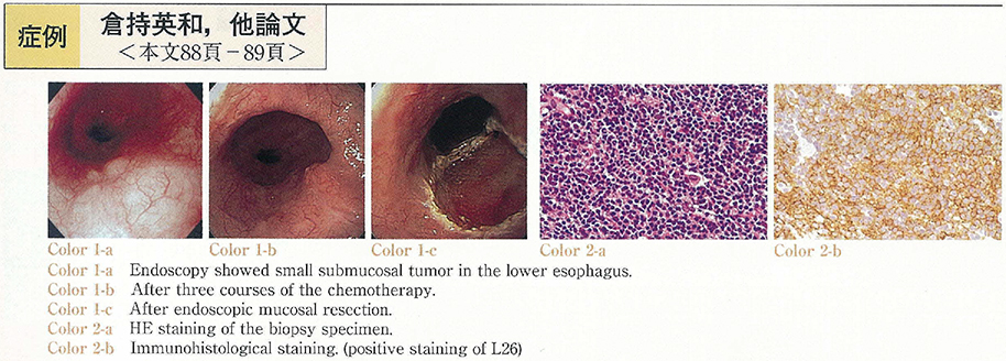
    2000 年 57 巻 2 号 p. 88-89
    発行日: 2000/11/15
    公開日: 2014/11/04
    ジャーナル フリー
    A 69-years old man was diagnosed as esophageal malignant lymphoma by regular endoscopic GI check up. Endoscopy showed small submucosal tumor in lower esophagus. EUS revealed low echoic tumor in lpm-sm layer. Three courses of chemotherapy (CHOP) was given from June 1999 to September 1999. The tumor showed no remarkable change in size by endoscopy and EUS after chemotherapy. We performed EMR of the lesion and the pathological finding showed that most of this elevated lesion was consisted of fibrosis and inflamatoric cell, but still there existed a certain amount of viable tumor cells at the edge of the specimen thus we added radiation therapy. In this case, EMR seems to be critical for not only treatment but also the accurate evaluation of chemotherapy.
  • 千原 直人, 内藤 英二, 重田 明子, 小峯 修, 駒崎 敏昭, 栗原 雄司, 松本 光司, 伊藤 正秀, 森山 雄吉
    2000 年 57 巻 2 号 p. 90-91
    発行日: 2000/11/15
    公開日: 2014/11/04
    ジャーナル フリー
    Although esophageal lesion of pemphigus vulgalis is rare, its incidence has been increasing with the spread of endoscopes. A 61-year-old man was admitted to our hospital because of idiopatic necrotic head of femur, pyrexia, stomatitis, exanthema and ascites when we followed up esophageal varices and alcoholic cirrhosis. The patient could not ingest because of oral pain. By a biopsy specimen of the erosive skin the patient was diagnosed as pemphigus vulgalis and received local treatment with steroid. It was hard to treat esophageal varices with endoscopy because of detachment of almost entire esophageal mucosa with fresh blood. Therefore, it is important in the follow-up of patients with pemphigus vulgalis to take esophageal lesion into consideration. Furthermore, we suggest that careful attension to endoscopic examination in selected cases of pemphigus vulgalis may reveal a higher incidence of this entity.
  • 田原 久美子, 三橋 利温, 山崎 いづみ, 田辺 聡, 今泉 弘, 木田 芳樹, 大井田 正人, 西元寺 克禮
    2000 年 57 巻 2 号 p. 92-93
    発行日: 2000/11/15
    公開日: 2014/11/04
    ジャーナル フリー
    We reported a 58-year-old female with a double pylorus, who had been treated with prednisolone because of rheumatoid arthritis. The first endoscopic examination showed a gastric ulcer on the posterior wall of the prepylorus. Two years later, the second examination showed an accessory opening and gastric ulcer on the posterior wall of the prepylorus. The third examination performed two years later showed an enlarged accessory opening and gastric ulcer on the posterior wall of the prepylorus. One year later at the fourth examination (five years after the initial examination) the double pylorus fused and formed a large single pylorus. Eight years after the initial examination, a gastric ulcer was observed on the same position. Of 118 cases of double pylorus reported in Japan, 15 cases developed a single pylorus. The fistula was closed in 4 cases and fused in 11 cases of 15 cases.
  • 野澤 宏彰, 津久井 元, 喜納 五月, 高倉 裕一, 橋本 直明, 岸田 由起子, 薬丸 一洋, 加賀美 尚, 関川 敬義
    2000 年 57 巻 2 号 p. 94-95
    発行日: 2000/11/15
    公開日: 2014/11/04
    ジャーナル フリー
    A 53-year-old woman was admitted to our hospital for evaluation and treatment of the abnormality in the gastric antrum revealed by barium fluorography. Endoscopic examination showed a semi-pedunculated submucosal tumor-like lesion with slight central erosion, 1 cm in diameter, in the greater curvature of the antrum. Furthermore, two similar polyps of 5 mm and 3 mm in diameter were found in the anterior wall of the angle. All the biopsy specimen of these polyps showed benign findings (Group I) . Since she was asymptomatic, only the polyp in the antrum was removed by endoscopic mucosal resection. Histopathologically, spindle-shaped cells proliferated chiefly in the submucosal space, which were positive for CD34 and vimentin. The polyp was diagnosed as inflammatory fibroid polyp (IFP) that exhibited unusual histological features ; typical“onion-skin pattern”was not observed, and infiltration of eosinophils was mild. Interestingly, the other polyps remained disappeared by endoscopy 6 months later. The observation seems to support that IFP may be developed through inflammation.
  • 新保 泉, 山口 武人, 尾高 健夫, 山口 和也, 関谷 武司, 中島 賢二, 小出 明範, 駒 嘉宏, 小山 秀彦, 石原 武, 露口 ...
    2000 年 57 巻 2 号 p. 96-97
    発行日: 2000/11/15
    公開日: 2014/11/04
    ジャーナル フリー
    77-year-old woman was referred to our hospital for further evaluation of the stomach lesion. A subpedunculated tumor, 15mm in diameter, was found at the upper anterior part of gastric body by the gastrointestinal endosopic examination. The tumor was well demarcated from the surrounding mucosa, and the surface of the tumor was papillary and lobulated with no ulcer and erosion. The tumor was diagnosed papillary adenocarcinoma by the biopsy. With the upper gastrointestinal radiologic examiniation, the tumor was depicted a round radiolucant mass without wall deformity. From the results of gastrointestinal endoscopy and radiologic examiniation, the tumor was considered to be early gastric carcinoma. However, the low-echoic mass located from the mucosal layer through the proper muscle layer was demonstrated by the endoscopic ultrasonography (EUS) , indicating the carcinoma in advanced stage. Total gastrectomy was performed and the histology of the resected specimen showed gastric carcinoma of the gastric type. In the detailed histopathological examination, differentiation of the tumor was varied ; well differentiated adenocarcinoma in superficial layer and poorly differentiated adenocarcinoma in deep layer which infiltrated the serosa. EUS appeared to be accurate in terms of tumor staging compared with gastrointestinal endosopy or upper gastrointestinal radiologic examination in this case.
  • 別府 加寿子, 武本 憲重, 小泉 浩一, 甲斐 俊吉, 山尾 剛一, 坂井 雄三, 陳 勁松, 丸山 雅一
    2000 年 57 巻 2 号 p. 98-99
    発行日: 2000/11/15
    公開日: 2014/11/04
    ジャーナル フリー
    A 55-year-old-woman who had a small discolored lesion of the rest stomach, revealed by an endoscopy performed by a local doctor, was admitted to our hospital in May, 1999. Endoscopic examination revealed a small discolored lesion with irregular margin of the rest stomach. The pathological diagnosis was undifferentiated type gastric cancer. Upper GI X-ray examination showed a small irregular depressed lesion of the greater curvature of the rest stomach.
    Then, we diagnosed a minute undifferentiated type gastric cancer of the rest stomach and performed endoscopic mucosal resection.
    The lesion was 3×3mm in size and undifferentiated adenocarcinoma, which located in the mucosal layer. In conclusion, It is important to look for discovered discolored lesions and discolored lesions with very small reddish granules of the middle part of gastric body.
  • 萩原 廣明, 吉永 輝夫, 樋口 次男
    2000 年 57 巻 2 号 p. 100-101
    発行日: 2000/11/15
    公開日: 2014/11/04
    ジャーナル フリー
    A 69-year-old woman who had underwent endoscopic polypectomy of stomach before 4 years was admitted to my clinic for endoscopy. In endoscopic examination, five polyps classified Yamada's type II were located on greater curvature of antrum, anterior wall of angulus and greater curvature of upper body. Histological findings of biopsied specimens from these polyps revealed hyperplasia of the foveolar epithelium with moderate infiltration of inflammatory cells which was consistent with hyperplastic polyp. These polyps were unchanged during 2 years and Helicobacter pylori (H. pylori) was found by the CLO test and cultures of biopsied specimens.
    She was treated with one week course of lansoprazole, amoxicillin and clarithromycin. About 14 months later, all polyps were disappeared and eradication of H. pylori was confirmed by biopsy, cultures and Urea breath test.
    These result suggests that eradication of H. pylori may be expected as a useful treatment to hyperplastic polyp of stomach.
  • 大野 隆, 水口 澄人, 北村 匡, 川添 一哉, 橘田 輝雄, 梶原 博, 朴 勝春, 佐伯 威男, 西元寺 克禮
    2000 年 57 巻 2 号 p. 102-103
    発行日: 2000/11/15
    公開日: 2014/11/04
    ジャーナル フリー
    We reported a rare case of adenoma of the papilla of Vater. A 63-year-old female had been diagnosed to adenoma of the papilla of Vater on ERCP and biopsy before 6 years. The papilla was followed up by endoscopy every 6 months. Recently, the papilla was enlarged and deformed with granular surface. ERCP showed normal findings in biliary system and main pancreatic duct, however accessory pancreatic duct was narrow. We performed endoscopic papillectomy for completely biopsy under the informed consent. Since endoscopic papillectomy is a safe treatment and useful in the final pathological diagnosis of the adenoma diagnosed by biopsy. The tumor was resected completely and the final pathological diagnosis was adenoma. However acute pancreatits, one of the complication of endoscopic papillectomy was occurred. This acute pancreatitis recovered by conservative therapy. Six months after papillectomy there was no sign of recurrence. This case suggested that endoscopic papillectomy is useful for adenoma of the papilla of Vater and that method of prevention from the complication of endoscopic papillectomy will further be investigated.
  • 鶴井 光治, 工藤 拓, 荻原 正示, 森田 重文, 大野 博之, 河合 隆, 三坂 亮一, 川口 実, 森安 史典
    2000 年 57 巻 2 号 p. 104-105
    発行日: 2000/11/15
    公開日: 2014/11/04
    ジャーナル フリー
    Shönlein-Henoch purpura (SHP) is a common vasculitis affecting the skin, gastrointestinal tract, joints and, occasionally other organs. Gastrointestinal manifestation, such as abdominal pain is common.
    We present a 23-year-old Japanese male with SHP. He was admitted to our hospital with abdominal pain, and purpuric rash on his extremities. Endoscopic examination of upper gastrointestinal tract revealed multiple erosion at the 2nd portion of duodenum. Colonoscopic findings were multiple small ulcers and erosions. We diagnosed SHP from physical findings and examinations, and started to treat with steroid medicine. Gradually, he recovered and discharged our hospital.
  • 小野里 康博, 佐川 俊彦, 阿部 毅彦, 石川 武志, 下田 隆也, 石原 弘, 池谷 俊郎, 安東 立正, 小川 哲史, 棚橋 美文, ...
    2000 年 57 巻 2 号 p. 106-107
    発行日: 2000/11/15
    公開日: 2014/11/04
    ジャーナル フリー
    A 44-year-old male complained of abdominal pain was admitted to our hospital. He had been diagnosed as Peutz-Jeghers syndrome and operated three times due to ileus caused by intestinal polyps. His diagnosis was invagination which was treated in the operation with minor wound. Sixty-six polyps ranging in size from 5mm to 40mm were polypectomized or ligated with detachable snare by intraoperative laparoscope-assisted enteroscope.
  • 関川 憲一郎, 松浦 広, 松川 雅也, 高倉 裕一, 関根 昌子, 古畑 総一郎, 小林 克也, 河原 弘規, 橋本 直明, 熊谷 隆志, ...
    2000 年 57 巻 2 号 p. 108-109
    発行日: 2000/11/15
    公開日: 2014/11/04
    ジャーナル フリー
    A 50-year-old woman showed a positive result for fecal occult blood test on her general health check-up. Colonoscopic examination at a local hospital showed a Borrmann type 1-like tumor on her ileocecal valve sizing 1.5 cm with a small top depression. Then she was referred to our hospital. Our examination on 12 days later revealed that it had grown to a Borrmann type 2-like lesion having a large ulcer. Histological examination on biopsy specimen showed dense submucosal infiltration of medium to large atypical lymphoid cells, which were positive for LCA and L-26 but negative for CD3 and UCHL-1. No other lymphoid lesions were observed. Thus diagnosis of diffuse medium to large-sized B cell lymphoma originated on the ileocecal valve was made. Ileocecal resection was performed on 50 days after initial examination. However no tumor was observed on the surgical specimen without localized microscopical aggregation of small lymphocytes which were positive for LCA and CD3 but negative for L-26. The result of immunohistochemical stain suggested that they were reactive T cells. Endoscopic and histological observation of disintegration and regression of intestinal malignant lymphoma is quite rare. The clinical course of our patient is unique in view of the spontaneous regression of intestinal MALT lymphoma and reactive lymphoid hyperasia.
  • 丸田 和夫, 小田 常人, 川上 浩平, 植田 健治, 棗 雅子, 堀 高史朗, 安田 有利, 平良 悟, 二木 修司, 渡辺 浩一, 土屋 ...
    2000 年 57 巻 2 号 p. 110-111
    発行日: 2000/11/15
    公開日: 2014/11/04
    ジャーナル フリー
    The patient was a 71-year-old woman. She became aware of her own exertional dyspnea in June 1999, and was referred to our hospital for admission. The patient underwent colonoscopic examination because iron deficiency anemia and positive occult blood reaction in the feces were observed. In the colonoscopic examination, angiodysplasia with an uneven surface was seen in the cecum, which was thought to be the source of bleeding. In addition, abdominal angiography of the patient revealed early venous filling in the region of the cecum., leading to a diagnosis of colonic arteriovenous malformation. Thereafter she was followed up as an ambulatory patient. However, form around November 1999, anemia became pronounced and the patient was again hospitalized.
    Colonoscopic examination conducted during hospitalization showed bleeding from the cecum with arteriovenous malformation. We unsuccessfully performed a hemostatic procedure with a heat probe, resulting in the adoption of laparoscopic ileocecal resection. The postoperative course of the patient has been satisfactory, and no progression of the anemia has been observed. We report our experience of a case with lower gastrointestinal bleeding due to colonic arterioveous malformation.
  • 戸塚 統, 棚橋 美文, 松本 広志, 竹吉 泉, 大和田 進
    2000 年 57 巻 2 号 p. 112-113
    発行日: 2000/11/15
    公開日: 2014/11/04
    ジャーナル フリー
    A 76-year-old woman visited the emergency room complaining of anal discomfort. Yellowish mucosa was prolapsed from the anus, and it was easily reductable with a hand manipulation. She admitted the hospital to do further examination four months later because of repeated. Barium enema roentogenograms revealed a smooth and well defined tumor, about 4cm in size, in the sigmoid colon 35cm from the anus. Colonoscopic examination showed a smooth and yellowish submucosal tumor with a positive cushion sign. These findings were compatible with a lipoma of the sigmoid colon, and endoscopic mucosal resection of the tumor should not be indicated. Under general anesthesia with muscle relaxation, we successfully drew the tumor to the outside the anus by using a colonoscope, and resected directly without laparotomy or laparoscopy. The patient resumed diet from the next day after surgery. This method may be applicable to a benign lower colonic tumor under general anesthesia with muscle relaxation.
  • 粟井 俊成, 貝瀬 満, 鈴木 伸明, 三輪 純, 中尾 國明, 松本 正廣, 岩田 滉一郎, 籏原 照晶, 太田 裕彦
    2000 年 57 巻 2 号 p. 114-115
    発行日: 2000/11/15
    公開日: 2014/11/04
    ジャーナル フリー
    A 58-year-old man with advanced cholangiocarcinoma used diclofenac sodium suppository up to 200 mg in order to relieve right hypochondralgia and fever. One month after continuous NSAID usage, he was hospitalize to our hospital because of bloody diarrhea followed by massive rectal bleeding. Urgent endoscopic examination revealed irregular shaped multiple rectal ulcers, indicating a diagnosis of sodium suppository-induced acute hemorrhagic rectal ulcer. Bowel rest and GeloformTM tamponade achieved temporal hemostasis. However, rectal bleeding recurred and total amount of hemorrhage reached to 6,000 ml. We performed colonoscopy-guided argon plasma coagulation (APC) to the hemorrhagic rectal ulcers two times and complete hemostasis was achieved. Since APC is able to coagulate digestive mucosa of target widely and safely, APC is considered as an effective endoscopic hemosatatic procedure to acute hemorrhagic rectal ulcer.
  • 清水 健, 葛谷 嘉久, 畔田 浩一, 真坂 彰, 毛利 勝昭, 飯塚 一郎
    2000 年 57 巻 2 号 p. 116-117
    発行日: 2000/11/15
    公開日: 2014/11/04
    ジャーナル フリー
    Bullous pemphigoid is known to be one of the paraneoplastic skin manifestation associated with internal malignancies.
    A 60-year-old male patient suffering from erythema and bulla of the body was diagnosed as bullous pemphigoid in 1991. In August, 1997 he was admitted to our hospital because of bloody stool. Endoscopic examination revealed rectal cancer. Thoracic CT and abdominal CT showed metastatic tumour both in the left lung and the liver. After rectal cancer was resected by Miles'method, both metastatic tumours were also resected. Dermatological symptoms were remarkably reduced after these treatment. Four months later, again bulla appeared simultaneously with the reccurence of liver metastasis. Resection of this lesion lead to an improvement in the skin manifestation.
    It is reported that internal malignancies are found in 5 to 20% of bullous pemphigoid patients. Including this case, only 9 cases of rectal cancer associated with bullous pemphigoid have been reported domestically. The characteristics of these patients are that they are comparatively aged and that the bullas are detected before the cancer is found. Also, 5 out of 9 patients showed improvement in their dermatological symptoms after resecting or chemotherapy treatment against the cancer, as in this case too.
    In conclusion, it is important to examine thoroughly for internal malignancies including lower gastrointestinal tracts, in bullous pemphigoid patients.
  • 丸山 祥司, 安藤 昌之, 大沼 忍, 坂庭 信行, 只友 秀樹, 原田 佳明, 田中 浩一, 山下 晋矢, 鈴木 和信, 関口 令安, 瀧 ...
    2000 年 57 巻 2 号 p. 118-119
    発行日: 2000/11/15
    公開日: 2014/11/04
    ジャーナル フリー
    A rare case of primary malignant lymphoma of the rectum is reported. An 83-years-old woman complained of hemorrhage on defecation. She was diagnosed as malignant lymphoma of the rectum with the colonoscopy and barium enema. Abdominoperineal resection of the rectum was performed. Histological examination of the resected specimen indicated a malignant lymphoma of the rectum, invaded to the proper muscle. Several immunohistochemical studies proved that the lymphoma was diagnosed as the diffuse large B cell type in REAL classification.
  • 吉田 誠, 猿谷 真也, 湯浅 和久, 小林 恒太, 鷲田 雄二, 岡村 信一, 森 昌朋
    2000 年 57 巻 2 号 p. 120-121
    発行日: 2000/11/15
    公開日: 2014/11/04
    ジャーナル フリー
    A 84-year-old man suffering from aspiration pneumonia on pulumonary emphysema who had taken home oxygen therapy was admitted to our hospital. Because of positive result of the fecal occult blood test on admission, colonoscopy was performed without oxygen administration after the cure of the pneumonia. A sigmoid colon polyp of 10 mm in diameter (ISP type) was easily removed in a short time by the method of saline injection in submucosal layer. On that night a light melena was found without abdominal pain, so sigmoidscopy was performed in the next day. Longitudinal mucosal redness and erosions were found in the sigmoid colon from the anal side of the resected site. Pathological findings of biopsy specimen were compatible with those of ischemic colitis. There were no pathogenic bacterium from liquid stool at sigmoidscopy and no recent history of antibiotics use. Endoscopic findings were improved one week later only by fasting and parenteral nutrition. We think this ischemic colitis was caused by circulatory disturbance due to temporal pressure and stretch of the colon in the colonoscopy procedure. In case colonoscopy or polypectomy is performed to a patient with hypoxemia, we think oxygen supplementation should be considered with extra care to explosion.
  • 安井 信隆, 亀山 哲章, 戸泉 篤, 田村 洋一郎, 影山 隆久
    2000 年 57 巻 2 号 p. 122-123
    発行日: 2000/11/15
    公開日: 2014/11/04
    ジャーナル フリー
    We report our experience with four cases of acute hemorrhagic rectal ulcer in recent years. The patients included three men and a woman. Age of the patients ranged from 76 to 86. All of them had some serious underlying disorder, including cerebral vascular disease, senile dementia, renal failure, respiratory failure and myelodysplastic syndrome. They complained of sudden fresh bloody stool without abdominal pain. Emergency colonoscopic examination revealed rectal ulcer localized in the lower rectum in all cases. Two patients were cured of anal bleeding with treatment in the lateral position. The others required treatment to stop bleeding, for example endoscopic hemostasis. Emergency colonoscopic examination is necessary for the diagnosis and rational treatment of acute hemorrhagic rectal ulcers.
feedback
Top