Spine Surgery and Related Research
Online ISSN : 2432-261X
ISSN-L : 2432-261X
Volume 4, Issue 4
Displaying 1-16 of 16 articles from this issue
ORIGINAL ARTICLE
  • Takashi Hirai, Toshitaka Yoshii, Satoru Egawa, Kenichiro Sakai, Hiroyu ...
    2020 Volume 4 Issue 4 Pages 294-299
    Published: October 27, 2020
    Released on J-STAGE: October 27, 2020
    Advance online publication: March 19, 2020
    JOURNAL OPEN ACCESS

    Introduction: Anterior decompression and fusion have shown favorable neurologic outcomes in patients with cervical myelopathy. However, implant migration sometimes occurs immediately after multilevel anterior cervical corpectomy with fusion (ACCF). Risk factors associated with early bone graft migration have not been precisely documented. The study aimed to investigate how frequently bone graft subsidence occurs after ACCF and to determine the factors affecting implant migration.

    Methods: Forty-seven consecutive patients who underwent ACCF for ossification of the posterior longitudinal ligament at our hospital between 2007 and 2015 and were able to complete 1 year of follow-up were enrolled. Patients treated with hybrid fixation were excluded. Data on demographics and radiographic findings, namely, fused segment angle and fused segment height (FSH), were collected. Implant migration was defined as subsidence of >3 mm. The patients were divided into 2-segment (2F), 3-segment (3F), and ≥4-segment (4F) groups. Results were compared between the groups using one-way analysis of variance, the Mann-Whitney U test, and the chi-square test.

    Results: Mean age was 61.6 years in the 2F group (n = 17), 62.1 years in the 3F group (n = 21), and 69 years in the 4F group (n = 9). There were no significant between-group differences in demographics or clinical characteristics. Implant subsidence occurred in 3 cases (17.6%) in the 2F group, 4 (19%) in the 3F group, and 3 (33.3%) in the 4F group. Revision surgery was required in 2 cases (1 patient each in the 3F and 4F groups). Logistic regression analysis showed a significant association of increased FSH and increased risk of postoperative implant subsidence.

    Conclusions: A postoperative increase in FSH may affect graft stability and lead to early implant migration.

    Download PDF (430K)
  • Ahmed Bakhsh, Ali Hassan Aljuzair, Hany Eldawoody
    2020 Volume 4 Issue 4 Pages 300-304
    Published: October 27, 2020
    Released on J-STAGE: October 27, 2020
    Advance online publication: March 19, 2020
    JOURNAL OPEN ACCESS

    Introduction: The World Report on Road Traffic Injury Prevention indicates that by 2020, road traffic injuries will be a major killer, accounting for half a million deaths and 15 million disability-adjusted life years. The Kingdom of Saudi Arabia (KSA) has one of the highest rates of spinal cord injuries in the world, with 62 people injured per 1 million, and the injuries are mostly due to traffic accidents.

    Methods: All polytrauma patients associated with spinal injuries admitted to Prince Mohammed bin Abdul Aziz Hospital (PMAH), Riyadh, from January 2017 to June 2018, were included in this study. Patients with old spinal injuries, any previous spinal surgery, spine infection, or concomitant diagnosed malignancies or osteoporotic collapse with or without falls were excluded. All patients underwent whole-spine computed tomography scan and, in selective cases, magnetic resonance imaging of the spine.

    Results: Of the 230 patients, 90.0% were male, and 60% were in the second and third decades. Motor vehicle accidents were responsible for 83% of the cases, of which 50% of the victims were the drivers, and 80% were passengers with no seatbelt on. Nearly 50% of the spinal injuries were associated with injuries in the other body parts. Cervical spine injury accounted for 44% of the cases, followed by the lumbar spine injury. Twenty five percent of the patients presented with fixed neurologic deficit in the form of quadriplegia or paraplegia (ASIA-A). The mortality rate was 1.3%.

    Conclusions: This study revealed that motor vehicle accidents are a major cause of spinal injuries in the KSA. One-fourth of the spinal Injuries are associated with complete spinal cord injuries. Therefore, in order to prevent lifelong disability in the young population, a nationwide program should be initiated to prevent road traffic accidents.

    Download PDF (68K)
  • Roop Singh, Sarita Magu, Arvinth Baskar, Rajesh Kumar Rohilla, Kiranpr ...
    2020 Volume 4 Issue 4 Pages 305-313
    Published: October 27, 2020
    Released on J-STAGE: October 27, 2020
    Advance online publication: July 10, 2020
    JOURNAL OPEN ACCESS

    Introduction: Many types of research are being carried out in the fields of understanding of the pathogenesis, early recognition, and improving the outcomes after spinal cord injury (SCI). Diffusion tensor imaging (DTI) is one of the modalities used in vivo microstructural assessment of SCI. The aim of the present study is to evaluate the role of DTI imaging and fiber tractography in acute spinal injury with clinical profile and neurological outcome.

    Methods: The study was carried out on twenty-five patients of acute spinal cord injury who presented within 48 hours of injury and completed minimum of six months follow-up.

    Results: The mean age of patients was 37.32±13.31 years and male & female ratio of 18:7. Total MIS score was 91.64±6.0 initially which improved to 96.92±3.68 after 3 months and 99.4±1.35 after 6 months. Total SIS score was similar at all the time intervals i.e. 224±0. Maximum subjects 14 (56%) were classified into AIS C and 5 (20%) into AIS D whereas only 6 (24%) subjects were having no deficit (AIS E). At the end of 6 months, 13 (52%) subjects had no deficit (AIS E). Mean fractional anisotropy (FA) initially was 0.451 (± 0.120) but after 6 months, it increased to 0.482 (± 0.097) (p<0.001). The mean apparent diffusion coefficient (ADC) initially was 3.13 (± 2.68) but after 6 months, it decreased to 3.06 (± 2.68) and this change was found to be statistically highly significant (p<0.001). Mean anisotropy index (AI) initially was 0.420 (± 0.245) but after 6 months, it increased to 0.430 (± 3.41) and this change was found to be statistically significant (p<0.01).

    Conclusions: DTI is a sensitive tool to detect neurological damage in SCI and subsequent neurological recovery. FA correlated with ASIA impairment scale. It can be useful as an adjunct to conventional MRI for better evaluation and predicting prognosis in SCI patients.

    Download PDF (622K)
  • Zachary T. Sharfman, Yaroslav Gelfand, Priyam Shah, Ari J. Holtzman, J ...
    2020 Volume 4 Issue 4 Pages 314-319
    Published: October 27, 2020
    Released on J-STAGE: October 27, 2020
    Advance online publication: March 31, 2020
    JOURNAL OPEN ACCESS

    Introduction: The effect of pelvic fixation on postoperative medical complications, blood transfusion, length of hospital stay, and discharge disposition is poorly understood. Determining factors that predispose patients to increased complications after spinopelvic fusion will help surgeons to plan these complex procedures and optimize patients preoperatively.

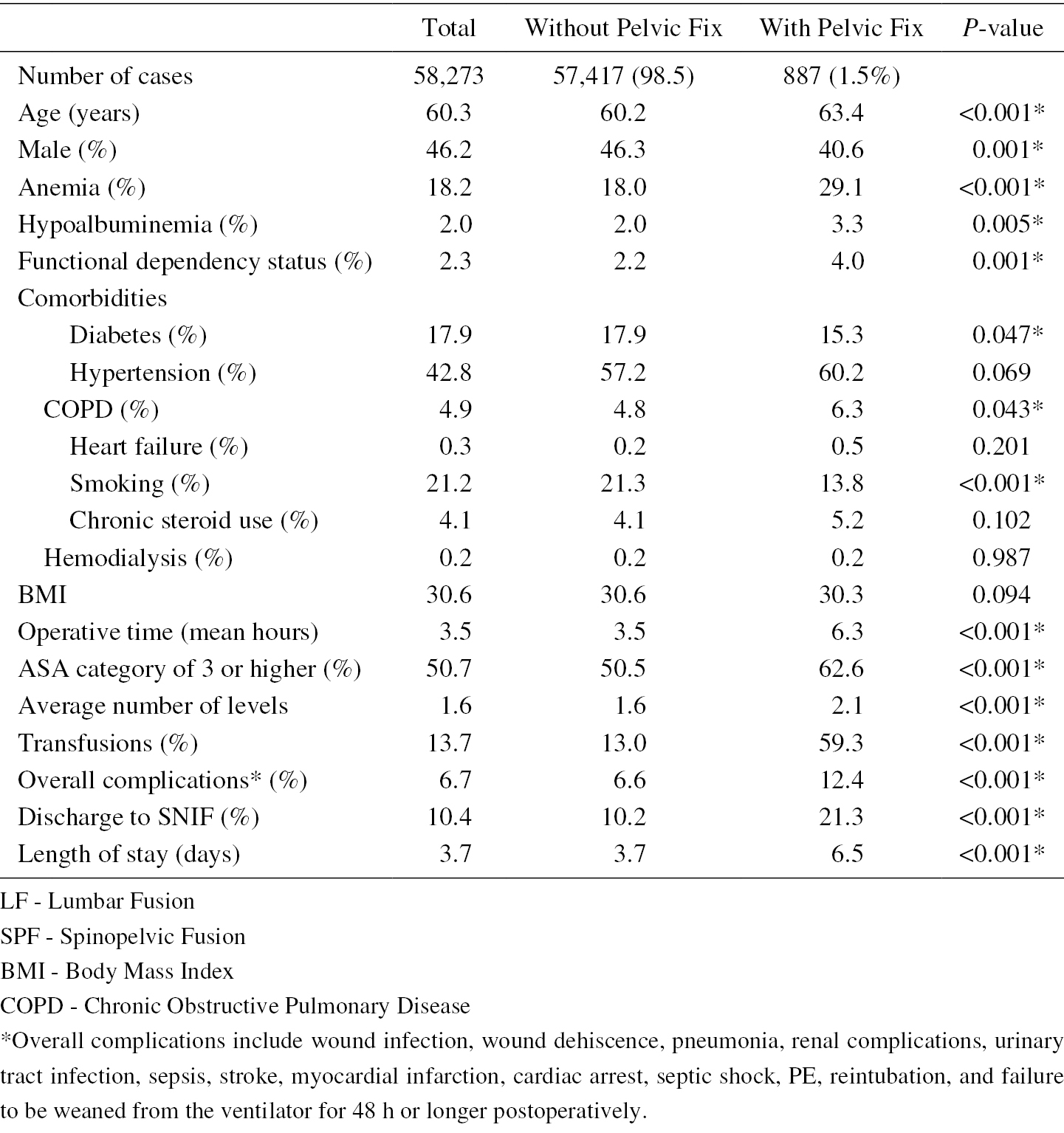
    Methods: We conducted a retrospective cohort study using data from the ACS-NSQIP database between 2006 and 2016 of patients who underwent lumbar fusion with and without spinopelvic fixation. Data regarding demographics, complications, hospital stay, and discharge disposition were collected.

    Results: A total of 57,417 (98.5%) cases of lumbar fusion without spinopelvic fixation (LF) and 887 (1.5%) cases of lumbar fusion with spinopelvic fixation (SPF) were analyzed. The transfusion rate in the SPF group was 59.3% vs 13% in the LF group (p < 0.001). The mean length of stay (LOS) and discharge to skilled nursing facility (SNF) were significantly different (LOS: SPF 6.5 days vs LF 3.5 days p < 0.001; SNF: SPF 21.3% vs LF 10.4% p < 0.001). After controlling for demographic differences, the overall complication rates were not significantly different between the groups (p = 0.531). The odds ratio for transfusion in the SPF group was 2.9 (p < 0.001). The odds ratio for increased LOS and increased care discharge disposition were elevated in the SPF group (LOS OR: 1.3, p < 0.012, Discharge disposition OR: 1.8, p < 0.001).

    Conclusions: Patients who underwent SPF had increased complications, transfusion rate, LOS, and discharge to SNF or subacute rehab facilities as compared with patients who underwent LF. SPF remains an effective technique for achieving lumbosacral arthrodesis. Surgeons should consider the implications of the associated complication profile for SPF and the value of preoperative optimization in a select cohort of patients.

    Download PDF (79K)
  • Hidetoshi Nojiri, Takatoshi Okuda, Kei Miyagawa, Nozomu Kobayashi, Tat ...
    2020 Volume 4 Issue 4 Pages 320-327
    Published: October 27, 2020
    Released on J-STAGE: October 27, 2020
    Advance online publication: June 18, 2020
    JOURNAL OPEN ACCESS

    Introduction: Pyogenic spondylitis of the lumbar spine markedly decreases the ability to perform activities of daily living and causes severe low back pain. The challenge is to improve low back pain and activities of daily living performance earlier and prevent post-infection sequelae, and conservative treatment with antibiotics is the mainstay of treatment.

    Methods: In the present study, patients who were unable to walk following lumbar pyogenic spondylitis even in the subacute phase after successful infection control, showing bone defects expanding from endplate to vertebral body in CT, were treated with posterior percutaneous short-range instrumentation and anterior autogenous bone grafting (group S, n = 10) or with conservative treatment alone (group C, n = 10). Acute cases of absolute surgical indication with paralytic symptoms and mild cases who could walk by antibiotics administration were excluded. The two groups were compared regarding the post-treatment change in C-reactive protein level, duration of bed rest, and post-infection local spinal deformities (local scoliosis angle in the coronal plane and local kyphosis angle in the sagittal plane).

    Results: Compared with group C, group S took a significantly shorter time for the C-reactive protein level to return to normal and required a significantly shorter duration of bed rest. Furthermore, surgery prevented the formation of kyphosis and scoliosis, while group C developed local kyphosis.

    Conclusions: The minimally invasive surgical method of posterior percutaneous short-range instrumentation and anterior autogenous bone grafting effectively enables early control of pain and maintenance of locomotive function and prevents spinal deformity in patients with lumbar pyogenic spondylitis in the subacute phase with advanced vertebral bone destruction.

    Download PDF (1056K)
  • Shunsuke Fujibayashi, Bungo Otsuki, Shimei Tanida, Ryoichi Nagahara, H ...
    2020 Volume 4 Issue 4 Pages 328-332
    Published: October 27, 2020
    Released on J-STAGE: October 27, 2020
    Advance online publication: July 10, 2020
    JOURNAL OPEN ACCESS

    Introduction: Intraspinal facet cysts resistant to conservative treatment are treated surgically. Surgical treatment was generally resection and decompression, but complications of dural tear and recurrence sometimes occurred. We present good clinical results and rapid spontaneous resolution following treatment of five cases of lumbar intraspinal facet cyst after lateral lumbar interbody fusion (LLIF).

    Methods: Multicenter series of five cases of lumbar intraspinal facet cyst with segmental instability treated with LLIF. The cross-sectional area (CSA) of the thecal sac and facet cyst on T2-weighted axial magnetic resonance imaging and the distance of facet joint (FJ) gap on axial computed tomography were measured preoperatively and postoperatively. Patient data and clinical and radiographic results were described.

    Results: Of five patients, one was male and four were female, with an average age of 72.6 (61-76) years. The mean preoperative CSA of facet cyst was 40.09 mm2. In all cases, intraspinal facet cyst resolved within two weeks after LLIF and good clinical results were obtained. The mean CSA of the thecal sac increased from 64.18 mm2 preoperatively to 95.72 mm2 postoperatively. The mean distance of FJ gap increased from 0.8 (0-1.5) mm preoperatively to 3.1 (0.5-6.0) mm postoperatively.

    Conclusions: LLIF may be indicated for intraspinal facet cysts with segmental instability.

    Download PDF (1272K)
  • Mitsuru Furukawa, Kunimasa Okuyama, Yusuke Kawano, Kentaro Kikuchi, Ta ...
    2020 Volume 4 Issue 4 Pages 333-340
    Published: October 27, 2020
    Released on J-STAGE: October 27, 2020
    Advance online publication: March 19, 2020
    JOURNAL OPEN ACCESS

    Introduction: When spinal fracture occurred in ankylosing spinal disorder (ASD) patients, it is important to evaluate not only the long lever arm but also bone density and bone quality for the determination of treatment strategies. This case-controlled study examined bone mineral density (BMD), bone metabolism markers, and pentosidine levels in patients with ASD.

    Methods: Subjects with bridging of minimum four contiguous vertebral bodies were classified into ASD group and the rest into non-ASD group. The former was further divided into two subgroups based on the presence/absence of sacroiliac joint ankylosis (SJA). We compared BMD, bone metabolism markers, and pentosidine levels in these groups.

    Results: The BMD T and Z scores of the femur proximal extremity were lower in the ASD with SJA group than those in the ASD without SJA group. When groups were matched for age, weight, and eGFR, compared with the non-ASD group, the ASD with SJA group had lower BMD of the lumbar spine and femur proximal extremity and the ASD without SJA group had significantly higher BMDs of the lumbar spine and femur proximal extremity. After matching, the ASD without SJA group showed a significantly higher pentosidine level than the non-ASD group.

    Conclusions: Patients with SJA have low femur proximal extremity BMD, whereas those with ASD without SJA have a higher BMD of the femur proximal extremity with high pentosidine level. Investigating the presence or absence of SJA is important for the determination of treatment strategies in fractured ASD patients.

    Download PDF (311K)
  • Shunsuke Kanbara, Ayato Nohara, Tetsuya Ohara, Toshiki Saito, Ryoji Ta ...
    2020 Volume 4 Issue 4 Pages 341-346
    Published: October 27, 2020
    Released on J-STAGE: October 27, 2020
    Advance online publication: March 19, 2020
    JOURNAL OPEN ACCESS

    Introduction: In patients with lumbosacral agenesis (SA), Renshaw type III or IV, lumbosacral instability is the primary cause of major clinical complications. Although they are usually treated with spinopelvic fusion, nonunion at the spinopelvic junction is a major complication due to the congenital sacropelvic abnormalities. The purpose of this study was to evaluate whether a combination of lumbosacral fixation and rigid fixation at the pubic symphysis could lead to postoperative bone union in patients with SA (Renshaw type III).

    Methods: Retrospective case series study. We present the cases of two patients with SA, Renshaw type III, who were surgically treated by lumbosacral fusion using a posterior approach, and they exhibited nonunion at the lumbosacral junction.

    Results: Case 1. A 10-year-old male underwent T8-S posterior fixation followed by multiple augmentations using allografts at the lumbosacral junction for delayed union. All additional procedures with bone graft using a posterior approach failed to achieve bone union; however, additional rigid fixation at the pubic symphysis resulted in a successful lumbosacral bone union.

    Case 2. A 6-year-old male underwent vertical expandable prosthetic titanium rib (VEPTR) surgery with multiple rod extension procedures. Subsequently, at the age of 10 years, a combined two-stage anterior (L1-3) and posterior (T8-iliac) fixation with T9 hemivertebrectomy was performed. As a result of subsequent nonunion with screw loosening, additional rigid fixation at the pubic symphysis was performed 1 month after posterior fixation. Bone union was finally achieved 1 year after all the surgical interventions.

    Conclusions: Rigid fixation at the pubic symphysis may play a significant role in achieving rigid bone union for unstable lumbopelvic connection, such as SA, Renshaw type III or IV.

    Download PDF (1462K)
  • Tomoka Endo, Tetsuro Ohba, Hiroki Oba, Kotaro Oda, Nobuki Tanaka, Hiro ...
    2020 Volume 4 Issue 4 Pages 347-353
    Published: October 27, 2020
    Released on J-STAGE: October 27, 2020
    Advance online publication: April 20, 2020
    JOURNAL OPEN ACCESS

    Introduction: There is a significant relationship between pulmonary function and degree of spinal deformity, location of apical vertebrae, and coronal imbalance in patients with childhood spinal deformity. By contrast, the pathophysiology, epidemiology, and influence of deformity on respiratory dysfunction in patients with adult spinal deformity (ASD) remain largely unknown. We sought to clarify and compare the prevalence of pulmonary function impairment in patients with ASD with that in patients with lumbar spinal stenosis (LSS), to determine radiographically which spinal malalignment parameters are associated with a risk of respiratory dysfunction, and to determine the association of respiratory dysfunction with corrective surgery.

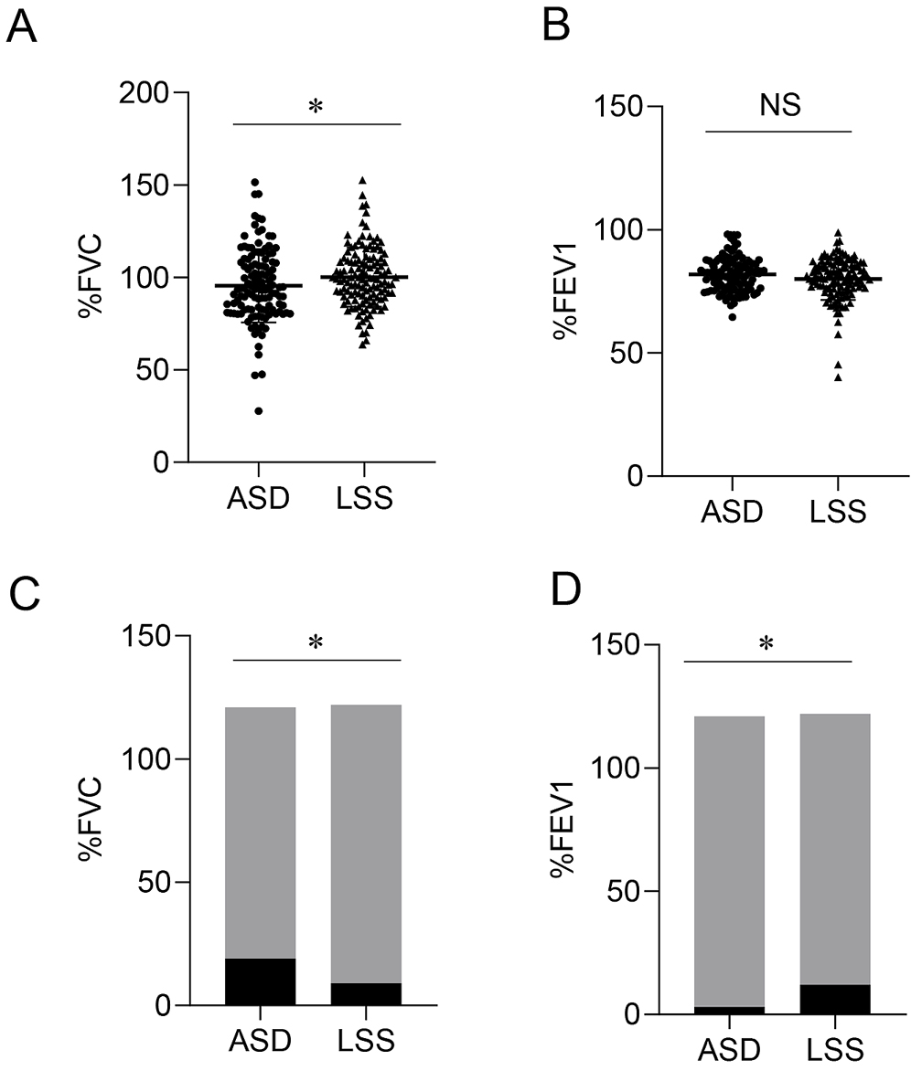
    Methods: We conducted a prospective study of consecutive patients with a diagnosis of ASD or LSS who underwent spinal surgery. We included data from 122 consecutive patients with ASD and 121 consecutive patients with LSS. Parameters were obtained from full-length lateral radiographs taken with the patients standing and in supine and prone positions. We compared respiratory dysfunction between a group of patients with ASD and LSS and determined correlations between respiratory dysfunction and spinopelvic parameters.

    Results: Preoperative % forced vital capacity (FVC) of patients with ASD was significantly lower than that of patients with LSS, and the frequency of restrictive ventilatory impairment was significantly higher in those with ASD (15.7%) than those with LSS (7.4%). Thoracolumbar kyphotic curvature (TK) while the patients were in supine position was significantly greater in the group with restrictive ventilatory impairment, and a significant negative correlation was found between %FVC and TK with the patients in supine position. We found no significant improvement of respiratory dysfunction 1 year after surgery.

    Conclusions: Spinal deformity is a potential risk factor for restrictive ventilatory impairment in the elderly. We propose that radiographs obtained when patients are in supine position are valuable for evaluating the flexibility of the TK. Rigid TK might be an etiology of restrictive ventilatory impairment in patients with ASD.

    Download PDF (329K)
  • Kazuhide Inage, Takeshi Sainoh, Takayuki Fujiyoshi, Takuma Otagiri, Ya ...
    2020 Volume 4 Issue 4 Pages 354-357
    Published: October 27, 2020
    Released on J-STAGE: October 27, 2020
    Advance online publication: July 10, 2020
    JOURNAL OPEN ACCESS

    Introduction: Mirogabalin should be equivalent to pregabalin, but with fewer incidences of adverse drug reactions (ADRs). To verify these benefits in actual clinical trials, our study investigated the frequency of ADRs and mirogabalin's analgesic effects during treatment of peripheral neuropathic pain.

    Methods: This study included 74 patients with lower limb pain. We surveyed patient reports of ADRs during the follow-up period as the primary endpoint and examined the visual analog scale (VAS) reported for lower limb pain as the secondary endpoint (before administration, and two and four weeks after administration).

    Results: The occurrence of ADR was 27.0%, like the frequency of ADRs in the clinical trials for other disorders. However, the discontinuation rate of administration was 10.8%, which was significantly lower than the frequency of ADR occurrences. When the analgesic effect was assessed, a significant decrease in the temporal change of VAS for lower limb pain was observed before administration, and two and four weeks after administration.

    Conclusions: In this study, the occurrence of ADRs reported by the patients was like the frequency of ADRs reported in the clinical trials for other disorders. When assessing the analgesic effect, the temporal change of VAS for lower limb pain was found to decrease significantly before administration, and two and four weeks after administration.

    Download PDF (90K)
TECHNICAL NOTE
  • Tomoaki Koakutsu, Toshimi Aizawa, Eiji Itoi
    2020 Volume 4 Issue 4 Pages 358-364
    Published: October 27, 2020
    Released on J-STAGE: October 27, 2020
    Advance online publication: February 26, 2020
    JOURNAL OPEN ACCESS

    Introduction: Cervical pedicle screw (CPS) fixation provides the strongest mechanical stability. It needs, however, wide soft tissue detachment to expose the entry point and carries the potential risk of iatrogenic damage to neurovascular structures. Malposition of the CPS cannot be completely avoided even using the navigation system.

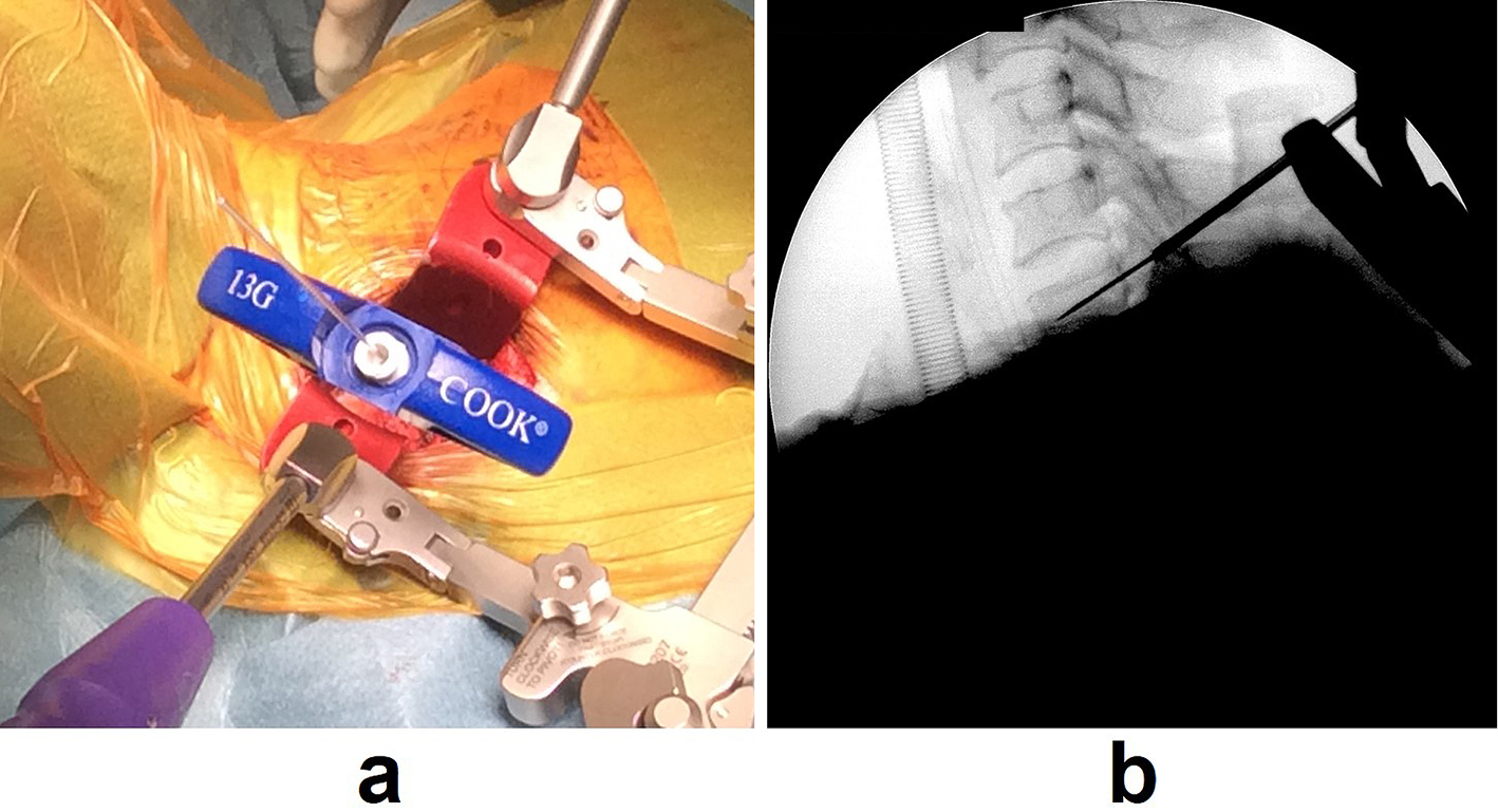
    Technical Note: Using the bone biopsy needle as drill guide, we developed a novel accurate CPS insertion technique. (1) The entry point of CPS was exposed using Southwick's technique for anterior fixation or Tokioka's technique for posterior fixation. (2) A 13G bone biopsy needle was inserted from the entry point established by the fluoroscopy-assisted pedicle axis view technique described by Yukawa et al. to within a few millimeters of the pedicle. (3) The external sleeve of the bone biopsy needle was left in place as a drill guide, and the 1.25 mm guidewire for a 4.0 mm cannulated screw was then inserted into the pedicle cavity. (4) The external sleeve of the bone biopsy needle was removed, and the screw trajectory was created by a 2.7 mm cannulated drill bit over the guidewire. (5) Tapping was conducted prior to CPS insertion.

    Using this method, 29 CPSs in nine patients were inserted. Postoperative computed tomography scans revealed that all the CPSs were placed accurately.

    Conclusions: Utilizing the bone biopsy needle as drill guide, our procedure enables accurate positioning of CPS without expensive instruments.

    Download PDF (609K)
CLINICAL CORRESPONDENCE
feedback
Top