Abstract
Purpose: To evaluate stress distribution around the marginal area of three full veneer crown restorations with different thicknesses.
Materials and Methods: Two types of premolar finite element model (normal and half thickness of crown; NC, HC) were made on the personal computer and three types of crown material (polyetheretherketone, PEEK, PK; hybrid composite resin, HR; and Ag-Pd-Cu-Au alloy, PD) were assumed. Then the magnitude of stress at the marginal area of crown was calculated.
Results: The magnitude of stress for PD-NC, HR-NC, PK-NC, PD-HC, HR-HC, PK-HC was 26.4, 21.3, 14.2, 50.6, 26.1, and 13.3 MPa respectively. The crown fabricated by PEEK prevented the stress concentration at the marginal area.
Conclusion: Crowns fabricated by PEEK prevent the concentration of stress at the crown margin, and there is a possibility that these crowns require less tooth preparation in this area, when compared to metallic crowns.
Introduction
Full coverage crowns are commonly indicated in cases of advanced tooth destruction, caused by caries or coronal fracture. Ceramics, composite resins, and dental alloys are used as restorative materials for full coverage crowns. Recently, the use of polyetheretherketone (PEEK) in dentistry has been explored, including construction of coronal restorations [1,2,3,4,5,6,7,8,9]. PEEK is a semi-crystalline thermoplastic polymer with favorable mechanical characteristics; high thermostability, high fatigue endurance, high impact resistance, and high creep resistance [1,2,3,4,5,6,7,8,9]. In addition, PEEK is metal-free; therefore, it could be beneficial for patients with a metal allergy.
One of the advantageous features of PEEK is its flexibility compared to dental alloys or other coronal restoration materials. Thus, there is potential that a PEEK crown could prevent the stress concentration at the crown margin that leads to the development of secondary caries. It is also possible that the marginal thickness of this crown may be reduced, relative to metal crowns, as a PEEK crown may require less tooth preparation because of its flexibility. The aim of this study was to evaluate the influence of crown materials including PEEK and crown thickness on stress distribution around crown margins susceptible to secondary caries.
Materials and Methods
Two types of three-dimensional nonlinear finite element model were fabricated on a personal computer, using finite element analysis software (MSC Marc Mentat 2013, MSC Software Corp., Santa Ana, CA, USA) [10,11]. The models were simulated endodontically treated premolar teeth, 18 mm long, with a diameter of 6 mm cervical dentin. The apical 12 mm of the root was surrounded with a 0.2-mm thickness of periodontal ligament and a 0.3-mm thickness of lamina dura. The length of the post was 8 mm, with the height of the coronal part being 6 mm. In addition, the height of the ferrule was 2 mm in all models. The margin design of the abutment teeth on this model was a rounded shoulder with 0.5-mm radius, and a taper of six degrees to the long axis of the tooth. (Normal crown model, NC). The remaining bone was modeled as cancellous bone and cortical bone. These models were analyzed using finite element analysis software in accordance with previous reports [10,11]. In this study, the hexahedral element was applied, instead of the tetrahedral element, to improve the precision of finite element analysis results. Two types of simulated crown model depending on the thickness of the crown were developed: one had a normal thickness of crown (NC), and the other had half the thickness of the normal crown in the axial wall (HC) (Fig. 1). In each model, three types of crown material were applied: Ag-Pd-Cu-Au alloy (PD), hybrid composite resin (HR), and PEEK (PK), with composite resin with a glass fiber post being used as a post and core system (Fig. 1). The main properties of each crown material in this research are shown in Table 1 [12,13,14,15,16,17,18,19,20,21,22]. Each material had unique elastic material properties, and linear elasticity was a feature of all materials, except for the periodontal ligament. Concern about the main property of the periodontal ligament, a nonlinear elastic property was applied in accordance with previous reports [10,11], due to its viscoelastic properties [13].
Table 1
Mechanical properties of materials
|
Elastic modulus (MPa) |
Poisson’s ratio |
Reference |
| Dentin |
15,000 |
0.31 |
[12] |
| Periodontal ligament |
Nonlinear |
Nonlinear |
[13] |
| Lamina dura |
13,700 |
0.30 |
[14] |
| Cancellous bone |
345 |
0.31 |
[15] |
| Cortical bone |
13,700 |
0.30 |
[14] |
| Gutta-percha |
0.69 |
0.45 |
[16] |
| Composite resin core material |
12,000 |
0.33 |
[17] |
| Glass fiber post |
29,200 |
0.30 |
[18] |
| Resin luting agent |
18,000 |
0.30 |
[19] |
| Ag-Pd-Cu-Au alloy |
86,000 |
0.33 |
[20] |
| Hybrid composite resin |
21,000 |
0.27 |
[21] |
| Polyetheretherketone (PEEK) |
4,100 |
0.40 |
[22] |
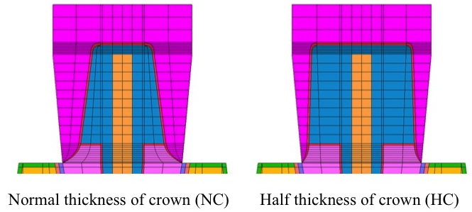
Fig. 1
Finite element model
Two kinds of finite element model of the mandibular premolar tooth were fabricated on the personal computer. One had normal thickness of crown model that had a taper of six degrees to the long axis of the tooth (NC), and the other had half thickness of the normal crown in the axial wall (HC).
For all models, the bottom of mandibular bone was restricted completely and the occlusal force, which was measured with a small 3-dimensional occlusal force meter during mastication of beef jerky, was applied to the center of the occlusal surface (lingual direction: 24 N, distal direction: 29 N, apical direction: 164 N) [10,11,23,24]. The analysis points (nodes) for this study were the top and bottom of the occlusal area of the crown, the marginal area of the crown (this node coincided with the luting agent), and the cervical area of root dentin (this node also coincided with the luting agent). Von Mises stress distribution within the crown and root dentin was subsequently calculated.
Results
The stress distribution of the abutment teeth and crowns in the frontal cross-section view, during mastication of a piece of beef jerky, is shown in Fig. 2. Stress distribution within the crowns is seen in Fig. 3, and Fig. 4 shows the stress distribution of the outer surface of crowns and abutment teeth. The magnitude of the stress at each analysis point is shown in Table 2. For premolars with the same thickness of crown, the magnitude of the stress at the margin of the PD crown was highest, while the lowest stress concentration was seen in the PK crown. Conversely, the magnitude of the stress at the bottom of the occlusal surface of the crown showed the opposite tendency: PK showed the highest stress concentration, and PD showed the lowest stress concentration. At the top of occlusal surface of the crown, HR showed the highest stress concentration and PD showed the lowest stress concentration, and at the cervical area of the dentin, PD showed the highest stress concentration and PK showed the lowest stress concentration, as seen at the crown margin.

Fig. 2
Stress distribution within the abutment teeth (Frontal plane)
The equivalent stress distribution in a buccolingual cross-section of the abutment tooth in two types of crown thickness model depending on the crown materials
Gray and yellow represent the high stress concentration area as indicated by the color legend.
Stress concentration was observed at the top of crown and cervical area of abutment tooth.
Fig. 3
Stress distribution within the crown
The equivalent stress distribution in a buccolingual cross-section of the crown in two types of crown thickness model depending on the crown materials
Gray and yellow represent the high stress concentration area as indicated by the color legend.
Stress concentration was observed at the top of crown and at the cervical area of crown, especially in the case of PD-HC.
In the comparison of the two crown thickness models, the magnitude of stress at the PD-HC crown margin showed considerably higher stress concentration than the PD-NC margin did. Conversely, the magnitude of stress at the PK-HC crown margin showed lesser stress concentration than the PK-NC margin. The magnitude of stress at the HR-HC crown margin was slightly higher than that of the stress of HR-NC, and in addition, the magnitude of stress at the cervical dentin showed almost the same tendency as the crown margin.
At the top of the crown, the magnitude of stress was unrelated to the thickness of the crown, however at the bottom of the crown, the magnitude of stress of PD and HR was increased with the reduced thickness of crown axial wall. Only the stress concentration of PK decreased with reduced crown thickness.
Fig. 4
Stress distribution within the crowns and abutment teeth
The equivalent stress distribution of the crown and abutment tooth in two types of crown thickness model depending on the crown materials
Gray and yellow represent the high stress concentration area as indicated by the color legend. Stress concentration was observed at the top of crown and at the cervical area of crown and abutment tooth, especially in the case of PD-HC.
Table 2
Stress value in MPa within crown depending on the crown material and shape
| Name |
Crown type |
Material of the crown |
Stress value (Crown) |
Stress value (Dentin) |
| Marginal area |
Bottom of occlusal surface |
Top of occlusal surface |
Cervical area |
| PD-NC |
Normal |
Ag-Pd-Cu-Au alloy |
26.4 |
20.2 |
340.1 |
17.5 |
| HR-NC |
Normal |
Hybrid composite resin |
21.3 |
26.8 |
382.6 |
16.0 |
| PK-NC |
Normal |
PEEK |
14.2 |
38.8 |
362.6 |
14.4 |
| PD-HC |
Half |
Ag-Pd-Cu-Au alloy |
50.6 |
25.5 |
334.9 |
23.2 |
| HR-HC |
Half |
Hybrid composite resin |
26.1 |
28.7 |
381.8 |
16.9 |
| PK-HC |
Half |
PEEK |
13.3 |
35.1 |
362.4 |
14.9 |
Discussion
One of the reasons for crown failure is the development of secondary caries at the crown margin. Stress concentration around the marginal area of the crown and dentin can lead to failure of luting agents, microfractures of the dentin can occur, and finally secondary caries can develop. Therefore, reduction of stress concentration around the marginal area of the crown and dentin seems useful in the prevention of secondary caries. Stress concentration is related to the type of luting agent, post and core system, and crown material [10,11,22,23,24,25,26]. In addition, there is a possibility that the thickness of the crown may have an influence on the stress distribution around the crown margin. Thus, we focused on crown materials and the thickness of the crown axial wall in this study. Until now, the elastic modulus of most crown materials was the same or higher than that of dentin. However, PEEK has a lower elastic modulus and higher flexibility, compared to conventional crown materials, and therefore, there is a possibility that a crown fabricated from PEEK can reduce marginal stress concentration.
There are two methods of establishing a finite element model: model fabrication applying the morphological data derived from computerized tomography (CT), and a hand-made model. In this research, hand-made finite element models were fabricated. One advantage of using a CT scan to construct teeth models for stress analysis is the close resemblance to the shape of the actual teeth. However, it is very difficult to construct each part to a uniform size and thickness, especially the crown and luting agents; this may influence stress distribution around the cervical area. Fabrication of the hand-made model enabled to optimize the detail of the luting agent and crown, based on the requirements of the experiment. However, there are also some disadvantages to the hand-made model; it is difficult to simulate the morphology of the actual root canal, and it is difficult to rebuild the tooth shape with cusps, ridges, and grooves on the occlusal surface.
The aim of this study was to reveal the influence of crown thickness and different crown materials on stress distribution around the cervical area of the crown. Therefore, hand-made finite element model was fabricated in this study after careful consideration of these advantages and disadvantages. For models with the same crown thickness, the magnitude of stress around the crown and dentin margin, from highest to lowest, were PD, HR, and PK, and this order coincided with the value of the elastic modulus. Conversely, at the level of the central occlusal groove, the order was reversed. This arose from the deformation of the crowns under loading; PK deformed easily and the occlusal force was transmitted to the top of core almost directly, through the intermediary of the luting agent. On the contrary, the occlusal force of PD dispersed not only to the top of the core through the luting agent but also the marginal area of the crown because of its inelasticity. Therefore, the magnitude of stress at the margin of PD was higher than that of PK.
At the margin, the magnitude of stress on the crown was higher than that of dentin in all models, except for PK-HC. This indicates that luting agents require a higher level of adhesion to crown materials than to dentin, to prevent failure and the resultant formation of secondary caries. It was found in the case of PD that crown thickness affected the magnitude of stress; a reduction in crown thickness by half lead to an increase in stress concentration two-fold at the crown margin. This indicated that a decrease in the amount of tooth preparation at the abutment axial wall was not appropriate because a reduced crown thickness might lead to an increase in stress at the crown and dentin margin. Conversely in this regard, PEEK can reduce the marginal thickness of the crown; PEEK application as a crown material can reduce the amount of tooth preparation required. Therefore, there is a possibility that the use of PEEK as a crown material can reduce the amount of tooth preparation required, when compared to PD or HR.
Although PEEK crowns can prevent the concentration of stress at the margin, its flexibility results in stress concentration at the core of the abutment and basal area of the post, rather than being distributed within the crown; resulting in less stress being transmitted to the core, as seen in PD or HD. Further studies regarding stress distribution at these areas are required [27]. Also, the magnitude of stress at the luting agent must be considered. One of the reasons for the development of secondary caries at the crown margin is the failure of the luting agent. In this study, the node for analysis of the crown marginal area was located at the edge of the crown, coincident with the top of the luting agent layer, and the node for analysis of the dentin marginal area was located at the edge of the dentin, coincident with the bottom of the luting agent layer. This means that the magnitude of the stress at the marginal area of crown and dentin reflected the magnitude of the stress within the luting agent. Among the three types of crown material, the PD crown margin had the most concentrated stress levels at the luting agent level, and the use of PEEK as a crown material did most to prevent stress concentration in this area.
In conclusion, crowns fabricated by PEEK prevent the concentration of stress at the crown margin, and there is a possibility that these crowns require less tooth preparation in this area, when compared to metal crowns.
Conflicts of Interest
The authors declare that there are no conflicts of interest related to the manuscript.
References
- 1) Tetelman ED, Babbush CA. A new transitional abutment for immediate aesthetics and function. Implant Dent 2008; 17: 51-8.
- 2) Schmidlin PR, Stawarczyk B, Wieland M, Attin T, Hammerle CH, Fischer J. Effect of different surface pre-treatments and luting materials on shear bond strength to PEEK. Dent Mater 2010; 26: 553-9.
- 3) Bayer S, Komor N, Kramer A, Albrecht D, Mericske-Stern R, Enkling N. Retention force of plastic clips on implant bars: a randomized controlled trial. Clin Oral Implants Res 2012; 23: 1377-84.
- 4) Santing HJ, Meijer HJ, Raghoebar GM, Özcan M. Fracture strength and failure mode of maxillary implant-supported provisional single crowns: a comparison of composite resin crowns fabricated directly over PEEK abutments and solid titanium abutments. Clin Implant Dent Relat Res 2012; 14: 882-9.
- 5) Tannous F, Steiner M, Shahin R, Kern M. Retentive forces and fatigue resistance of thermoplastic resin clasps. Dent Mater 2012; 28: 273-8.
- 6) Stawarczyk B, Beuer F, Wimmer T, Jahn D, Sener B, Roos M, et al. Polyetheretherketone—a suitable material for fixed dental prostheses? J Biomed Mater Res B Appl Biomater 2013; 101: 1209-16.
- 7) Stawarczyk B, Keul C, Beuer F, Roos M, Schmidlin PR. Tensile bond strength of veneering resins to PEEK: impact of different adhesives. Dent Mater J 2013; 32: 441-8.
- 8) Stawarczyk B, Eichberger M, Uhrenbacher J, Wimmer T, Edelhoff D, Schmidlin PR. Three-unit reinforced polyetheretherketone composite FDPs: influence of fabrication method on load-bearing capacity and failure types. Dent Mater J 2015; 34: 7-12.
- 9) Sturz CR, Faber FJ, Scheer M, Rothamel D, Neugebauer J. Effects of various chair-side surface treatment methods on dental restorative materials with respect to contact angles and surface roughness. Dent Mater J 2015; 34: 796-813.
- 10) Okada D, Miura H, Suzuki C, Komada W, Shin C, Yamamoto M, et al. Stress distribution in root restored with different kinds of post systems with composite resin. Dent Mater J 2008; 27: 605-11.
- 11) Luo S, Okada D, Bakhit M, Shin C, Ogura R, Miura H. Stress distribution in luting agents with different post and core systems. Asian Pac J Dent 2017; 17: 15-22.
- 12) Rees J, Jacobsen P. Elastic modulus of the periodontal ligament. Biomaterials 1997; 18: 995-9.
- 13) Pini M, Wiskott H, Scherrer S, Botsis J, Belser U. Mechanical characterization of bovine periodontal ligament. J Periodont Res 2002; 37: 237-44.
- 14) Borchers L, Reichart P. Three-dimensional stress distribution around a dental implant at different stages of interface development. J Dent Res 1983; 62: 155-9.
- 15) Rees J, Jacobsen P. Elastic modulus of the periodontal ligament. Biomaterials 1997; 18: 995-9.
- 16) Asmussen E, Peutzfeldt A, Sahafi A. Finite element analysis of stresses in endodontically treated, dowel-restored teeth. J Prosthet Dent 2005; 94: 321-9.
- 17) Lanza A, Aversa R, Rengo S, Apicella D, Apicella A. 3D FEA of cemented steel, glass and carbon posts in a maxillary incisor. Dental Materials 2005; 21: 709-15.
- 18) Komada W, Miura H, Okada D, Yoshida K. Study on the fracture strength of root reconstructed with post and core: alveolar bone resorbed case. Dent Mater J 2006; 25: 177-82.
- 19) Nakamura T, Ohyama T, Waki T, Kinuta S, Wakabayashi K, Takano N, et al. Finite element analysis of fiber-reinforced fixed partial dentures. Dent Mater J 2005; 24: 275-9.
- 20) Matsuo S, Watari F, Ohata N. Fabrication of a functionally graded dental composite resin post and core by laser lithography and finite element analysis of its stress relaxation effect on tooth root. Dent Mater J 2001; 20: 257-74.
- 21) Nakamura T, Ohyama T, Waki T, Kinuta S, Wakabayashi K, Takano N, et al. Finite element analysis of fiber-reinforced fixed partial dentures. Dent Mater J 2005; 24: 275-9.
- 22) Schwitalla AD, Abou-Emara M, Spintig T, Lackmann J, Muller WD. Finite element analysis of the biomechanical effects of PEEK dental implants on the peri-implant bone. J Biomech 2015; 48: 1-7.
- 23) Suzuki C, Miura H, Okada D, Komada W. Investigation of stress distribution in roots restored with different crown materials and luting agents. Dent Mater J 2008; 27: 229-36.
- 24) Suzuki C, Miura H, Okada D, Komada W, Miyasaka M, Yamamoto M, et al. The Investigation of distortions around the cervical area of teeth restored with two kinds of crown materials. Dent Mater J 2009; 28: 142-52.
- 25) Ma J, Miura H, Okada D, Yusa K. Photoelastic stress analysis of endodontically treated teeth restored with different post systems: normal and alveolar bone resorption cases. Dent Mater J 2012; 30: 806-13.
- 26) Oshima F, Okada D, Ogura R, Shin C, Ueda Y, Inagaki T, et al. A finite element analysis of stress distribution in roots with different types of post systems. Asian Pac J Dent 2016; 16: 1-7.
- 27) Chiba A, Hatayama T, Kainose K, Nakajima M, Pashley DH, Wakabayashi N, et al. The influence of elastic moduli of core materials on shear stress distributions at the adhesive interface in resin built-up teeth. Dent Mater J 2017; 36: 95-102.