Abstract
The basilar part of the occipital bone is occasionally partially divided by lateral notches, termed transverse basilar clefts (TBCs). In the present paper, we describe the condition of the TBC in six skulls from prehistoric Jomon sites in the Japanese archipelago and then compare TBC frequencies between the Jomon and recent Japanese cranial series. The frequencies of TBCs in Jomon skulls from the southern Hokkaido and Tohoku districts are higher than in recent Japanese specimens from the Tohoku district. Since the TBC is considered a variation that appears at an early developmental stage in the human cranium, it is worthwhile investigating this trait as one of the nonmetric cranial variants in various human population groups, including immature individuals.
Introduction
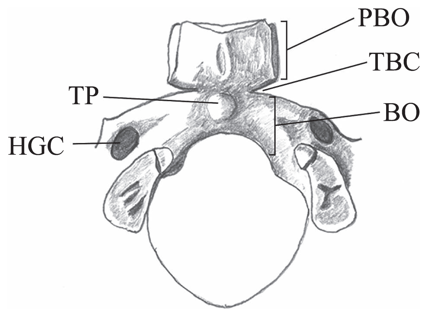
On rare occasions, the basilar part of the occipital bone is partially divided by lateral notches; in very rare cases, it is completely divided by a fissure into two portions (Bryce, 1915). Although these lateral notches have been referred to by various names, such as incisura basi-praebasioccipitalis (Osawa, 1894; Shima, 1941), sillon intermédiaire (Charpy and Nicolas, 1911), transverse fissure (Limson, 1932; Lombardi, 1961), transverse cleft (Kruyff, 1967; Shapiro and Robinson, 1976; Lang, 1995), basioccipital cleft (Johnson and Israel, 1979), basilar clefting (Anderson, 2000), and basilar transverse fissure (Hofmann and Prescher, 2012), the term ‘transverse basilar cleft’ (TBC) (Scheuer and Black, 2000) is used in the present paper. As shown in Figure 1, the portion anterior to the cleft is called ‘basiotic’ or ‘praebasioccipital,’ whereas the portion posterior to the cleft is termed ‘basioccipital’ (Osawa, 1894; Le Double, 1903; Shima, 1941; Kruyff, 1967; Hofmann and Prescher, 2012).
Although earlier researchers observed TBCs in skulls from pathological individuals (for details, see Le Double, 1903; Kruyff, 1967), these traits are now generally regarded as an anatomical or embryological variation. Various hypotheses have been proposed for the occurrence of the TBC. Several authors believe that it is a result of the retention of segmentation in embryological occipital sclerotomes (Lombardi, 1961; Shapiro and Robinson, 1976; Lang, 1995), whereas others assume that the TBC marks the border between two ossification centers in the basilar part of the occipital bone—one in the praebasioccipital and the other in the basioccipital (Bryce, 1915; Hofmann and Prescher, 2012). Lang (1995) believes that the TBC corresponds to the borderline between the embryological parachordal cartilage and occipital sclerotomes, which are fused together to form the basilar part of the occipital bone (Hamilton and Mossman, 1972; Carlson, 2009). Anderson (2000) claims that there appears to be a genetic component involved in the occurrence of the TBC, because it was detected in two fetal skulls recovered from the same grave.
In the present study, we aimed: (i) to report the presence of the TBC in six skulls from prehistoric Jomon sites in the Japanese archipelago; (ii) to compare the frequencies of the TBC between the Jomon and recent Japanese cranial series using both immature and adult skulls; (iii) to consider the manifestation of the TBC in terms of an embryological aspect; and (iv) to examine whether the TBC is regarded in nature as a nonmetric trait of developmental variation.
Materials and Methods
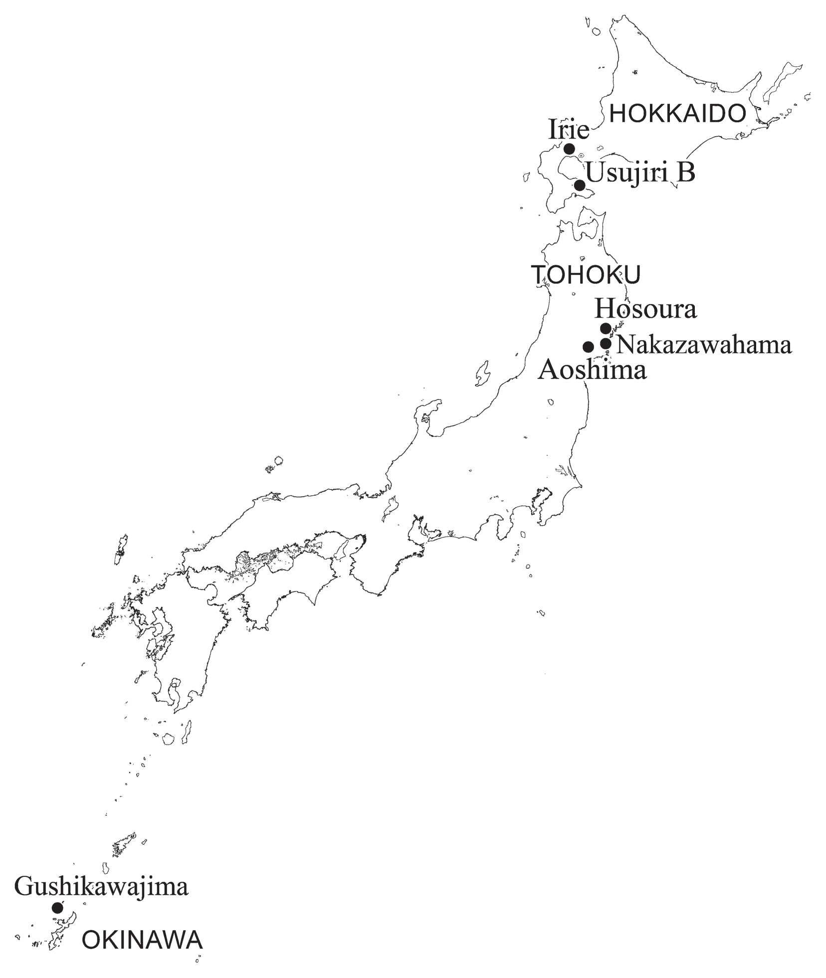
A total of 71 adult and 12 immature Jomon skulls of both sexes from various sites in the southern Hokkaido and Tohoku regions were investigated and subjected to frequency analyses for the presence of TBCs. These sites belong to the Early to Final Jomon periods (ca. 7000–2800 cal BP) and include the shell mounds of Kitakogane, Irie, Takasago, Usu-moshiri, Yakumo-kotan, and Usujiri B in southern Hokkaido as well as Miyako’ozuke, Kuma’ana, Miyano, Hosoura, Ohora, Nakazawahama, Ebishima (Kaitori), Tagara, Maehama, Minamizakai, Aoshima, and Satohama in the Tohoku district. The Jomon skulls are housed at the Sapporo Medical University, Date City Institute of Funkawan Culture, Iwate Prefectural Museum, Tohoku University, Tohoku History Museum, Historical Museum of Jomon Village OkuMatsushima, University of Tokyo, National Museum of Nature and Science, Tokyo, and Osaka University. In addition to these specimens, we described TBCs in an adult male skull from the Shiitachi-nishiku site in Gushikawajima, Okinawa (stored at the Okinawa Prefecture Archaeological Center) because we detected typical TBCs in a photograph of this specimen taken by Dr. N. Doi of the University of the Ryukyus.
A TBC was considered to be present when a definite transverse notch was observed in the basilar part of the occipital bone. The length of the TBC was ≥4 mm and all cases of this trait were photographed.
For comparative purposes, we examined the presence or absence of the TBC in a recent Japanese cranial series of both sexes. This series from the Tohoku district consists of 162 fetal skulls aged from 6 months to term, 31 juvenile skulls aged from 7 days to 19 years, and 156 adult skulls aged ≥20 years. These skulls are kept at the Tohoku University School of Medicine. The frequencies of TBCs were compared between the Jomon cranial series from the southern Hokkaido and Tohoku regions and the recent Japanese cranial series from the Tohoku district. The difference in TBC occurrence between these series was statistically tested by calculating the Fisher’s exact probability using the statistical software package HALWIN (Gendaisugakusha).
Results
Description of TBC
The Jomon sites yielding skulls with TBCs include the Irie shell mound and Usujiri B site in southern Hokkaido; the Hosoura, Nakazawahama, and Aoshima shell mounds in the Tohoku district; and the Shiitachi-nishiku site from Gushikawajima in Okinawa. The site locations are shown in Figure 2.
Nakazawahama No. 2 skull
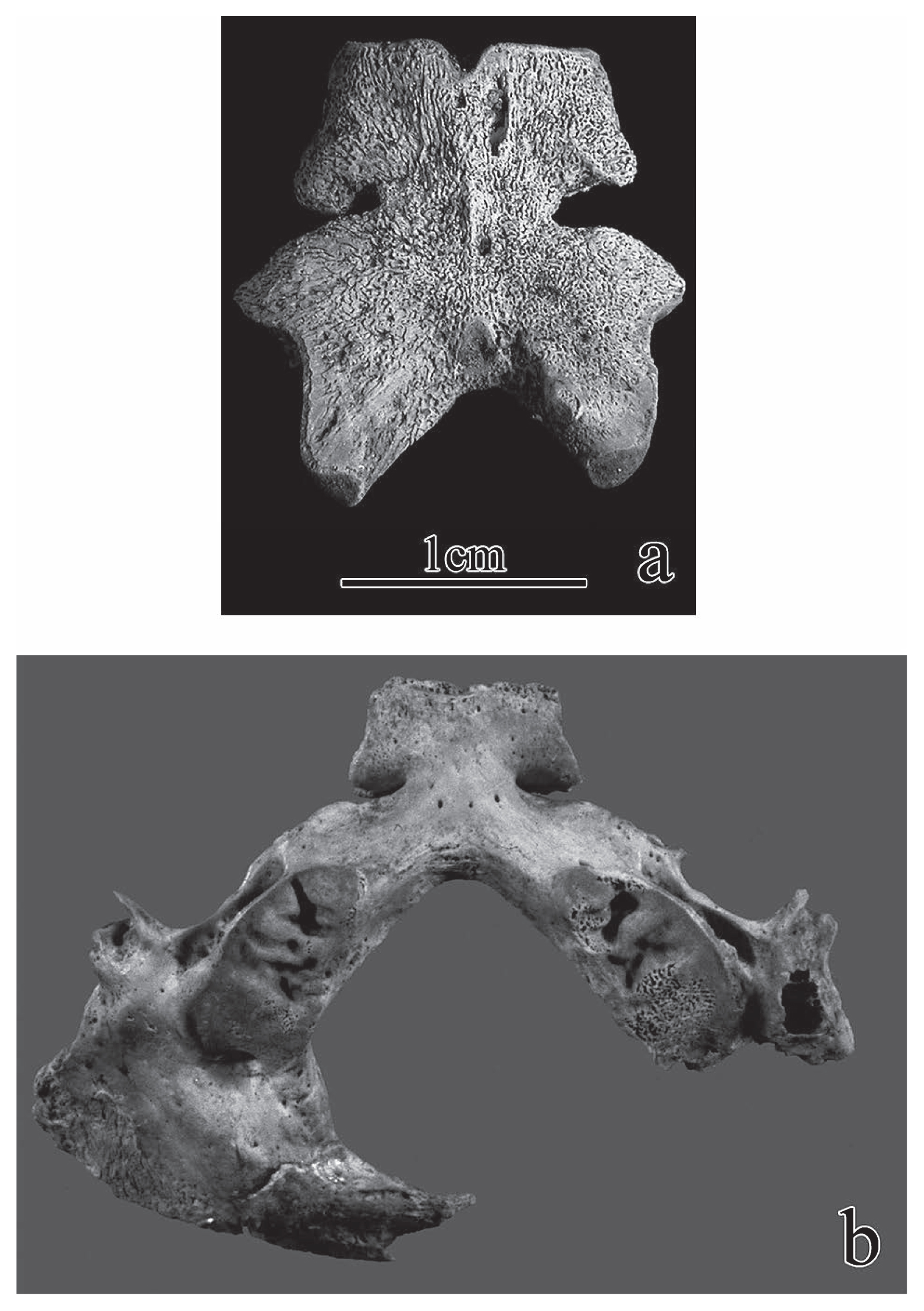
Skeletal remains from 18 individuals were unearthed at the Nakazawahama shell mound in Rikuzentakata, Iwate Prefecture, Tohoku District, by the Rikuzentakata City Board of Education between 1984 and 1988 (Editorial Board of Rikuzentakata City History, 1994). These remains included 2 fetal and 10 adult skulls. The No. 2 fetal skull from the Final Jomon period (ca 3300–2800 cal BP) showed TBCs on both sides of the occipital basilar part (Figure 3a). This fetal specimen’s age was estimated at approximately 37 weeks based on the lengths of associated limb bones.
Irie No. 7 skull
Skeletal remains of 15 individuals (5 juveniles, 10 adults) were unearthed at the Irie shell mound in Toyako-cho, southern Hokkaido, by the Abuta-cho Board of Education in 1966 and 1967 (Mitsuhashi, 1968). TBC was observed bilaterally in the No. 7 juvenile skull from the Middle Jomon period (ca. 5500–4500 cal BP), as shown in Figure 3b. This individual’s age was estimated at 9 years based on the developmental stage of dentition.
Usujiri B No. 1 skull
Skeletons of a young adult female (No. 1) and a juvenile aged approximately 7 years (No. 2) from the Middle Jomon period (ca. 5500–4500 cal BP) were excavated from a burial pit at the Usujiri B site in Hakodate, southern Hokkaido, by the Minamikayabe-cho Board of Education in 1978 (Dodo and Yamaguchi, 1980). The No. 1 adult female skull showed TBCs on both sides of the basilar part of the occipital bone (Figure 4a).
Hosoura H-IX skull
Nine skulls from the Middle to Late Jomon periods (ca. 5500–3300 cal BP) were excavated by K. Ogushi in 1917 at the Hosoura shell mound in Ofunato, Iwate Prefecture, Tohoku District. They included five well-preserved adult specimens (Kanda, 1964). A TBC was observed on the left side of the occipital basilar part of the H-IX adult female skull (Figure 4b).
Aoshima No. 10 skull
Skeletal remains of 15 individuals (3 juveniles, 12 adults) of the Middle to Late Jomon periods (ca. 5500–3300 cal BP) were unearthed by H. Matsumoto in 1919 at the Aoshima shell mound in Tome, Miyagi Prefecture, Tohoku District (Matsumoto, 1920). A TBC was observed on the left side of the occipital basilar part of the No. 10 middle-aged female skull (Figure 4c).
Shiitachi-nishiku 2006 skull
The Shiitachi-nishiku site in Gushikawajima, Okinawa, was archaeologically surveyed between 1989 and 1992 by the Izena-son Board of Education/Okinawa Prefectural Board of Education and between 2006 and 2009 by the Okinawa Prefecture Archaeological Center (2011). Skeletal remains from more than 25 individuals were uncovered during excavations (Matsushita and Ota, 1993; Okinawa Prefecture Archaeological Center, 2011). As shown in Figure 4d, TBCs on both sides of the occipital basilar part were clearly observed in the photograph of a well-preserved adult male skull of the Late Jomon period (ca. 4500–3300 cal BP) discovered in 2006 (Doi et al., 2007).
Frequency of TBC
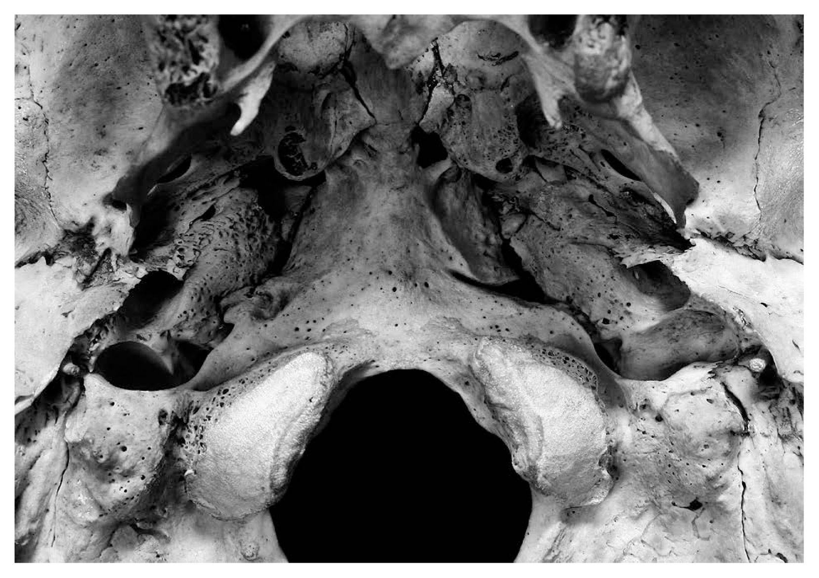
Table 1 shows frequencies of TBCs in a recent Japanese cranial series of fetal, juvenile, and adult specimens from the Tohoku district. TBC was observed on the left side of the occipital basilar part in one adult male skull aged 27 years (Figure 5), with an overall TBC frequency of 1/156 (0.64%) in the adult series. None of the fetal or juvenile skulls showed the TBC, resulting in a total cranial series frequency across all age groups of 1/349 (0.29%). Table 2 presents the frequencies of the TBC in the Jomon cranial series, including immature and adult specimens from the southern Hokkaido and Tohoku regions. The frequency in the adult series was 3/71 (4.23%) and that in the immature series was 2/12 (16.67%). Therefore, the total frequency in the Jomon cranial series across all age groups was 5/83 (6.02%).
Table 1
Frequency of occurrence of the transverse basilar cleft (TBC) in the recent Japanese cranial series from the Tohoku district
| Age group |
n |
Occurrence of TBC |
% |
| Fetal (6–10 months) |
162 |
0 |
0.00 |
| Juvenile (7 days–19 years) |
31 |
0 |
0.00 |
| Adult (≥20 years) |
156 |
1 |
0.64 |
| Total |
349 |
1 |
0.29 |
Table 2
Frequency of occurrence of the transverse basilar cleft (TBC) in the Jomon cranial series from the southern Hokkaido and Tohoku regions
| Age group |
n |
Occurrence of TBC |
% |
| Immature (perinatal–subadult) |
12 |
2 |
16.67 |
| Adult |
71 |
3 |
4.23 |
| Total |
83 |
5 |
6.02 |
Although the frequency of the TBC in the adult Jomon series (4.23%) was higher than that in the recent adult Japanese series (0.64%), the difference was not statistically significant at the 5% level (Fisher’s P = 0.0918). The immature Jomon cranial series showed a significantly higher frequency than the immature Japanese (16.67% vs. 0.00%; Fisher’s P = 0.0032). The total frequency in the Jomon cranial series across all age groups (6.02%) was significantly higher than that in the recent Japanese series (0.29%) at the 1% level (Fisher’s P = 0.0012).
Discussion
Embryological aspects of the TBC
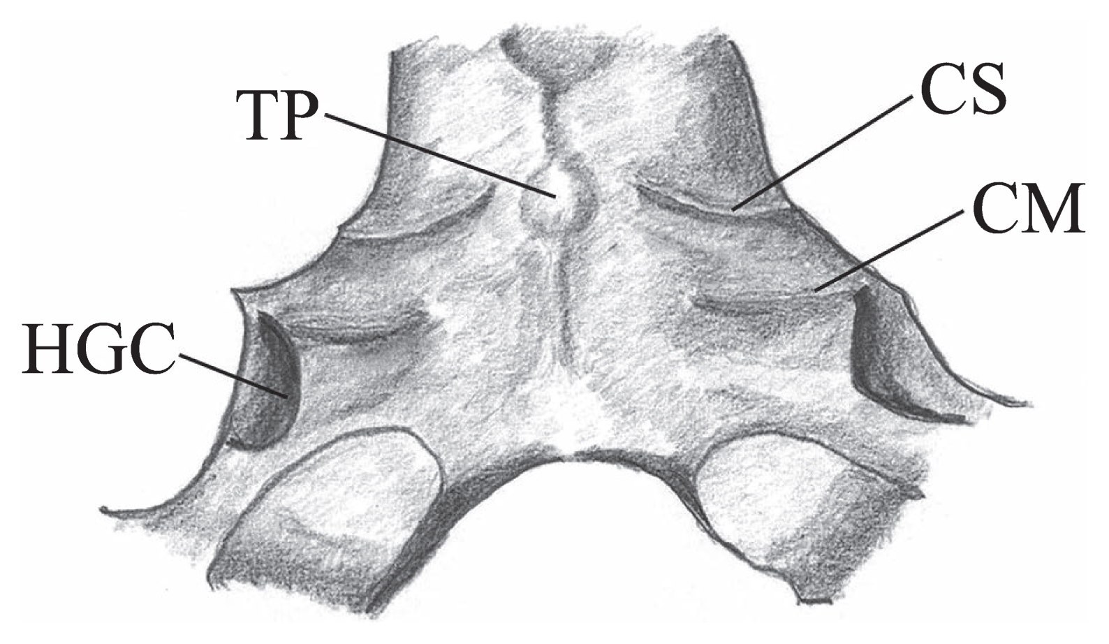
Figure 6 depicts the lower surface of the basilar part of the occipital bone. Two bony ridges are visible lateral to the tuberculum pharyngeum. The anterior ridge, termed the crista synostotica, marks the border between the praebasioccipital and basioccipital portions, whereas the formation of the posterior ridge, termed the crista muscularis, results from the insertion of the rectus capitis anterior muscle (Hofmann and Prescher, 2012). All the TBCs detected in adult skulls from the present study appear to correspond to the transverse ridge along the crista synostotica (Figure 4, Figure 5).
The basilar part of the occipital bone is formed by fusion of the parachordal cartilage and four occipital sclerotomes, as shown in Figure 7 (Hamilton and Mossman, 1972; Lang, 1995; Carlson, 2009). Since the occipital sclerotomes are strongly related to the hypoglossal canal as well as the hypoglossal nerve roots, they form the posterior portion of the basilar part of the occipital bone (O’Rahilly and Müller, 1984). Therefore, the gap between the most anterior segment of the occipital sclerotomes and posterior end of the parachordal cartilage likely corresponds to the borderline (crista synostotica) between the praebasioccipital and basioccipital portions of the basilar part.
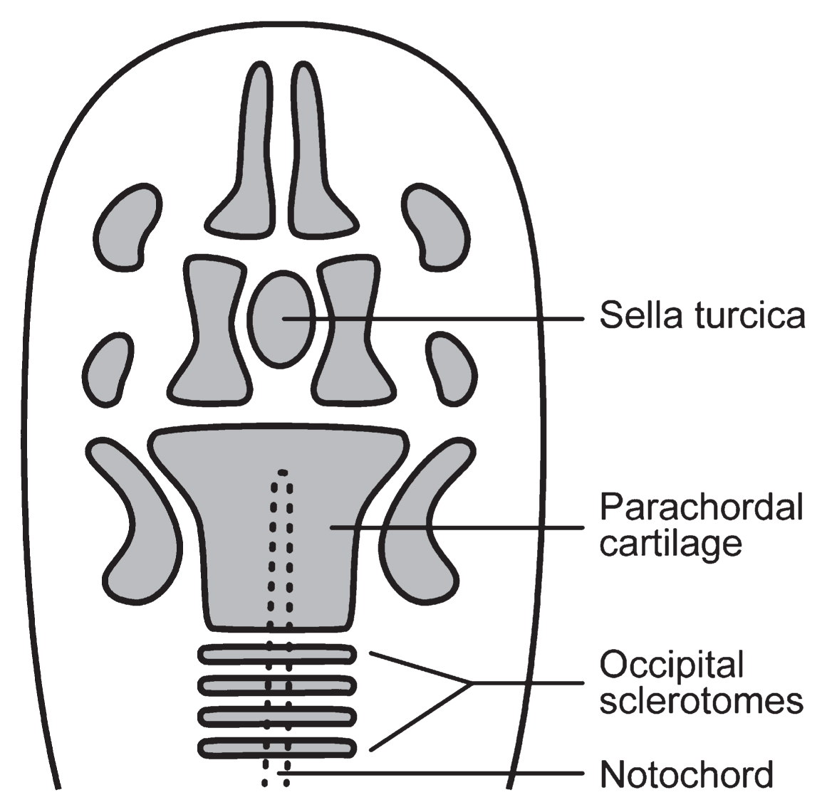
Although current human anatomy textbooks (e.g. Breathnach, 1965; Trotter and Peterson, 1966; Hamilton, 1976; Williams et al., 1989; Scheuer and Black, 2000) describe a single ossification center in the basilar part of the occipital bone, Hofmann and Prescher (2012) believe that, in rare cases, two ossification centers might occur—one anterior (praebasioccipital) and the other posterior (basioccipital). The borderline between the anterior and posterior centers appears to correspond to the gap between the parachordal cartilage and occipital sclerotomes. Thus, irrespective of whether one or two ossification centers occur, it can be postulated that the TBC develops along this borderline (crista synostotica), as suggested by Lang (1995) and our present findings. Moreover, the TBC is observed in both fetal and juvenile skulls; therefore, it might be regarded as a developmental variation appearing at an early growth stage of the human cranium.
Anthropological implications of the TBC
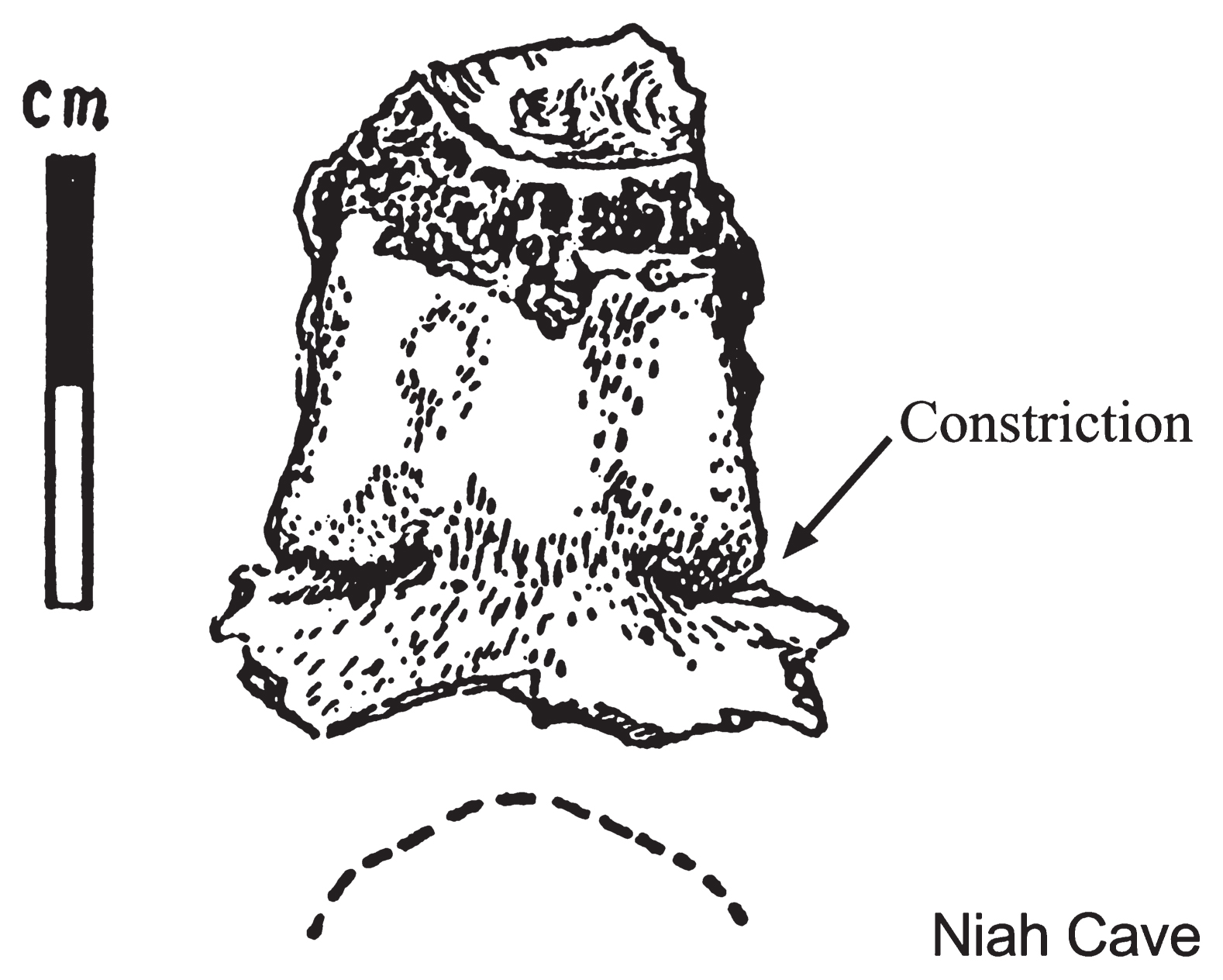
The Late Pleistocene juvenile skull from Niah Caves in Sarawak, Malaysia, is widely accepted as one of the earliest anatomically modern human fossils in East/Southeast Asia (Brothwell, 1960; Kennedy, 1977). Brothwell (1960) described the presence of a TBC in this skull (Figure 8), but considered it to be morphologically insignificant.
In his examination of 1100 Egyptian skulls, including specimens from 62 children, Smith (1912) detected a TBC in only one juvenile skull aged 12 years. Therefore, the frequency of TBC is 1/1100 (0.09%) in the Egyptian sample. Limson (1932) noted that 3/163 (1.84%) fetal and infant skulls from Euro- and Afro-Americans exhibited TBCs. Osawa (1894) and Shima (1941) reported frequencies of 1/162 (0.62%) and 2/193 (1.04%) in recent Japanese and recent Mongolian adult cranial series, respectively.
With regard to adult specimens, the frequency of 0.62% (1/162) in a recent Japanese cranial series reported by Osawa (1894) is almost identical to the frequency of 0.64% (1/156) in the recent Japanese cranial series from the Tohoku district assessed in the present study. As described above, although the frequency of the TBC in the adult Jomon skulls from southern Hokkaido and Tohoku was higher than that in the recent Japanese adult skulls from Tohoku, the difference was not statistically significant at the 5% level. However, when the data of Osawa (1894) and the present study are combined, the difference in the TBC frequency between the Jomon (3/71 or 4.11%) and recent Japanese adult series (2/318 or 0.63%) is statistically significant at the 5% level (Fisher’s P = 0.0443).
Among the total cranial samples from all age groups (fetal, juvenile, and adult), the frequency of TBC in the Jomon series from the southern Hokkaido and Tohoku regions was significantly higher at the 1% level than that in the recent Japanese series from the Tohoku district (Fisher’s P = 0.0012). Therefore, although the number of immature Jomon skulls examined in the present study was very limited, it might be postulated that the TBC occurs more frequently among the prehistoric Jomon population than among the current Japanese population.
Since it is believed that TBC occurrence involves a genetic component (Anderson, 2000), and its presence might be a developmental variation, this trait is worth investigating as a nonmetric cranial variant in various human population groups, including immature individuals.
Acknowledgments
For their permission to study the Jomon skeletal remains under their care, we express our sincere thanks to the staff and curators of the following universities and museums: Sapporo Medical University; Date City Institute of Funkawan Culture; Iwate Prefectural Museum; Tohoku History Museum; Historical Museum of Jomon Village OkuMatsushima; University of Tokyo; National Museum of Nature and Science, Tokyo; and Osaka University. Dr. N. Doi from the University of the Ryukyus provided a photograph of the Shiitachi-nishiku 2006 skull and the Okinawa Prefecture Archaeological Center permitted us to publish it. Dr. Y. Nagura of Tohoku University assisted with the black-and-white line drawings. This study was financially supported by a Grant-in-Aid for Scientific Research (Kakenhi: 25440250) from the Japan Society for the Promotion of Science.
References
- Anderson T. (2000) Basilar clefting: a familial condition? Annals of Anatomy, 182: 583–587.
- Breathnach A.S. (1965) Frazer’s Anatomy of the Human Skeleton, 6th edn. J. & A. Churchill, London.
- Brothwell D.R. (1960) Upper Pleistocene human skull from Niah Caves, Sarawak. Sarawak Museum Journal, New Series, 9(15–16): 323–349.
- Bryce T.H. (1915) Osteology and arthrology. In: Schäfer E.A., Symington J., and Bryce T.H. (eds.), Quain’s Elements of Anatomy, 11th edn. Longmans-Green, London, Vol. IV, Part I, pp. 1–329.
- Carlson B.M. (2009) Human Embryology and Developmental Biology, 4th edn. Mosby, Philadelphia.
- Charpy A. and Nicolas A. (1911) Poirier-Charpy Traité d’Anatomie Humaine, Vol. 1. Masson, Paris.
- Dodo Y. and Yamaguchi B. (1980) Human skeletal remains recovered from No. 10 house pit at the Usujiri B site. [Usujiri B iseki dai 10gou jukyo-shi yori hakken sareta jinkotsu ni tsuite.] In: Usujiri B Iseki. Minamikayabe-cho Board of Education, Hakodate, Hokkaido, pp. 114–122 (in Japanese).
- Doi N., Takenaka M., and Katagiri C. (2007) Additional Jomon skull excavated from the Shitach-nishiku site, Gushikawa Island, Okinawa. Anthropological Science, 115: 252 (abstract).
- Editorial Board of Rikuzentakata City History (1994) The History of Rikuzentakata City, Vol. 2: Geology and Archaeology [Rikuzentakata shishi, dai 2kan, chishitsu/koko hen.] Rikuzentakata City (in Japanese).
- Hamilton W.J. (1976) Textbook of Human Anatomy, 2nd edn. Macmillan, London.
- Hamilton W.J. and Mossman H.W. (1972) Human Embryology, 4th edn. Macmillan, London.
- Hofmann E. and Prescher A. (2012) The clivus: anatomy, normal variants and imaging pathology. Clinical Neuroradiology, 22: 123–139.
- Johnson G.F. and Israel H. (1979) Basioccipital clefts. Radiology, 133: 101–103.
- Kanda S. (1964) On the excavated skulls from the Hosoura shell-mound in Iwate Prefecture. Medical Journal of Osaka University, 15: 123–135.
- Kennedy K.A.R. (1977) The Deep Skull of Niah: an assessment of twenty years of speculation concerning its evolutionary significance. Asian Perspectives, 20: 32–50.
- Kruyff E. (1967) Transverse cleft in the basi-occiput. Acta Radiologica, 6: 41–48.
- Lang J. (1995) Skull Base and Related Structures: Atlas of Clinical Anatomy. Schattauer, Stuttgart.
- Le Double A.F. (1903) Traité des Variations des Os du Crane de l’Homme. Vigot Frères, Paris.
- Limson M. (1932) Observations on the bones of the skull in white and negro fetuses and infants. Contributions to Embryology, 23: 205–233.
- Lombardi G. (1961) The occipital vertebra. American Journal of Roentgenology, 86: 260–269.
- Matsumoto H. (1920) Customary tooth evulsions in some Jomon sites. [2, 3 sekki-jidai iseki ni okeru basshi fushu no umu oyobi yoshiki ni tsuite]. Jinruigaku Zasshi, 35: 61–83 (in Japanese).
- Matsushita T. and Ota J. (1993) Human skeletal remains unearthed at Gushikawajima sites in Okinawa. [Okinawa-ken Gushikawajima iseki-gun shutsudo no kojinkotsu.] In: Gushikawajima Sites. Izena-son Board of Education, Izena-son, Okinawa, pp. 215–244 (in Japanese).
- Mitsuhashi K. (1968) A report on the second season excavation of the Irie shell mound site, Abuta-cho, Hokkaido. [Hokkaido Abuta-cho Irie kaizuka dai 2ji hakkutsu hokoku.] Hokkaido Jinruigaku Kyokai Tsushin, 11: 45–46 (in Japanese).
- Okinawa Prefecture Archaeological Center (2011) A Preliminary Report on the Excavation of Archaeological Sites in Gushikawajima, Okinawa. [Okinawa-ken Izena-son Gushikawajima iseki-gun hakkutsu chosa gaiyo hokokusho.] Nishihara-cho, Okinawa (in Japanese).
- O’Rahilly R. and Müller F. (1984) The early development of the hypoglossal nerve and occipital somites in staged human embryos. American Journal of Anatomy, 169: 237–257.
- Osawa G. (1894) On the basilar part of the occipital bone. [Kotokotsu kisobu ni tsuite.] Tokyo Igakkai Zasshi, 8: 642–645 (in Japanese).
- Scheuer L. and Black S. (2000) Developmental Juvenile Osteology. Academic Press, London.
- Shapiro R. and Robinson F. (1976) Anomalies of the craniovertebral border. American Journal of Roentgenology, 127: 281–287.
- Shima G. (1941) A study on the Mongolian skulls. [Mokojin tokotsu no kenkyu.] Jinruigaku Sokan, Ko, Jinruigaku, 2: 1–108 (in Japanese).
- Smith H.D. (1912) Observations on the occipital bones in a series of Egyptian skulls with especial reference to the persistence of the synchondrosis condylo-squamoza (Zaaijer; synchondrosis intraoccipital posterior, BNA). Biometrika, 8: 257–261.
- Trotter M. and Peterson R.R. (1966) Osteology. In: Anson B.J. (ed.), Morris’ Human Anatomy, 12th edn. McGraw-Hill, New York, pp. 133–315.
- Williams P.L., Warwick R., Dyson M., and Bannister L.H. (1989) Gray’s Anatomy, 37th edn. Churchill Livingstone, Edinburgh.