Abstract
This article reports eight new humeral, ulnar, and radial fragments of Nacholapithecus kerioi collected from Nachola, Kenya during the 1998/1999 field seasons. The study refines the description of its forelimb bones, which was mostly based on a single partial skeleton. The most distinctive feature of the distal humerus is a large, globular, medially tilted capitulum. The groove between the capitulum and the zona conoidea is quite deep. The medial part of the humeral trochlea is also diagnostic in showing a less salient medial border. The medial epicondyle is moderately long and more posteriorly reflected than was previously presumed. The coronoid process of the ulna is quite wide. Its medial portion is distinctly concave. The ulnar shaft is anteroposteriorly deep in its proximal half, slender, straight in frontal view, and weakly anteriorly bowing. The elbow of Nacholapithecus exhibits a primitive functional pattern as a hominoid, including lack of universal stability of the humeroulnar joint through full extension and flexion, restriction of hyperextension of the elbow, and relatively anteroposteriorly oriented loading at the proximal ulna. On the other hand, it is derived in terms of enhanced rotational mobility and stability of the radius, incipiently increased stability at the humeroulnar joint, and more frequent maximum extension of the elbow compared to proconsulids. This mosaic morphology is different from both early Miocene proconsulids and later suspensory or orthograde European fossil apes. Although Nacholapithecus was neither suspensory nor orthograde, its forelimbs may have played a greater role for body support or balance maintenance, more frequently reaching to and exploiting overhead supports than in early Miocene proconsulids.
Introduction
Nacholapithecus kerioi is a Middle Miocene (16–15 Ma) hominoid (kenyapithecine: Alba et al., 2012) discovered at Nachola, northern Kenya (Ishida et al., 1999; Nakatsukasa and Kunimatsu, 2009; Kunimatsu et al., 2019). The majority of Nacholapithecus specimens, including the holotype KNM-BG 35250 partial skeleton, have been collected at site BG-K at Nachola (Nakatsukasa et al., 1998; Ishida et al., 1999). Since KNM-BG 35250 includes many skeletal elements, comparative anatomical study has largely relied on it. However, unfortunately, most fossils from BG-K, including KNM-BG 35250, suffer from plastic deformation and/or the cracking due to compression during fossilization. Due to this taphonomic issue, proper characterization of Nacholapithecus anatomy is not easy (e.g. Takano et al., 2018).
After the discovery of KNM-BG 35250, excavation continued at the fossiliferous site BG-K, and additional fossil specimens, including several forelimb bones, were unearthed during the 1998/1999 field seasons. This article reports on those specimens to amend the current description of the forelimb long bones of Nacholapithecus (Takano et al., 2018). Notably, this collection includes a well-preserved proximal portion of ulna with relatively minor deformation. A morphometric study of Nacholapithecus ulnae is being prepared as a separate analytical paper.
Materials and Methods
The material reported here consists of three humeral, four ulnar, and one radial fragment (Table 1). All specimens are stored at the Earth Science Department, the National Museums of Kenya (KNM), and are now fully accessible for study. Nacholapithecus is highly dimorphic in body size (Kikuchi et al., 2018). Sexing of new specimens was done based on their size using KNM-BG 35250 as the male reference. All of these specimens comfortably fall in the male category due to their large size.
Table 1
New forelimb specimens of
Nacholapithecus
| Accession no. |
Part |
side |
Collection information |
| KNM-BG 38384 |
distal humerus |
R |
BG-K in situ (Trench 99-2) |
| KNM-BG 37303 |
humeral trochlea fragment |
R |
BG-K surface collection (1998) |
| KNM-BG 38610A |
humeral shaft |
R |
BG-K in situ (Trench 99-2) |
| KNM-BG 38391B |
proximal ulna |
R |
BG-K in situ (Trench 99-2) |
| KNM-BG 38610B |
proximal ulna without olecranon |
R |
BG-K in situ (Trench 99-2) |
| KNM-BG 37352 |
proximal ulna without olecranon |
L |
BG-K surface collection (1998) |
| KNM-BG 38387 |
ulnar shaft |
R |
BG-K in situ (1999) |
| KNM-BG 40021 |
proximal radius |
R |
BG-K in situ (Trench 99-2) |
A digital sliding caliper was used to take linear measurements to a tenth of millimeter. Measurements and indices are the same as those published by Takano et al. (2018) (Table 2, Table 3, Table 4. Table 5). Because most of these specimens are subject to plastic deformation and/or cracking, damage was evaluated from computed tomography (CT) images captured with a CT scanner (X-CT Research SA+, Stratec Medizintechnik GmbH, Germany) with a resolution of 0.1 mm. Comparative extant primate specimens used in this study are the same as those used in Takano et al. (2018). For illustrative purposes (visual comparison and virtual restoration), some original fossil specimens were surface-scanned with a NextEngine 3D scanner (Nextengine Inc., Santa Monica, CA, USA) using the Macro and HD (high point density) settings.
Table 2
Humeral measurements. Comparative data are from
Takano et al. (2018).
| Measurement |
KNM-BG 38384 |
KNM-BG 37352 |
KNM-BG35250M |
Definition |
| Bicondylar breadth |
(>43.9)1 |
— |
(<53.4)1 |
(c) in Rose et al. (1992) |
| Capitular height |
16.2 |
— |
(<16.9) |
(f) in Rose (1988) |
| Articular width |
31.3 |
— |
(<38.7) |
(a) in Rose (1988) |
| Capitular + zona width |
(17.4) |
— |
18.9 |
(d) in Rose (1988) |
| Trochlear width (anterior) |
(>13.9) |
|
(<18.9) |
(e) in Rose (1988) |
| Capitular width |
12.7 |
— |
14.1 |
(i) in Rose (1988) |
| Medial trochlear rim height |
16.1 |
15.02 |
— |
(c) in Rose (1988) |
| Lateral trochlear rim height |
— |
13.72 |
(13.9) |
(j) in Rose (1988) |
| Trochlear notch height |
(>12.1) |
10.52 |
(11.1) |
(k) in Rose (1988) |
| Posterior breadth of distal articulation |
19.1 |
— |
(15.8) |
(1) in Harrison (1982) |
1 Values in parentheses represent estimates or values affected by deformation.
2 Due to damage, measured in the left counterpart.
Table 3
Humeral ratios. Comparative data are from
Takano et al. (2018).
| Taxon |
Specimen |
Articular width/bicondylar width |
Capitular + zona width/trochlear width |
Capitular height/capitular width |
Medial trochlear rim height/lateral trochlear rim height |
| Nacholapithecus |
KNM-BG 38384 |
(0.71) |
(1.25>) |
1.28 |
— |
| Nacholapithecus |
KNM-BG 35250M |
(0.73)1 |
(1.0) |
1.2 |
(1.09)2 |
| ?Rangwapithecus |
KNM-SO 31232 |
0.72 |
0.98 |
1.19 |
1.25 |
| Ekembo heseloni |
KNM-RU 2036AH |
0.81 |
0.91 |
1.11 |
1.23 |
| Kenyapithecus wickeri |
KNM-FT 2751 |
0.79 |
0.97 |
1.17 |
1.25 |
| Pan troglodytes |
n = 11 |
0.73 |
0.99 |
1.14 |
1.22 |
| (range) |
|
(0.70–0.76) |
(0.86–1.06) |
(0.92–1.20) |
(1.17–1.39) |
| hylobatids3 |
n = 5 |
0.74 |
1.27 |
1.33 |
1.15 |
| (range) |
|
(0.72–0.77) |
(1.16–1.34) |
(1.19–1.42) |
(1.08–1.29) |
| Cercopithecus mitis |
n = 12 |
0.77 |
1.42 |
1.16 |
1.46 |
| (range) |
|
(0.74–0.82) |
(1.24–1.60) |
(0.99–1.26) |
(1.28–1.75) |
| Colobus guereza |
n = 9 |
0.81 |
1.76 |
0.98 |
1.33 |
| (range) |
|
(0.77–0.85) |
(1.58–2.09) |
(0.85–1.07) |
1.05–1.53) |
| Alouatta seniculus |
n = 10 |
0.71 |
1.57 |
0.95 |
1.29 |
| (range) |
|
(0.65–0.79) |
(1.33–1.89) |
(0.87–1.02) |
(1.04–1.45) |
1 Values in parentheses are affected by deformation.
2 Measured on the left counterpart (KNM-BG 35250N).
Table 4
Ulnar measurements. Comparative data are from
Takano et al. (2018).
| Measurement 1 |
KNM-BG 38391B
Nacholapithecus |
KNM-BG 38610B
Nacholapithecus |
KNM-BG 37352
Nacholapithecus |
KNM-BG 35250V
Nacholapithecus |
KNM-RU 1786
Ekembo nyanzae |
KNM-RU 2036CF
Ekembo heseloni |
KNM-WK 16950R
Turkanapithecus |
Definition |
| Sigmoid notch depth (SND) |
14.4 |
— |
(>16.0) |
17.0 |
20.0 |
11.5 |
9.8 |
Begun (1992), Richmond et al. (1998) |
| Anteroposterior thickness at distal beak of trochlear notch (PAAP) |
(>26.1) |
— |
30.1 |
29.5 |
— |
19.3 |
16.2 |
Begun (1992) |
| Proximal shaft anteroposterior thickness (PAP) |
18.8 |
— |
21.1 |
(<22.1) |
26.5 |
14.8 |
11.8 |
Begun (1992), Richmond et al. (1998) |
| Olecranon process mediolateral breadth (OPML) |
15.9 |
— |
— |
(14.9) |
19.0 |
11.5 |
10.5 |
Richmond et al. (1998) |
| Sigmoid notch mediolateral width (SML) |
11.4 |
— |
— |
12.8 |
14.1 |
9.7 |
8.5 |
Richmond et al. (1998) |
| Trochlear articular mediolateral breadth (TAB) |
16.6 |
(>16.3) |
19.6 |
17.5 |
18.6 |
11.4 |
9.9 |
Begun (1992), Richmond et al. (1998) |
| Sigmoid notch proximodistal length (NPD)3 |
(16.6) |
|
— |
— |
— |
15.0 |
11.5 |
Richmond et al. (1998) |
1 Values in parentheses represent values affected by damage.
2 Distance between tips of the olecranon beak and coronoid process projected to the shaft axis. This definition differs from the original definition by
Richmond et al. (1998).
Table 5
Ulnar ratios. Comparative data are from
Takano et al. (2018).
|
|
PAAP/SND |
TAB/SML |
TAB/SND |
| Nacholapithecus |
KNM-BG 38391B |
>1.81 |
1.46 |
|
| Nacholapithecus |
KNM-BG 37352 |
1.88> |
— |
|
| Nacholapithecus |
KNM-BG 35250V |
1.73 |
1.37 |
1.03 |
| Nacholapithecus |
KNM-BG 35250C |
1.75 |
— |
— |
| Ekembo nyanzae |
KNM-RU 1786 |
— |
1.30 |
0.93 |
| Ekembo heseloni |
KNM-RU 2036CF |
1.63 |
1.18 |
0.99 |
| ?Rangwapithecus |
KNM-KT 38000B1 |
1.632 |
— |
|
| Turkanapithecus |
KNM-WK 16950R |
1.65 |
1.16 |
1.01 |
| Pan troglodytes |
n = 11 |
1.64 |
— |
(1.20)3 |
| range |
|
1.46–1.73 |
|
|
| Hylobatids |
n = 5 |
2.18 |
— |
— |
| range |
|
2.0–2.39 |
|
|
| Cercopithecus |
n = 12 |
1.83 |
— |
— |
| range |
|
1.68–2.0 |
|
|
| Colobus |
n = 9 |
1.91 |
— |
(0.94)3 |
| range |
|
1.81–2.10 |
|
|
| Alouatta |
n = 10 |
1.74 |
— |
— |
| range |
|
1.62–1.85 |
|
|
3 Calculated from mean values of TAB and SND in
Begun (1992).
Descriptions
KNM-BG 38384 (distal portion of right humerus)
This specimen is a c. 8 cm long distal portion of a right humerus (Figure 1). The size of this specimen is comparable to that of the previously described KNM-BG 35250M (Table 2).
The shaft is smashed anteroposteriorly and displays an exaggeratedly wide appearance. The olecranon fossa is damaged and perforated. Thus, the anterior surface where the coronoid fossa would have been is broken. The distal joint is better preserved than that of KNM-BG 35250M although the trochlea is cracked proximomedially to distolaterally on the anterior surface (arrows in Figure 1a) and the articular surface proximal to the crack is displaced posteriorly (arrow in Figure 1f).
Due to the severe damage, the original cross-sectional shape of the shaft is unknowable. The lateral supracondylar ridge is slightly eroded. It lacks a prominent lateral flare. The lateral epicondyle is massive and proximodistally long. It is distorted and deflected slightly anteriorly (Figure 1c). The height of the lateral epicondyle from the distal surface of the capitulum is 23.5 mm. The radial fossa is shallow.
The capitulum is large, tall, and globular (Table 2, Table 3). The proximodistal height is 16.2 mm. The capitulum is tilted medially in anterior view (Figure 1a). In this view, the lateral and medial borders of the capitulum are almost parallel. They start converging gradually as they turn to the distal side. The capitulum and the zona conoidea form a distinctly deep groove (Figure 1e, f). This groove is accentuated by the medial tilt of the capitulum. The lateral trochlear border is blunt and difficult to define precisely. However, the zona is apparently wide.
The trochlear groove is wide and shallow. The medial portion (slope) is much wider than the lateral one. The medial portion draws a convex curve (instead of a straight line or concave curve) mediolaterally in distal view. The blunt lateral trochlear border spirals posteriorly to approach the lateral keel of the olecranon fossa.
On the posterior aspect, the medial border of the trochlear surface runs proximolaterally into the olecranon fossa. The attachment area of the posterior band of the ulnar collateral ligament is not deep (asterisk in Figure 1b) unlike the previously reported specimen (Takano et al., 2018). The olecranon fossa is deep. The lateral keel of the olecranon fossa is prominent although its distal part is anteriorly compressed. The capitulum and the shaft surface lateral to the olecranon fossa are flattened due to compression.
The medial epicondyle is intact. It is thick and moderately long and projects posteromedially with a retroflexion angle of 55°. Bi-epicondylar width is 43.9 mm. This might be lower than the original dimension due to the distortion of the lateral epicondyle.
KNM-BG 38384 resembles KNM-BG 35250M in many of aforementioned features. Although the former exhibits a narrower joint than the latter, it is an effect caused by deformation in KNM-BG 35250M (see below). Takano et al. (2018) noted damage (cracking) on the trochlea of KNM-BG 35250 (p. 136). This damage considerably altered the trochlear width beyond what was expected. Thus, KNM-BG 38384 differs from KNM-BG 35250M in having the articular surface for the radius (capitulum and zona) wider than that for the sigmoid notch (Table 3). We think that the condition in KNM-BG 38384 is typical for Nacholapithecus in this regard.
KNM-BG 37303 (right humeral trochlea)
This specimen is a fragment of right humeral trochlea. It also preserves a large portion of the zona conoidea. It is parasagittally cut off from the capitulum. The break runs at the capitulum–zona boundary anteriorly (Figure 2a). The proximolateral part of trochlear articular surface is missing on the posterior aspect (Figure 2d). The anterior articular surface of the trochlea is extensively polished and cancellous bone is exposed (Figure 2a). However, there is no marked plastic deformation. The medial epicondyle is missing (Figure 2c, d).

The trochlear groove is only moderately deep but deeper than that of other Nacholapithecus specimens (Figure 2b). The medial portion of the trochlear surface is wide and convex mediolaterally. The lateral portion is much narrower. The lateral trochlear border is blunt and difficult to define. In posterior view, the medial trochlear rim runs distomedially to proximolaterally. The most distal part on the trochlea is located a few millimeters lateral to the medial trochlear border (arrow in Figure 2d). Medial to the trochlea is a depression in which the ulnar collateral ligament attached (Figure 2d). The olecranon fossa is mostly lost.
KNM-BG 38610A (right humeral shaft)
This is a c. 15 cm long right humeral shaft that is severely damaged by mediolateral compression (Figure 3). The medial cortex is crushed and adhered to the cortex on the opposite (= lateral) side. Proximally, a flat deltoid plane (Figure 3, DLT) accompanying a sharp crest laterally (arrows) is observed. Distally, the deltoid plane ends around the midpart of this fragment. The maximum width of the deltoid plane is 19.5 mm. Distally, the proximal part (c. 3 cm long) of a sharp lateral supraepicondylar ridge (SER) is preserved.
KNM-BG 38610A was unearthed from Trench 99-2 at BG-K together with a right ulnar specimen (KNM-BG 38610B) that is described below. This specimen is also associated with a left scapular fragment described by Senut et al. (2004).
KNM-BG 38391B (proximal portion of right ulna)
This ulna was recovered from Trench 99-2 in association with several other postcranial elements, including a proximal portion of a femur (Kikuchi et al., 2018) and a detached proximal epiphysis of a tibia. This femur (KNM-BG 38391A) is one of the smallest specimens among adult males (Kikuchi et al., 2018). The detached tibial epiphysis may suggest that this individual was a young adult so that the epiphysis had not fully fused with the metaphysis.
This specimen is a 58 mm long proximal portion of right ulna (Figure 4). It is broken c. 15 mm below the radial notch. Plastic deformation is relatively minor. The tip of the coronoid process is squashed. Its remnant clings beneath the process (Figure 4a: circumscribed by a dotted line). The medial border of the coronoid process is partially broken posteriorly (bracket in Figure 4c). Although two cracks run horizontally across the sigmoid notch, the broken surfaces are tightly fitted.

The olecranon is high and projects proximally with no retroflexion. The proximal surface of the olecranon is slanted posteriorly. The anteromedial part of the olecranon forms a triangular (in side view: Figure 4c) eminence proximally projecting. On the medial side of the olecranon, the attachment site for the posterior and oblique bands of the ulnar collateral ligament forms a large rugose depression. The height of the olecranon process measured from the olecranon beak (to the tip of the triangular eminence) is 15.8 mm (10.4 mm when measured to the center of the proximal surface). The olecranon beak is long and projects anteriorly, measuring 25.9 mm anteroposteriorly from its tip to the posterior surface of the bone. The sigmoid notch is c. 17 mm proximodistally. Because of the damage on the coronoid process, this measurement cannot be accurate. The surface of the sigmoid notch is slightly raised centrally but no median ridge is formed. The proximolateral part of the articular surface expands on the lateral side of the olecranon. The coronoid process is wide. It is divided into a wide and concave medial portion and a flat and narrow band-like lateral portion, although the boundary between these two cannot be clearly defined. There is a small eminence on the anterior border of the coronoid process, which is also a remnant of its crushed anterior beak. The coronoid process projects anteriorly with a downward inclination angle of 24°. The anteroposterior thickness at the coronoid process is >26.1 mm.
The radial notch is crescentic (Figure 4b). It consists of a proximodistally long posterior portion and narrow anterior portion due to an encroachment by a deep concavity on the anterolateral surface that is probably an attachment for the annular ligament of the radius. The radial notch measures c. 14 mm anteroposteriorly and c. 12 mm proximodistally. The surface is concave as a whole, facing laterally and slightly proximally (Figure 4a). From the most posterior part of the radial notch, the supinator crest runs distally (arrows in Figure 4b). It is not sharp, possibly as a result of weathering, and becomes faint before it reaches the distal break. On the medial side of the shaft, a sharp ridge starts beneath the sigmoid notch (asterisk in Figure 4c) and runs distally behind the insertion of the brachialis muscle. The cross section of the proximal shaft is diamond-shaped with the anterior triangular portion longer than the posterior portion (Figure 4e). The shaft near the break is c. 16 mm thick anteroposteriorly and c. 12 mm mediolaterally. The anterior border is more acute than the posterior one (Figure 4f). The cortex is thick anteriorly and posteriorly, and thinner at the medial and lateral sides.
KNM-BG 38610B (proximal half of right ulna)
This is a proximal half of a right ulna (Figure 5). The proximal end (olecranon and sigmoid notch) is broken off at the level of the coronoid process. The preserved length is 121 mm. The shaft is badly damaged by mediolateral compression and shows an exaggeratedly narrow (or anteroposteriorly thick) appearance in anterior view. The medial cortex is entirely crushed and adhered to the lateral cortex. The shaft bows posteriorly in its proximal part unlike other ulnae of Nacholapithecus (e.g. KNM-BG 35250C: see also the description of KNM-BG 38387). Given that this specimen consists of two large pieces glued together, the longitudinal curvature is most likely artificial due to a narrow contact area between the two fragments. The general size of the coronoid process is close to that of KNM-BG 38391B (Table 4).

The coronoid process and the radial notch are relatively well preserved. However, the band-like bony protuberance beneath the border of the coronoid process (Figure 5: dotted lines) is likely a remnant of an original sharp border that was smashed postmortem. The coronoid process is broad (>16.3 mm) and projects anteriorly. The downward inclination angle is 21° with respect to the proximal part of the shaft. However, since the shaft itself is crushed and deformed, this angle is tentative. The medial part of the articular surface on the coronoid process is wide and concave mediolaterally. Along the lateral border, a narrow flat lateral portion (c. 4 mm) is displayed. Unlike previously described specimens, the anterior border of the coronoid process appears relatively symmetric mediolaterally (Figure 6), with the most projecting part being positioned centrally (arrow in Figure 6). However, this condition may not be original as the anteromedial border is eroded (see Figure 6, bracket).

The radial notch is crescentic in shape. It is c. 14 mm anteroposteriorly and 12.6 mm proximodistally and is concave in both directions. It faces laterally and slightly proximally. Anteriorly, a narrowing band extends along the border of coronoid process. There is an excavated area anterodistal to the radial notch, which is probably an attachment site of the annular ligament. While KNM-BG 38610B is one of the smallest specimens among the Nacholapithecus ulnar specimens based on the size of the coronoid process, the radial facet appears the largest among the four specimens (Figure 6). Probably, erosion of the sharp facet border caused an optical illusion in other specimens.
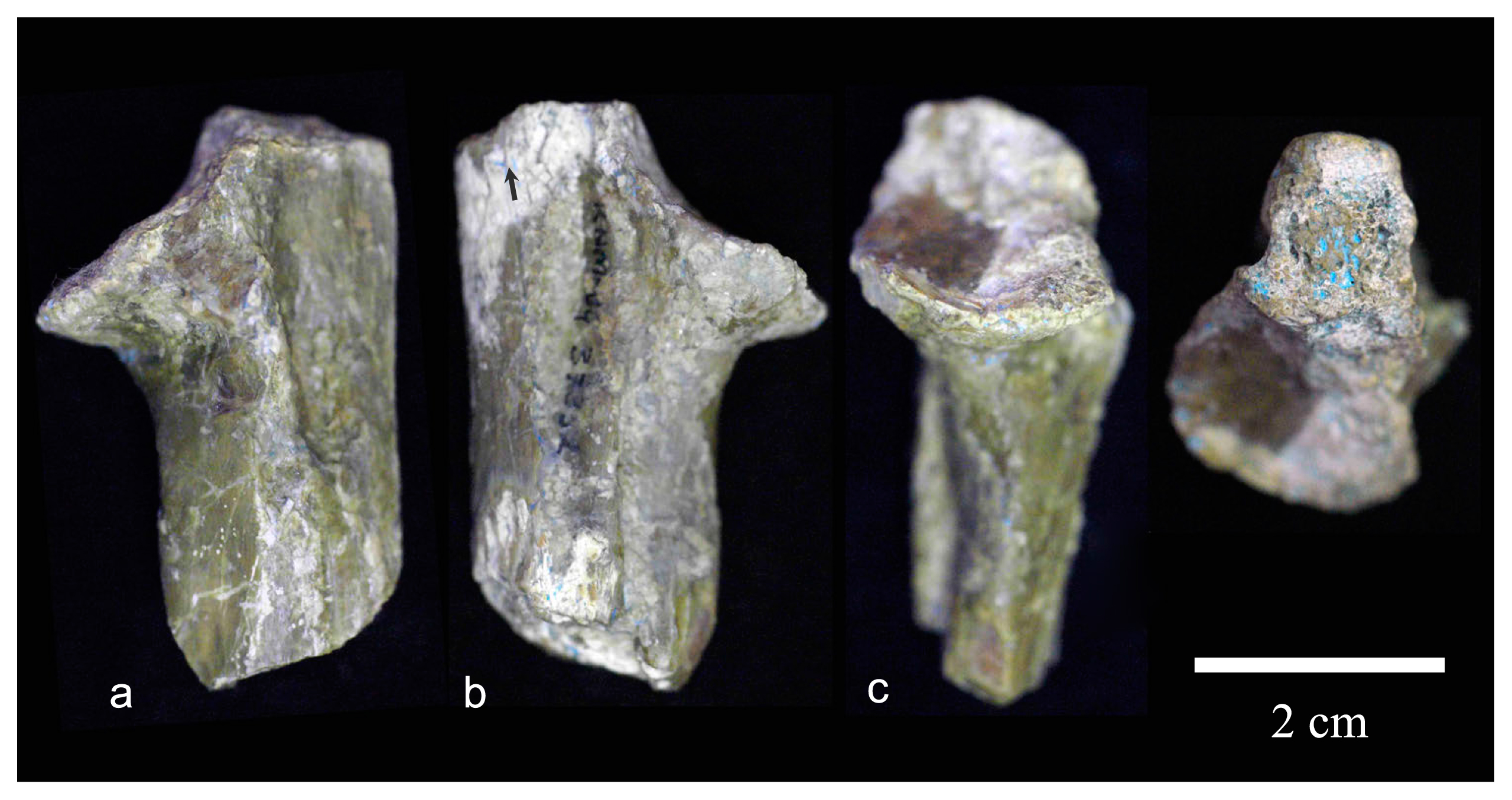
KNM-BG 37352 (left proximal ulna without olecranon process)
This specimen is a c. 5 cm long proximal fragment of left ulna (Figure 7). The olecranon is broken off at around the middle of the sigmoid notch and c. 3 cm below the radial notch. The border of the radial facet is widely eroded, and the posterior part of the shaft is squashed mediolaterally. The lateral portion of the articular surface of the coronoid process is weathered and the cancellous bone is exposed (Figure 6, Figure 7c).
This specimen is remarkably large in overall size, being the largest among Nacholapithecus ulnar specimens (Figure 6, Table 4). Its trochlear articular breadth is 12% larger than that of the second largest specimen (KNM-BG 35250V).
The coronoid process is relatively well preserved. Its anterior border is intact and sharp, unlike the other specimens described above. The superior surface of the coronoid process is wide. Its medial portion is strongly concave. The inclination angle of the coronoid process is larger (36°) than that of other specimens. However, weathering of the lateral portion of the superior articular surface might exaggerate somewhat the distal angulation.
The radial notch appears small relative to the overall size due to the erosion of the articular border. It faces laterally and proximally. Concavity for the annular ligament is not developed. Likewise, the insertion of the brachialis muscle is not marked.
KNM-BG 38387 (right ulnar shaft)
This specimen is a c. 16 cm long right ulnar shaft (Figure 8). Both proximal and distal ends are missing. Although this specimen was found in situ in Trench 99 of site BG-K, where many postcranial specimens were excavated, its association with other ulnar specimens is uncertain. Based on the size (see below), this specimen was assigned to a male.

At the distal break, the pronator crest (c. 3 cm long) is pronounced on the medial side of the shaft (arrows in Figure 8). This suggests that the distal break is close to the metaphysis expansion. The proximal portion of the shaft (c. 6 cm from the proximal break) shows cortical fractures on the medial and lateral sides. Near the proximal break, a sharp crest is observed on the medial side (asterisks in Figure 8). This is the crest that descends behind the insertion of the brachialis muscle (Figure 4c, asterisk). This crest becomes faint c. 3 cm below the radial notch in another proximal ulnar fragment of a Nacholapithecus male (KNM-BG 17824: Rose et al., 1996). The supinator crest is not observed laterally (Figure 8; cf. Figure 4b). Therefore, the proximal break is probably c. 2.5–3 cm distal from the radial facet. The anteroposterior thickness of the shaft at the break is >16 mm.
In anterior view, the shaft is slender and almost straight. In lateral and medial side views, it shows weak anterior bowing. The proximal portion of the shaft is thicker anteroposteriorly rather than mediolaterally. Distally, the shaft becomes thicker mediolaterally at the level of the pronator crest. On the lateral side, the attachment of the interosseous membrane can be traced (Figure 8, dotted line), though is not sharp, in the distal portion.
KNM-BG 40021 (proximal portion of right radius)
This is a 63 mm long proximal fragment of a right radius (Figure 9). It is broken c. 1 cm below the radial tuberosity. The specimen is compressed anterolaterally to posteromedially. The shaft is flattened, and the head is tilted so that the proximal surface faces proximolaterally.
The posterior side of the head is compressed and shows an almost linear contour. The minimum diameter of the head is 16.8 mm and the perpendicular diameter (approximately equivalent to the mediolateral diameter) is 19.5 mm. The fovea is deep although it is exaggerated by compression. Since the beveling area surrounding the fovea is wide, the fovea itself is proportionally narrow. It is c. 13 mm mediolaterally. The beveling surface is slightly wider anteriorly and laterally than medially. The width of its posterior part is unknowable. The articular surface for the ulnar notch measures 8.4 mm in height anteriorly. No useful information is obtained from the shaft.
Discussion
In this report we give descriptions of eight forelimb fragments of N. kerioi. This expanded sample helps us to distinguish true anatomical characteristics from taphonomic damage and to comprehend individual variation in Nacholapithecus (see Figure 6). Table 6 summarizes the revised morphology of the elbow joint (and ulnar shaft) of Nacholapithecus. The most diagnostic feature of the distal humerus is a large, globular, medially tilted capitulum. Due to this medial rotation, the groove between the capitulum and the zona conoidea is notably deep. The medial part of the humeral trochlea is also diagnostic in showing a less salient medial border and convex (neither concave nor linear) profile in anterior view. The medial epicondyle is moderately long, and more posteriorly reflected than was previously presumed. Posteriorly, the development of a groove-like depression medial to the articular surface is actually variable among individuals. The capitular morphology in Nacholapithecus is reminiscent of that in extant great apes, especially orangutans, in its globularity combined with proximodistally long shape (Figure 10). On the other hand, the deep groove between the capitulum and the zona conoidea is similar to that of African apes. The medial part of the humeral trochlea shows a convex profile in anterior view in Nacholapithecus and extant great apes (Figure 10). However, unlike the condition in extant apes where this area is more or less constant in breadth all around, it is remarkably wide in the distal aspect in Nacholapithecus. This point may suggest that the stability of the Nacholapithecus humeroulnar joint changes according to the degree of elbow flexion–extension. The posteriorly reflected medial epicondyle (unlike that of extant great apes) may also be related to this feature.
Table 6
Diagnostic traits of the
Nacholapithecus kerioi elbow and ulnar shaft with comparison to
E. africanus and
Kenyapithecus wickeri
| N. kerioi |
E. africanus |
K. wickeri |
| Humerus |
| Radial fossa reduced and shallow |
? |
+ |
| Coronoid fossa rounded and deep |
? |
shallower |
| Capitulum proximodistally tall, large, and globular |
? |
tall but less globular |
| Capitulum mediolaterally tilted |
? |
less strong |
| Zona conoidea well defined |
+ |
+ |
| Groove between the capitulum and zona deep |
? |
shallower |
| Capitulum and zona wider than or same as trochlear width |
? |
similar in width |
| Trochlear groove wide and shallow |
+ |
+ |
| Medial part of the trochlear surface convex mediolaterally |
concave |
+ |
| Medial epicondyle moderately long and reflected posteriorly |
+ |
reduced in length |
| Massive and proximodistally long lateral epicondyle |
? |
+ |
| Deep olecranon fossa with a prominent lateral wall |
? |
+ |
| Ulna |
| Olecranon high and non-retroflexed |
+ |
? |
| triangular lateral eminence on the olecranon present |
less marked |
? |
| Olecranon beak anterior projection strong |
? |
? |
| Olecranon beak mediolaterally symmetric |
+ |
? |
| Sigmoid notch articular surface mediolaterally weakly concave |
+ |
? |
| Proximolateral extension of the sigmoid notch articular surface remarkable |
less well developed |
? |
| Coronoid process less slanted distally |
+ |
? |
| Articular surface on the coronoid process wide and concave |
(not concave) |
? |
| Radial notch facing laterally and slightly proximally |
+ |
? |
| Proximal shaft mediolaterally narrow |
+ |
? |
| Mid-to-distal shaft slender with weak anterior bowing |
? |
? |
+, same as N. kerioi; ?, unknown; ( ), not definitive.

The proximal ulna exhibits features of the coronoid process corresponding to the humeral trochlea. It is quite wide. Its wide medial portion is concave. Its anterior projection is variable, but on average relatively prominent. Attachment sites of muscles or ligaments are variably developed. Insertion of the brachialis muscle is not always very deep. Likewise, the attachment of the annular ligament may or may not be distinctly deep. This variability is not size related since it is least developed in the largest specimen. The coronoid process of extant great apes is wide and strongly projecting anteriorly (Figure 11). However, because of the presence of the median keel of the sigmoid notch, the lateral portion of the coronoid articular surface is better developed. Furthermore, as the anterior projection of the coronoid process is stronger, the sigmoid notch, as a whole, faces more proximally in extant great apes (Figure 11). The olecranon is much reduced and retroflexed, which increases the range of elbow extension. This olecranon morphology contrasts with that of Nacholapithecus.

The elbow of Nacholapithecus shows some similarities and dissimilarities to that of the contemporary Equatorius africanus and Kenyapithecus wickeri. The capitulum is less expanded and globular, less tilted medially, and the groove between the capitulum and the zona conoidea is shallower in K. wickeri (KNM-FT 2751) (Figure 10, Figure 12) (there is no humeral specimen of Equatorius in which this part can be observed). However, the medial part of the humeral trochlea shows a convex profile in anterior view in K. wickeri as in Nacholapithecus. On the other hand, it is almost linear in Equatorius (KNM-TH 28860G). The retroflection of the medial epicondyle is strong in all these fossil apes. It is reduced in length in K. wickeri compared to Nacholapithecus and Equatorius (Figure 12). The ulnae of Nacholapithecus and Equatorius (KNM-TH 28860K) resemble each other in showing relatively strong anterior projection. However, the superior surface of the coronoid process appears flat rather than concave in Equatorius, unlike Nacholapithecus, although the medial portion of the coronoid process is largely missing in this specimen (Figure 11). This observation is concordant with the difference of the medial portion of the humeral trochlear morphology between these species (see above). Other differences are a less extensive proximolateral expansion of the sigmoid articular surface and the absence of a marked triangular eminence on the anteromedial part of the olecranon proximal surface in Equatorius. Unfortunately, no ulnar specimen of K. wickeri is known. There is one proximal radius on Equatorius (KNM-TH 28860J) (Ward et al., 1999). However, since its head suffers from major damage, no comparison is possible.

This is the first report on the morphology of the whole ulnar shaft in Nacholapithecus. It is anteroposteriorly deep in its proximal half, slender, straight in frontal view, and weakly anteriorly bowing (Figure 8). This gave us an opportunity to compare ulnar shaft morphology between Nacholapithecus and another kenyapithecine, Griphopithecus suessi (11.6–11.1 Ma: Casanovas-Vilar et al., 2011), from Klein Hadersdorf, Austria (Zapfe, 1960; Begun, 1992). The ulna of Nacholapithecus males is similar to the Klein Hadersdorf ulna (supposedly belonging to a female) in overall size, as provisionally estimated from the proximal portion of the ulna (Takano et al., 2018). The proximal ulnae of Nacholapithecus and G. suessi differ from each other in the morphology of the coronoid process and the retroflexion of the olecranon (narrower coronoid process and presence of weak retroflexion in G. suessi: Takano et al., 2018). The humerus of the G. suessi male is chimpanzee sized (Zapfe, 1960). The congeneric species G. alpani (14–13 Ma, Turkey) was smaller and Nacholapithecus sized based on phalangeal specimens (Ersoy et al., 2008). The size increase in the genus Griphopithecus 2–3 million years ago might have introduced such morphological difference although this possibility needs to be tested by a comparison between Nacholapithecus and G. alpani.
The elbow of Nacholapithecus exhibits primitive functional hominoid signs: lack of universal stability of humeroulnar joint through full extension and flexion (wide and shallow trochlear groove of the humerus, sigmoid notch with no median ridge); restricted hyperextension of the elbow (long and unretroflexed olecranon); and relatively anteroposteriorly oriented loading at the proximal ulna (anteroposteriorly thick and mediolaterally narrow proximal ulnar shaft). On the other hand, derived (= extant hominoid-like) functional characteristics are: enhanced rotational mobility and stability of the radius (proximodistally high and remarkably globular capitulum, broad capitulum relative to the trochlea, deep gutter between the capitulum and zona, a wide zona conoidea, radial notch facing laterally and slightly proximally); incipient stability at the humeroulnar joint (mediolaterally rounded medial trochlear surface, wide and concave superior articular surface of the coronoid process); and more frequent maximum extension of the elbow (sigmoid notch with a proximolaterally expanding surface, anteriorly projected coronoid process, ulnar shaft with weak anterior bowing instead of posterior bowing) (a virtual three-dimensional restoration of the proximal ulna is available as online supplementary material). This morphological mosaic is different from both early Miocene proconsulids and some later (c. 10–8 Ma) European fossil apes (e.g. Hispanopithecus laietanus, Rudapithecus hungaricus, Oreopithecus bambolii: Knussmann, 1967; Morbeck, 1983; Alba, 2012; Alba et al., 2012). Although Nacholapithecus is not categorized as a suspensory or orthograde ape, its forelimbs may have played a more important role for body support or balance maintenance, more frequently reaching for and exploiting overhead supports than in Miocene proconsulids (Nakatsukasa et al., 2007a, b; Nakatsukasa and Kunimatsu, 2009).
It is also interesting to note that Nacholapithecus differs from other kenyapithecines in some specialized elbow features (Figure 10, Figure 11, Figure 12), which may suggest that the elbow of the stem kenyapithecine was more conservative than that of Nacholapithecus. This is likely since Nacholapithecus differs markedly from the contemporaneous Equatorius (whose postcranial anatomy is the second best known among kenyapithecines) in the robusticity of forelimb long bones, indicating more enhanced forelimb dominance in its positional behavior. However, our interpretation of the elbow anatomy of other kenyapithecines relies on a very small sample as exemplified above. Individual variation and taphonomic factors need to be taken into account along with additional specimens to reach a firm conclusion.
If Nacholapithecus (or broadly kenyapithecines) is placed after the split of the hylobatid lineage (Alba, 2012; Begun et al., 2012; Kunimatsu et al., 2019), its forelimb morphology is important to construct evolutionary scenarios of the forelimb anatomy of extant great apes and the acquisition of their specialized locomotor behaviors, such as vertical climbing and suspension. Based on this phylogenetic scheme, it is reasonable to think that the hylobatid basal lineage had rather primitive forelimb anatomy compared to other living hominoids. A small-bodied (4–5 kg) ape, Pliobates cataloniae, from the late Middle Miocene (11.6 Ma) of Spain is especially relevant here. It is inferred to be a stem hominoid that is more derived than Proconsul but less derived than hylobatids regarding some aspects of its forelimb anatomy (Alba et al., 2015). Features of the Pliobates elbow include a long and medially projecting medial epicondyle, a globular but laterally less extensive capitulum, a deep (but shallow compared to Nacholapithecus) groove between the capitulum and zona, a conical humeral trochlea (lacking the trochlear groove) with a relatively prominent posteromedial border, virtual lack of coronoid fossa, weak tilt of the radial head, a less elongated radial head with well-defined beveled area for the humeral zona, and a narrow and distally slanted coronoid process of ulna (Alba et al., 2015; also M. Nakatsukasa, personal observation). Alba et al. (2015) proposed that a suite of features in the humeroradial and wrist joints in Pliobates might be homologous with those in extant apes. Interestingly, the humeroulnar joint of Pliobates is quite primitive among the Hominoidea despite some skeletal features (e.g. limb elongation) that suggest some degree of below-branch forelimb suspension within its positional behavior (not as specialized as the hylobatid brachiation though; Alba et al., 2015). Among its humeroradial joint features, expansion of the capitulum and deepening of the gutter between the capitulum and zona are observed also in kenyapithecines. Unfortunately, the radial head is not well represented in kenyapithecines so that no comparative remarks are possible. In other postcranial skeletal elements, Pliobates and Nacholapithecus exhibit different mosaics of anatomy to be specialized towards ‘modern’ orthograde climbing or suspension. The elbow of Pliobates is a combination of a primitive catarrhine-like humeroulnar joint, a more derived or modern ape-like (though less so compared to kenyapithecines so far as the capitulum is concerned) humeroradial joint, and a more derived wrist joint (reduction of the pisostyloid contact). On the other hand, Nacholapithecus had a more derived elbow joint (especially the humeroradial joint) but retains a generally Ekembo-like primitive wrist joint (with the pisostyloid contact: Ogihara et al., 2016). Although the body size difference between Pliobates and Nacholapithecus (>5-fold) should be taken into account, their different mosaic postcranial patterns indicate that distinctive evolutionary paths can arise when cautious and eclectic climbing/clambering arboreal quadrupeds become specialized towards modern ape-like orthograde vertical climbing/suspension, and also stress the high degree of homoplasy among the different hominoid lineages.
Acknowledgments
We wish to thank NACOSTI for permission to carry out this research in Kenya and are grateful to the directors and other staff of the National Museums of Kenya for their support of and collaboration with this project. We thank Sergio Almécija and an anonymous reviewer for their thoughtful comments on the early versions of this article. The JSPS office in Nairobi provided us with logistic support as well as great assistance to obtain research permits. This study was supported by JSPS Grants-in-Aid KAKENHI to H.I. (10041164, 13375005), M.N. (16H02757) and M.P. (17F17394).
References
- Alba D.M. (2012) Fossil apes from the Valles-Penedés Basin. Evolutionary Anthropology, 21: 254–269.
- Alba D.M., Almécija S., Casanovas-Vilar I., Méndez J.M., and Moyà-Solà S. (2012) A partial skeleton of the fossil great ape Hispanopithecus laietanus from Can Feu and the mosaic evolution of crown-hominoid positional behaviors. PLoS One, 7: e39617.
- Alba D.M., Almécija S., DeMiguel D., Fortuny J., Pérez de los Ríos M., Pina M., Robles J.M., and Moyà-Solà S. (2015) Miocene small-bodied ape from Eurasia sheds light on hominoid evolution. Science, 350: aab2625.
- Begun D.R. (1992) Phyletic diversity and locomotion in primitive European hominids. American Journal of Physical Anthropology, 87: 311–340.
- Begun D.R., Nargolwalla M.C., and Kordos L. (2012) European Miocene hominids and the origin of the African ape and human clade. Evolutionary Anthropology, 21: 10–23.
- Casanovas-Vilar I., Alba D.M., Garcés M., Robles J.M., and Moyà-Solà S. (2011) Updated chronology for the Miocene hominoid radiation in Western Eurasia. Proceedings of the National Academy of Sciences of the United States of America, 108: 5554–5559.
- Ersoy A., Kelley J., Andrews P., and Alpagut B. (2008) Hominoid phalanges from the middle Miocene site of Paşalar, Turkey. Journal of Human Evolution, 54: 518–529.
- Gebo D.L., Malit N.R., and Nengo I.O. (2009). New proconsuloid postcranials from the early Miocene of Kenya. Primates, 50: 311–319.
- Harrison T. (1982) Small-bodied apes from the Miocene of East Africa. Ph.D. Ddssertation, University College London.
- Ishida H., Kunimatsu Y., Nakatsukasa M., and Nakano Y. (1999) New hominoid genus from the middle Miocene of Nachola, Kenya. Anthropological Science, 107: 189–191.
- Kikuchi Y., Nakatsukasa M., Tsujikawa H., Nakano Y., Kunimatsu Y., Ogihara N., Shimizu D., Takano T., Nakaya H., Sawada Y., and Ishida H. (2018) Sexual dimorphism of body size in an African fossil ape, Nacholapithecus kerioi. Journal of Human Evolution, 123: 129–140.
- Knussmann R. (1967) Das proximale Ende der Ulna von Oreopithecus bambolii und seine Aussage über dessen systematische Stellung. Zeitschrift für Morphologie und Anthropologie, 59: 57–76.
- Kunimatsu Y., Nakatsukasa M., Shimizu D., Nakano Y., and Ishida H. (2019) Loss of the subarcuate fossa and the phylogeny of Nacholapithecus. Journal of Human Evolution, 131: 22–27.
- Morbeck M.E. (1983). Miocene hominoid discoveries from Rudabánya. In Ciochon R.L. and Corruccini R.S. (eds.), New Interpretations of Ape and Human Ancestry. Plenum Press, New York and London, pp. 369–404.
- Nakatsukasa M. and Kunimatsu Y. (2009) Nacholapithecus and its importance for understanding hominoid evolution. Evolutionary Anthropology, 18: 103–119.
- Nakatsukasa M., Yamanaka A., Kunimatsu Y., Shimizu D., and Ishida H. (1998) A newly discovered Kenyapithecus skeleton and its implications for the evolution of positional behavior in Miocene East African hominoids. Journal of Human Evolution, 34: 657–664.
- Nakatsukasa M., Kunimatsu Y., Nakano Y., and Ishida H. (2007a) Vertebral morphology of Nacholapithecus kerioi based on KNM-BG 35250. Journal of Human Evolution, 52: 347–369.
- Nakatsukasa M., Kunimatsu Y., Nakano Y., Egi N., and Ishida H. (2007b) Postcranial bones of infant Nacholapithecus: ontogeny and positional behavioral adaptation. Anthropological Science, 115: 201–213.
- Ogihara N., Almécija S., Nakatsukasa M., Nakano Y., Kikuchi Y., Kunimatsu Y., Makishima H., Shimizu D., Takano T., Tsujikawa H., Kagaya M., and Ishida H. (2016) Carpal bones of Nacholapithecus kerioi, a Middle Miocene hominoid from northern Kenya. American Journal of Physical Anthropology, 160: 469–482.
- Richmond B.G., Fleagle J.G., Kappelman J., and Swisher C.C., III (1998) First hominoid from the Miocene of Ethiopia and the evolution of the catarrhine elbow. American Journal of Physical Anthropology, 105: 257–277.
- Rose M.D. (1988) Another look at the anthropoid elbow. Journal of Human Evolution, 17: 193–224.
- Rose M.D., Leaky M.G., Leaky R.E.F., and Walker A.C. (1992). Postcranial specimens of Simiolus enjiessi and other primitive catarrhines from the early Miocene of Lake Turkana, Kenya. Journal of Human Evolution, 22: 171–237.
- Rose M.D., Nakano Y., and Ishida H. (1996) Kenyapithecus postcranial specimens from Nachola, Kenya. African Study Monographs, Supplement 24: 3–56.
- Senut B., Nakatsukasa M., Kunimatsu Y., Nakano Y., Takano T., Tsujikawa H., Shimizu D., Kagaya M., and Ishida H. (2004) Preliminary analysis of Nacholapithecus scapula and clavicle from Nachola, Kenya. Primates, 45: 97–104.
- Takano T., Nakatsukasa M., Kunimatsu Y., Nakano Y., Ogihara N., and Ishida H. (2018) Forelimb long bones of Nacholapithecus (KNM-BG 35250) from the middle Miocene in Nachola, northern Kenya. Anthropological Science, 126: 135–149.
- Ward S., Brown B., Hill A., Kelley J., and Downs W. (1999) Equatorius: a new hominoid genus from the middle Miocene of Kenya. Science, 285: 1382–1386.
- Zapfe H. (1960) Die Primatenfunde aus der miozänen Spaltenfüllung von Neudorf an der March (Děvinská Nová Ves), Tschechoslowakei. Mit Anhang: Der Primatenfund aus dem Miozän von Klein Hadersdorf in Niederoesterreich. Schweizerishce Palaeontologische Abhandlungen, 78: 1–293.