Regular Article
Effects of Gamma Radiation on Mitosis and Meiosis Anomalies and Agronomic Features in M2 Plants of Vicia faba cv. Saraziri
2020 Volume 85 Issue 2 Pages 135-140
Details
2020 Volume 85 Issue 2 Pages 135-140
Investigation of aberrations caused by gamma radiation in mitosis and meiosis might be fundamental to elucidate the interaction between radiations and biological systems. To identify the effects of the various doses of gamma radiation (25, 35, 45, 55 Gy) on mitotic and meiotic traits and agronomical features, a field experiment and microscopy assays were conducted on M2 generation of Vicia faba cv. Saraziri. Various types of mitotic and meiotic anomalies including laggard chromosomes, precocious movement of chromosome, anaphase and telophase bridge, chromosome fragments, chromosome stickiness, micronuclei, disturbed anaphase, vagrant chromosomes, the trinucleate status in telophase and tripolar anaphase were observed in root meristem cells and pollen mother cells (PMCs). The meiosis of PMCs showed less sensitivity towards gamma radiation than mitosis of root meristem cells. Among different stages of mitosis and meiosis, the most anomalies in terms of frequency were attributed to anaphase in mitosis and telophase II in meiosis. The recorded data of different anomalies confirmed that the micronuclei in telophase of mitosis and meiosis had the most frequencies in all gamma ray doses. The effects of different doses of gamma rays on agronomic traits including the number of pods per plant, number of seeds per pod, number of seeds per plant, and seed yield per plant were significant and the values decreased as the irradiation dose increased. Moreover, increasing irradiation doses caused a delay in flowering, pod setting, and pod ripening period. The highly negative correlations between mitosis and meiosis anomalies and agronomical traits were reported in this research.
Radiation application in agricultural breeding, especially creating genetic diversity and producing mutants as the main change in reforming plants has attracted a lot of attention. The mutation is a hereditary change that affects genes and/or chromosomes. Gamma ray is one of the main physical mutagens for mutation studies in plants (Melki and Dahmani 2009, Kumar and Srivastava 2010). Gamma ray is a part of ionizing rays and produces free radicals when colliding with atoms and molecules. These radicals can do damage or make important morphological, anatomical, biochemical, or physiological changes, depending on the type of the plant and the radiated dose, to the plant cell ingredients (Wi et al. 2005). Also, this ray because of easy application, replicability, the high frequency of mutation and causing fewer problems, is used more often in radiation programs (Chung et al. 2006, Badr et al. 2014). Gamma ray can have positive or harmful effects on the chromosome in the plants. Cytogenetic studies are necessary to acquire information about the role and effect of mutagen and also to show the response of genotypes to a specific mutagen (Bharathi et al. 2014, Omidi et al. 2014). A lot of studies have been done on the effect of gamma rays on many plant species, and a variety of mitotic and meiotic anomalies has been reported e.g., presence of chromosome laggard in anaphase (Viccini and De Carvalho 2001, Tripathi and Kumar 2010), presence of pole in anaphase I/II (Golubinova and Gecheff 2011, Girija et al. 2013, Ramesh and Verma 2015), a trinucleate condition in telophase II (Khursheed and Khan 2014), presence of chromosome fragments in metaphase I/II (Sinha and Godward 1972, Khursheed and Khan 2014), disturbed polarity in anaphase I/II and tripolar anaphase (Soheir et al. 1969).
Five genotypes of faba bean were exposed to two gamma ray doses (25 and 50 Gy), the genotypes varied for sensitivity to gamma radiation, and abnormal leaflets, flowers and pollen grains were observed among mutants (Nurmansyah et al. 2018). In a study on novel inflorescence architecture in gamma radiation-induced faba bean mutant populations, the determinant and indeterminant mutants were characterized by Nurmansyah et al. (2019) and it was reported that determinant mutants had shorter plant height, earlier maturity, lower biological and seed yield than indeterminant mutants. The potency of gamma radiation on increasing genetic diversity of morphological and agronomical traits was investigated in faba bean and demonstrated the successful program of induced mutagenesis in this species (Nurmansyah et al. 2020)
The chromosomes of faba bean because of the easy accessibility, big size, small number (2n=12), and recognizable properties are suitable for research purposes such as mutation breeding and studying chromosome anomalies (Sharma et al. 2009, Asthana and Kumar 2014). Although the application of gamma rays to create a population with a large genetic diversity and access to mutants with appropriate properties has been accepted by researchers, the presence of various chromosomal anomalies in the population is inevitable. To the best of our knowledge, the researches comparing the anomalies in mitosis and meiosis in term of type and frequency and their relations with agronomic traits is unavailable. The present study aims to examine the different types and frequencies of mitotic and meiotic anomalies under different doses of gamma radiation and agronomical features of mutants in V. faba.
The seeds of V. faba cv. Saraziri obtained from Agriculture and Natural Resources Research and Education Center of Safi Abad Dezful, Iran were exposed to radiation under four doses of 25, 35, 45, and 55 Gy with the dose rate of 0.159 Gy s−1 using the gamma cell device model Gammacell 220 Excel at the Agricultural, Industrial, and Medical Institute of the Nuclear Energy Organisation of the Islamic Republic of Iran, Karaj. Dose zero was used as control. The radiated seeds were grown to produce M1 generation in the experimental field of the Faculty of Agriculture, Shahid Chamran University of Ahvaz, Iran. The farm was located in 31°20′ northern latitude, and 48°41′ eastern longitude with an altitude of 20 m above sea level. The harvest of the M2 seeds of each plant for every dose of gamma ray was done separately.
Field studyTo evaluate M2 plants, an experiment was conducted in the frame of a completely randomized design with four replications. Each experimental plot consisted of four stacks of 2 m long and 75 cm apart, with 30 cm spacing between the plants. Ten plants were randomly selected from each treatment in each replicate to measure the traits of pods per plant, number of seeds per pod, number of seeds per plant, seed yield per plant, pod sterility percentage, and weight of 100 seeds. For a better comparison of mutant plants with control plants, the control plants were cultivated on the borders of the plots.
Data analysis was performed using SAS software. Before the analysis of variance (ANOVA), two main assumptions underlying ANOVA, including the normal distribution of data and the homogeneity of variance in experimental errors, were examined and, if necessary, data transformation was carried out. Duncan’s multiple range test was used to compare means.
Cytogenetic studyBecause of the harmful effects of gamma irradiation especially in high doses, a lot of plants in M1 generation were unable to enter in germinating, budding, flowering and/or seed setting stages. Therefore, the cytogenetic study was performed on M2 generation produced from 20 plants of M1 generation in every dose of the ray. Approximately, 100 cells of the root meristem and 100 PMCs of anthers, and in total 2000 cells were examined using microscopic studies to detect the mitotic and meiotic anomalies for each dose of ray.
Some of the M2 generation seeds were soaked in distilled water for 24 h, and planted in cocopeat. After growing, the root tips for 2–3 cm were cut and pretreated in water in a refrigerator with 4°C for 24 h. Next, the root tips were fixed in fresh Carnoy solution (ethanol and acetic glacial acid with the ratio of 3 : 1) for 24 h in the refrigerator. After washing, hydrolysis was performed with 1M HCl for 25 min at 60°C. After the hydrolysis, the roots were washed with distilled water for 10 min and subsequently, were stained with 1% acetocarmine for 3–4 h. In the squash stage, one drop of 45% acetic acid was poured on a glass slide and after putting the coverslip, squashing of the cells was done under low heating and pressing and eventually, the prepared slides were studied under an Olympus BH2 microscope.
The unbloomed florets of the plants were collected during the flowering period. A sampling of the florets was done early in the morning (7–8 am). The collected florets were put in the Carnoy’s solution in the cold condition for 24 h. After fixation, the samples were washed with distilled water and kept in 70% ethanol under 4°C. To observe meiosis, a few anthers from each floret were separated, stained, squashed and transmitted to microscopic study as depicted in the preceding paragraph.
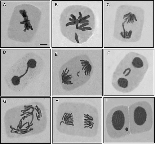
In M2 generation of irradiated plants, various mitotic anomalies were observed (Fig. 1). For all anomalies, with an increase in the ray dose, the anomaly rate also increased. Chromosomal stickiness in metaphase in all treatments was observed (Fig. 1A, Table 1). Precocious movements of chromosomes were another type of anomaly that was observed in the metaphase of all the treatments (Fig. 1B). The chromosome fragments were observed during anaphase and its frequency roughly increased depending on the gamma ray dose (Table 1). The chromosome bridges in anaphase were more than those in telophase. The frequency of this anomaly in the anaphase increased with an increase in gamma ray dose so that it had the highest frequency (28.57%) in the dose of 55 Gy. But the chromosome bridges in telophase were observed in all gamma ray doses except control and dose of 25 Gy (Fig. 1C, D). Laggard chromosomes appeared during anaphase and also telophase. In anaphase, the laggards were observed in all doses of gamma rays, and with an increase in the ray dose, the anomaly rate also increased. The most and least percentage of the laggards was related to the witness (0.52%) and 55 Gy dose (13.25%), but laggards in telophase were merely observed in 55 Gy dose with the frequency of 1.52% (Fig. 1E, F, Table 1). Disturbed anaphase was another anomaly observed in all the treatments. The most disturbed anaphase was related to 55 Gy dose (Fig. 1G). Vagrant chromosomes were one of the observed anomalies during the anaphase and only in doses of 25, 35, and 55 Gy were observed with the frequency of 0.50%, 0.53%, and 0.78%, respectively (Fig. 1H, Table 1). The presence of micronuclei in telophase stage was another anomaly which was observed frequently in all treatments. The frequency of micronuclei increased with an increase in gamma ray dose, and among the mitotic anomalies was the highest frequency (Fig. 1I, Table 1).
| γ-Ray dose (Gy) | Metaphase | Anaphase | Telophase | ||||||||||
|---|---|---|---|---|---|---|---|---|---|---|---|---|---|
| Cell No. | Anamalies (%) | Cell No. | Anamalies (%) | Cell No. | Anamalies (%) | ||||||||
| (1) | (2) | (3) | (4) | (5) | (6) | (7) | (8) | (9) | (10) | ||||
| 0 | 221 | 0.9 | 0.90 | 383 | 0.52 | 1.04 | — | — | — | 201 | — | — | 1.99 |
| 25 | 207 | 18.36 | 9.18 | 401 | 5.24 | 4.49 | 0.50 | 8.73 | 0.50 | 185 | — | — | 9.99 |
| 35 | 199 | 22.11 | 14.57 | 378 | 8.73 | 8.47 | 2.65 | 15.08 | 0.53 | 179 | — | 1.12 | 22.91 |
| 45 | 203 | 23.15 | 22.66 | 384 | 12.50 | 16.15 | 2.60 | 14.06 | — | 175 | — | 4.43 | 45.14 |
| 55 | 218 | 22.02 | 28.44 | 385 | 13.25 | 28.57 | 2.86 | 18.44 | 0.78 | 197 | 1.52 | 3.55 | 65.48 |
(1) Stickiness, (2) precocious chromosome, (3) laggard, (4) bridge, (5) fragment, (6) disturbed, (7) vagrant, (8) laggard, (9) bridge, (10) micronucleus. Total nomber of scored cells for each gamma ray dose was two thousands.
Different doses of gamma ray resulted in different varieties of anomalies such as stickiness, precocious chromosome movements, anaphase bridges, chromosome fragments, laggard chromosomes, tripolar, micronuclei and trinucleate in different meiotic stages of PMCs. In meiosis likewise mitosis the frequency of the anomalies increased with the increase in gamma ray doses. Chromosome stickiness, precocious movements of chromosome and chromosome fragments were observed in metaphase I and II. Precocious movement of the chromosome was more frequent than two other anomalies in metaphase I and II (Fig. 2A). The highest frequency of this anomaly related to 55 Gy dose with a frequency of 32.49% (Table 2). Chromosome fragment was observed in metaphase I and II in all treatments except the control and 25 Gy dose. The most chromosome fragment was related to the 55 Gy treatment with a frequency of 1.41% (Table 2). Bridges were observed in anaphase I and II (Fig. 2B). Also, in some cells, the chromosome bridge was observed along with laggard chromosomes in high doses (45 and 55 Gy) in anaphase II (Fig. 2C). Laggard chromosomes in the present study were observed in anaphase and telophase. Laggard chromosomes in anaphase I and II were observed in all treatments, the highest frequency was related to 55 Gy dose with the frequency of 8.83%. But in the telophase this anomaly was observed only in 35, 45, and 55 Gy doses. The highest frequency (5.25%) was detected in 55 Gy dose (Fig. 2D, Table 2).

| γ-Ray dose (Gy) | Metaphase I/II | Anaphase I/II | Telophase I/II | ||||||||||
|---|---|---|---|---|---|---|---|---|---|---|---|---|---|
| Cell No. | Anamalies (%) | Cell No. | Anamalies (%) | Cell No. | Anamalies (%) | ||||||||
| (1) | (2) | (3) | (4) | (5) | (6) | (7) | (8) | (9) | (10) | ||||
| 0 | 392 | — | — | — | 425 | 0.24 | — | — | — | 321 | 4.67 | — | — |
| 25 | 402 | 0.75 | 1.24 | — | 411 | 2.19 | 1.70 | — | — | 342 | 11.11 | — | 2.34 |
| 35 | 376 | 1.33 | 3.72 | 0.53 | 378 | 3.44 | 3.44 | — | 0.53 | 338 | 20.12 | 1.78 | 6.21 |
| 45 | 369 | 2.98 | 18.16 | 0.81 | 384 | 4.95 | 4.43 | 1.04 | — | 351 | 39.60 | 4.27 | 9.69 |
| 55 | 354 | 5.93 | 32.49 | 1.41 | 385 | 8.83 | 7.01 | 3.38 | 1.82 | 362 | 40.33 | 5.25 | 6.63 |
(1) Stickiness at metaphase I and II, (2) precocious chromosome at metaphase I, (3) fragment at metaphase I and II, (4) laggard at anaphase I and II, (5) bridge at anaphase I and II, (6) bridge and Laggard at anaphase II, (7) tripolar at anaphase I, (8) micronuclei, (9) laggard at telophase I, (10) trinucleate condition at telophase II. The total number of scored cells for each gamma ray dose was two thousand.
Disturbed polarity was observed during anaphase II and telophase II. Another anomaly observed in anaphase II was tripolar which was merely observed in 35 and 55 Gy doses with frequencies of 0.53% and 1.82%, respectively (Fig. 2E). Observing micronuclei in telophase I and II were another instance of anomalies which were observed in all doses of the gamma ray. With an increase in gamma ray dose from 25 to 55, the frequency of micronuclei increased from 11.11 to 40.33%. In the control, the presence of micronuclei with a frequency of 4.67% was observed (Table 2). A trinucleate condition during telophase II was another meiotic anomaly observed in the study which was observed in all gamma ray doses (Fig. 2F).
Study of agronomic traits in M2 generationThe results obtained for agronomic traits including fertility and seed production in plants and phenology of plants revealed significant differences among the treatments in all traits except the weight of 100 seeds (Table 3). The comparison of mean values of fertility and seed production showed that the number of pods per plant, number of seeds per pod, number of seeds per plant, and seed yield per plant were similarly affected by doses of irradiation; higher doses tended to reduce all of these traits as compared with the control (Table 4). All treatments except 25 and 35 Gy showed a significant difference compared with control in the number of seeds per pod. Regarding the pod sterility percentage, mean comparison of treatments showed an increase by increasing irradiation dose, so that the highest percentage of sterility was observed in 55 Gy treatment, and the lowest was found in control. The weight of 100 seeds was not significantly affected by irradiation doses, and the mean comparison placed treatments in one group. According to these results, it can be argued that irradiation has little effect on seed weight in the M2 generation (Table 4).
| S.O.V | DF | Mean of squares | ||||||||
|---|---|---|---|---|---|---|---|---|---|---|
| Number of pods per plant | Number of seeds per pod | Number of seeds per plant | Seed yield per plant | Percentage of sterility in pods | 100 seeds weight | Days to flowering | Days to pod setting | Days to ripening | ||
| Treatment | 4 | 4.61** | 0.38** | 38.63** | 43.94** | 0.057** | 78.6ns | 103.55** | 19.69* | 268.15** |
| Error | 13 | 0.013 | 0.039 | 0.388 | 0.69 | 0.001 | 40.6 | 6.79 | 4.71 | 8.04 |
| C.V. (%) | — | 4.29 | 8.14 | 9.39 | 12.6 | 14.75 | 6.5 | 4.04 | 2.48 | 1.91 |
* and **: Significant at 0.05 and 0.01 respectively; ns: non-significant.
| Treatment (Gy) | Number of pods per plant | Number of seed per pod | Number of seeds per plant | Seed yield per plant | Percentage of sterility in pods | 100 seeds weight | Days to flowering | Days to pod setting | Days to ripening |
|---|---|---|---|---|---|---|---|---|---|
| 0 | 4.17a | 2.64a | 10.67a | 10.93a | 0.065d | 103.39a | 59.25c | 86b | 136d |
| 25 | 3.29b | 2.62a | 8.59b | 8.75b | 0.18c | 100.97a | 61.5c | 86.5b | 145.75c |
| 35 | 2.25c | 2.60a | 5.57c | 5.55c | 0.23c | 95.17a | 63.5c | 86.5b | 150.5b |
| 45 | 1.17d | 2.04b | 3.54d | 3.24d | 0.33b | 92.23a | 68.5b | 89.25ab | 155.25a |
| 55 | 1.34e | 1.95b | 2.58d | 2.56d | 0.42a | 99.18a | 74.5a | 92.5a | 159a |
Different letters in each column show a significant difference (p<0.05).
Increasing gamma ray doses caused a delay in flowering initiation, pod setting, and plant ripening period. There was a significant difference between treatments and control in flowering initiation at high doses of irradiation such as 45 and 55 Gy. There were no significant differences among treatments and control in days to pod setting, except for 55 Gy treatment (Table 4). A delayed ripening was observed by increasing irradiation doses, and the difference between treatments and control was significant. Also, the results of the mean comparison revealed that the maximum (159) and the minimum (136) days to ripening were obtained in 55 Gy treatment and control, respectively.
All the detected anomalies in current project were observed in both mitosis and meiosis except multipolar cells which was only observed in meiosis. Chromosome stickiness during mitosis was observed with higher frequency compared to meiosis. The survival probability of the cells with stickiness is low because mainly the separation of these chromosomes in anaphase is aborted and consequent disturb nuclear division would be happened. The precocious movement of chromosome in mitotic metaphase happened with higher frequency (75.76%) compared to metaphase I and II (55.61%) of meiosis. Chromosome stickiness and unequal separation of chromosomes might cause to the failure of the migration or precociously initiated poleward movement of the chromosomes. As we observed, stickiness interfered in the order of the natural placing of chromosomes in metaphase plate and cause more inability to separate, which as a result led to the formation of sticky bridges in anaphase, hence the occurrence of chromosome bridges of both mitosis and meiosis was dominant in anaphase and observed with a higher frequency compared to telophase in mitosis.
A chromosome that did not overlap along with the spindles and underlay un-proper segregation might create laggard chromosomes in cell divisions. The laggard chromosomes mostly observed in anaphase and during mitosis with a higher frequency compared to meiosis. The laggard chromosomes can produce micronuclei if they cannot get to the poles. This confusion in meiosis can drive to aneuploidy gametes as reported by Kumar et al. (2011) and Souza et al. (2006) and sum up to forming defective gametes that have imbalanced chromosomes, that is an additional reason for the reduction in fertility-related traits.
In general, mutagens phenotypically create inhibition of generative growth, not forming the reproductive organs in a floret, forming infertile gametes, abortion of embryo and development of abnormal seed. Nevertheless, increasing gamma ray doses reduced fertility and seed production and increased sterility in plants. The low fertility following high doses radiation may be attributed to the phenomenon of chromosomal aberrations and/or change in the sequence of genes. Furthermore, significant negative correlations (p≤0.05) were estimated between mitosis and meiosis anomalies and all agronomic traits (table is not presented). The maximum negative correlation was observed between the number of seeds per plant and meiosis anomalies (−0.98) and the number of pods per plant and mitosis anomalies (−0.97). Anomalies in meiosis and mitosis divisions persuade abnormalities in gametes and bod meristems and disturb the agronomical traits likes the number of seeds and pod number per plants. On average, the mutants took retardation for phenological traits i.e., more days to heading, flowering and ripening while the gamma ray dose increased, indicating that applying radiations to evolve early maturing verities is still in question.
We intend to thank and appreciate Mr. Karim Sorkhe for his valuable technical help in performing cytogenetic studies. This study was funded by the Research Council of Shahid Chamran University of Ahvaz referred to the contract number of 96/03/02/16670.