Abstract
Purpose: The principle treatment of infectious aortic aneurysm is to remove the infected aneurysm and replace it with Rifampicin-soaked prosthesis by omentopecxy. This study aimed to clarify the efficacy of long-term antibiotics and subsequent thoracic endovascular aneurysm repair (TEVAR) for infectious thoracic aortic aneurysm.
Methods: Between July 2011 and December 2015, 213 TEVARs were performed at Hiroshima University Hospital. Six patients (2.8%) had infectious aneurysm and received long-term antibiotic therapy and secondary TEVAR. Long-term antibiotic therapy and subsequent TEVAR is paradoxical. This study aimed to clarify the timing of TEVAR for infectious thoracic aortic aneurysm.
Results: All patients presented with fever and back pain, and had positive blood cultures; five patients had significant co-morbidities. Bacteraemia was caused by Methicillin Sensitive Staphylococcus Aureus (MSSA) (2), Streptococcus sanguinis (1), Methicillin Resistant Staphylococcus Aureus (MRSA) (1), Chryseobacterium meningosepticum (1), and Enterococcus faecalis (1). Blood examination at admission revealed a WBC count ranging from 10,470 to 16,170/μl and CRP ranging from 7.9 to 16.4 mg/dl. Long-term antibiotic therapy was continued until WBC and CRP were within the normal range. TEVAR was performed emergently in 3 cases and electively in 3 cases. The time from admission to TEVAR ranged from 7 to 26 days. One stent-graft was deployed in all 6 cases. All patients survived and were followed for an average of 48 months; they were free from re-infection.
Conclusion: Long-term antibiotics and simple TEVAR may be a feasible treatment for infectious thoracic aortic aneurysms.
In cases of infectious aneurysm, open surgery allows for both wide debridement of infected tissue and extra-anatomical bypass3,4,15). In situ rifampin-soaked grafts, rather than extra-anatomical bypass, have been shown to reduce the risk of re-infection1; however, high mortality remains. Thoracic endovascular aneurysm repair (TEVAR) is a minimally invasive therapy for thoracic aortic aneurysms. However, the treatment potential of TEVAR for infectious thoracic aneurysm is controversial because of the risk of prosthesis re-infection and inability to remove the infected aneurysm5). This paper describes our early experiences with long-term antibiotic therapy and simple TEVAR for infectious thoracic aortic aneurysm according to the diagnosis of serum C-reactive protein (CRP) and white blood counts (WBC) and computed tomography (CT) angiography. We also discuss the feasibility of long-term antibiotics and simple TEVAR for infectious thoracic aortic aneurysm.
MATERIALS AND METHODS
This retrospective study was approved by the Institutional Review Board of the Hiroshima University Hospital. The study complied with the Declaration of Helsinki, and informed consent was obtained from each patient. Between 2011 and 2015, we performed 213 TEVARs for thoracic and thoracoabdominal aortic aneurysms. Six of the 213 cases (2.8%) involved thoracic aortic aneurysms with bacteraemia, including 2 impending ruptured aneurysms, which showed hematoma around abdominal aortic aneurysm and 4 growing aneurysms. No patients complained of haemoptysis or vomiting of blood, which excluded aorto-oesophageal or aorto-bronchial fistulae by CT angiography. The patients included 5 men and 1 woman. Their ages ranged from 64 to 84 years (mean, 74.0 years). Thoracic aortic aneurysms were located in the distal aortic arch in 3 patients and the descending thoracic aorta in 3 patients. Chief complaints included high fever (6 patients) and back pain (3 patients). Preoperative blood cultures revealed bacteraemia in all six cases. Bacteraemia was caused by MSSA (2), Streptococcus sanguinis (1), MRSA (1), Chryseobacterium meningosepticum (1), and Enterococcus faecalis (1). Preoperative blood examination at admission revealed a WBC count ranging from 10,470 to 16,170/μl (mean, 12,660/μl) and CRP ranging from 7.1 to 16.4 mg/dl (mean, 10.8 mg/dl). Comorbidities included autoimmune pancreatitis with steroid treatment (1), central venous port infection following treatment for oesophageal cancer, which occurred 1 month prior to TEVAR (1), ileocecal resection due to cancer, which was treated 3 months prior to TEVAR (1), chronic dialysis (1), and multiple sclerosis (1) (Table 1). The patients with oesophageal cancer and ileocecal cancer did not show infectious events before admission. All patients were treated with intravenous (i.v.) antibiotics after admission until CRP and WBC decreased to the normal level, but 3 patients underwent emergent TEVAR during i.v. antibiotic therapy. Causes of emergent TEVAR included doubt of sealed rupture of the aneurysm (1 patient), and two rapidly growing aneurysms (Table 1). Indication of emergent TEVAR for aneurysmal growth was defined by rapid growth of the maximum aneurysm diameter compared with that measured at admission. Elective TEVAR was performed after administration of i.v. antibiotics when CRP was confirmed to have decreased and WBC had normalized (3 patients). Time from admission to TEVAR ranged from 7 days to 26 days (mean, 14.5 days) according to infection control. The therapeutic sequence involves continuous antibiotic therapy and then TEVAR. If the aneurysm is truly infected and the diameter of aneurysm increases rapidly, we think the risk of rupture is very high and perform emergent TEVAR. If bacteraemia is thought to be resolved by antibiotic therapy, we select the clearance of infection before deploying TEVAR and first administer a high dose of antibiotics, and then perform TEVAR. Preoperative blood culture prior to TEVAR showed resolution of serum bacterium in all cases. Serial CT angiography every 2 days was performed to diagnose using the above criteria.
Table 1
Patient characteristics between emergent and elective TEVAR groups
Emergent TEVAR group
| |
M/F |
Age |
Site |
morphology |
Bacteria at admission |
WBC at admission→before TEVAR |
CRP at admission→before TEVAR |
Co-morbidity |
| 1 |
M |
84 |
distal arch |
sac |
MSSA |
13720 |
→ |
8680 |
16.4 |
→ |
3 |
autoimmune pancreatitis |
| 2 |
M |
75 |
descending |
sac |
MRSA |
16170 |
→ |
10500 |
10.1 |
→ |
4.7 |
esophageal cancer |
| 3 |
M |
74 |
descending |
pu |
Streptococcus sanguinis |
11010 |
→ |
6410 |
7.1 |
→ |
3.8 |
multiple sclerosis |
| Mean |
|
78 |
|
|
|
13633 |
→ |
8530 |
11.2 |
→ |
6.2 |
|
Elective TEVAR group
| El |
M/F Age |
Site |
morphology |
Bacteria at admission |
WBC at admission→before TEVAR |
CRP at admission → before TEVAR |
Co-morbidity |
| 3 |
M |
73 |
distal arch |
sac |
MSSA |
12120 |
→ |
7180 |
7.9 |
→ |
1.6 |
colon cancer |
| 4 |
M |
64 |
descending |
pu |
C. meningoseptium. |
10470 |
→ |
5090 |
14.4 |
→ |
0.9 |
= |
| 6 |
F |
79 |
distal arch |
sac |
Enterococcus faecalis |
12460 |
→ |
5870 |
8.7 |
→ |
2 |
cerebral infarction |
| Mean |
|
72 |
|
|
|
11683 |
→ |
6047 |
10.3 |
→ |
1.5 |
|
Em. Emergency cases, El, elective cases sac, saccular aneurysm, pu, penetrating ulcer
*MSSA, methicillin-sensitive Staphylococcus aureus;
MRSA, methicillin-resistant Staphylococcus aureus
C. meningoseptium., Chryseobacterium meningosepticum
=, no detection
RESULTS
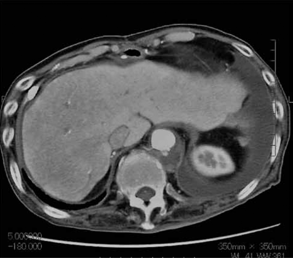
The average time between admission and TEVAR was 14.5 days (range, 7-26 days). Morphologically, CT angiography revealed saccular aneurysm (SA) in 4 cases and penetrating ulcer (PU) in 2 cases. Three patients underwent early TEVAR and 3 patients underwent elective TEVAR. Early TEVAR was performed on the 7th day after admission for a case where sealed rupture of aneurysm was doubted (Fig. 1a, pre-TEVAR; Fig. 1b, post-TEVAR) and for one rapidly growing aneurysm on day 12 of hospitalization (Fig. 2a, pre-TEVAR; Fig. 2b, post-TEVAR). The patient with sealed ruptured aneurysm presented with back pain and progressive anaemia. Preoperative blood examination showed a WBC count of 11,010/μl and CRP level of 7.1 mg/dl. Blood culture was positive for Streptococcus bacteria. CT angiography before TEVAR revealed a sealed rupture of a descending thoracic aortic aneurysm with hematoma surrounding the penetrating ulcer (Fig. 1a). The patient received administration of antibiotics for 7 days until the CRP level was less than 2.0 mg/dl. Simple TEVAR was performed using Gore-Tag to cover the descending aorta. Postoperative enhanced CT revealed complete coverage of the lower descending thoracic aorta (Fig. 1b), and the subsequent clinical course was uneventful. One patient presented with rapidly growing aneurysms (Fig. 2a, saccular aneurysm) and received early TEVAR on day 12 of hospitalization (Fig. 2b). This patient showed rapid aneurysm growth of more than 3 mm in diameter compared with CT at admission and CRP had decreased less than 3 mg/dl. Serial enhanced CT angiography was performed every week in all 6 cases.


We compared bacterial species and blood examinations (WBC count and CRP level) pre- and postoperatively. The patient with sealed rupture showed Streptococcus infection, with a WBC count of 11,010/μl and CRP level of 7.1 mg/dl preoperatively. After 7 days of antibiotic therapy, the blood culture showed no Streptococcus bacteria and the blood examination revealed that WBC count and CRP level decreased to 6410/μl and 2.0 mg/dl, respectively (Table 1). Another early case showed MSSA infection. The WBC count of these patients was 13,700/μl, which decreased to 8,686/μl post-antimicrobial therapy (Table 2). Because the CRP level decrease was significant (CRP reduced from 16.4 mg/dl to 3.0 mg/dl), we performed early TEVAR to prevent aneurysm rupture. Elective cases showed a significant decrease of WBC count and CRP level after antimicrobial therapy (mean, 11,683 to 6,047/μl in WBC count, 10.3 to 1.6 mg/dl in CRP level). After TEVAR, we continued antibiotic therapy; the final blood examination showed 5,800/μl for WBC count and 0.9 mg/dl for CRP level at discharge. The mean hospital stay was 38 days.
Table 2
Characteristics of procedure (comparison with emergent and elective TEVAR groups)
Emergent TEVAR group
| No. |
Em |
Aneurysm growth |
Device x number |
Bacteria at TEVAR |
Days to TEVAR |
Access |
Pro. landing |
AKA cover |
Hosp. after TEVAR |
Result |
|---|
| 1 |
Em |
3mm at 10th day |
C-TAGx1 |
no |
10 |
rt.FA |
Zone2 |
= |
34 |
good |
| 2 |
Em |
3mm at 11th day |
TAG x1 |
no |
12 |
rt.FA |
Th5 |
Th8 |
15 |
good |
| 3 |
Em |
sealed rupture |
TAG x1 |
no |
7 |
rt.FA |
Th9 |
Th12 |
17 |
good |
| Mean |
|
|
|
|
9.7 |
|
|
|
22 | |
Elective TEVAR group
| No. |
El |
Aneurysm growth |
Device x number |
Bacteria at TEVAR |
Days to TEVAR |
Access |
Pro. landing |
AKA cover |
Hosp. after TEVAR |
Result |
| 4 |
El |
no growth at 14th day |
TX2 x1 |
no |
18 |
rt.FA |
Zone3 |
= |
16 |
good |
| 5 |
El |
no growth at14th day |
TX2 x1 |
no |
19 |
lt.FA |
Th6 |
Th9 |
31 |
good |
| 3 |
El |
no growth at 21th day |
TAG x1 |
no |
26 |
abdominal aorta |
Zone2 |
= |
14 |
good |
| Mean |
|
|
|
|
21 |
|
|
|
20 |
|
*Em, emergency cases; El, elective cases; days to TEVAR, days from admission to TEVAR (Thoracic endovascular aneurysm repair); rt.FA, right femoral artery; Pro.landing, proximal landing zone; AKA, Adamkiewicz artery; Th, thoracic vertebrae; EVAR, endovascular aneurysm repair
*MSSA, methicillin-sensitive Staphylococcus aureus;
MRSA, methicillin-resistant Staphylococcus aureus
C. meningoseptium., Chryseobacterium meningosepticum
=, no detection
Several types of stent-grafts were used in the 6 cases: Gore-Tag (Gore corp.) in 3 cases, TX2 (Cook corp.) in 2 cases, and c-Tag (Gore corp.) in 1 case. One stent-graft (length, 10 to 20 cm; diameter; 28 to 36 mm) was deployed toward the aneurysm in all 6 cases. The proximal landing zone ranged from zone 2 to Th9. The artery of Adamkiewicz was identified by CT angiography preoperatively and covered in 3 patients, but no spinal cord injury was observed. Days to TEVAR were 12 days in the early TEVAR cases and 21 days in the elective TEVAR cases. There was a significant difference in days to TEVAR between groups (Table 2). However, postoperative stay was same between groups. All patients survived without any serious complications. The median post-procedural hospital stay was 22 days (range, 14-34 days).
Intravenous antibiotic treatment was tailored to the causative organism in all cases, and i.v. treatment lasted a mean 22 days. Cefazolin was used for 12 to 30 days in 4 patients. Vancomycin plus carbapenem was administered for 21 days in a patient with MRSA infection. Penicillin-G was used for 30 days in a patient with streptococcal infection. Oral antibiotics (oral penicillin or cefalosporin) were administered throughout treatment. At a median 48 months of follow-up (range, 26-68 months), all patients were alive with no clinical sign of re-infection (Table 2).
DISCUSSION
Infectious thoracic aortic aneurysm is among the most challenging aortic aneurysms to treat. Blood culture and CT angiography alone cannot differentiate between infectious aneurysms, and thoracic aortic aneurysms and bacteraemia. Although PET-CT can be used to differentiate these entities, it is unavailable for use in Japan. We have thus grouped both entities under ‘thoracic aneurysm with bacteraemia’. Because operative intervention is associated with high rates of morbidity and mortality1,2,8,10,11,14) in infected aneurysms, treatment with broad spectrum antibiotics is an essential therapy, as well as an early intervention. TEVAR is a less invasive treatment option, but previous reports have shown mixed results in patients with infectious aneurysms7,12,17). Early mortality following TEVAR and late re-infection have ranged from 0 to 50% and 0 to 100%, respectively1,7,8,10-12,14,17). Part of the reason for the wide range of results is that these studies included patients with aorto-oesophageal fistulae secondary to erosion of the aneurysm. Aorto-oesophageal and aorto-bronchial fistulae are very difficult to treat by simple TEVAR and are often fatal complications of infectious aneurysms, regardless of the treatment technique7,12,17). Our study did not include patients with aorto-oesophageal fistulae or aorto-bronchial fistulae. That is why we observed 100% survival and absence of re-infection. We performed 3 emergent TEVAR and 3 elective TEVAR procedures according to the patient’s condition. If the patient showed rapid growth of the aneurysm as evaluated by serial CT angiography (aneurysmal growth >3 mm compared with the diameter measured at admission), we performed emergent TEVAR. If the aneurysm diameter measured by serial CT angiography was unchanged after admission, we selected antibiotic therapy first until the clearance of bacteraemia was confirmed. The extended duration of antibiotic therapy might also explain the excellent outcomes we observed1,8,10,11). Stent-grafts are deployed and maintained in an infected field9), and long-term administration of antibiotics may decrease the risk of re-infection. Our experience suggests that oral antibiotic treatment should last for at least 6 months to prevent re-infection and continue indefinitely. We selected emergency TEVAR cases by establishing sealed rupture and rapid growth of the aneurysm (>3 mm growth in diameter compared to the diameter measured in preoperative CT angiography). We wondered why simple TEVAR and antibiotic therapy could eliminate aneurysm with bacteraemia without late infection. We suppose it is because the porosity of the TEVAR graft is large and can allow permeation of antibiotics between the blood and infected aneurysm. Thus, the antibiotics can easily diffuse through the stent-graft wall, reach the aneurysmal sac, and destroy the microorganisms. Long-term use of antibiotics may also control infection and prevent late recurrence of infection.
There are several reports of surgical intervention for infectious aneurysms from Southern-eastern Asian countries15,17). Some were performed by surgical exclusion of the aneurysm and prosthetic replacement with omental wrapping17). The disease-specific mortality was very high (31.25%) in infected abdominal aortic aneurysms when conventional surgery was performed13). Endovascular repair and long-term antibiotics may be feasible for both infected thoracic and abdominal aortic aneurysms18). Treatment strategies for prosthesis infection has evolved recently, but complete excision of the infected stent-graft and surrounding tissue is mandatory once stent-graft infection occurs6). Although antibiotic therapy alone for infectious aneurysm is dangerous due to the risk of rupture, we think simple TEVAR and antibiotic therapy is feasible for aneurysms with bacteraemia. The timing of TEVAR for infected aneurysms is controversial. Infection should be eliminated using antibiotics before TEVAR. However, some patients require emergent TEVAR because of impending rupture. Serial CT angiography is useful to determine if the aneurysm is dangerous. The morphology of the aneurysm (saccular or penetrating ulcer) and the growth rate of the aneurysm diameter as measured by CT angiography may indicate the risk of rupture of the aneurysm. Although the number of our patients was very small, this study suggests a treatment for thoracic aneurysm with bacteraemia using TEVAR and antibiotic therapy.
We used i.v. antibiotic therapy until the fever resolved and WBC count was normalized. In 3 cases, emergent TEVAR was performed during i.v. antibiotic therapy. In the other 3 cases, elective TEVAR was performed after resolution of bacteraemia. Bacteraemia resolved in all 6 cases and we observed no infection-related complications. While the incidence of TEVAR infection is rare, it can occur during the late phase. Once endograft infection occurs, the treatment should be performed with complete exclusion of the endograft and surgical replacement of the whole infected aorta6). Long-term oral antibiotic therapy may reduce the likelihood of the emergence and spread of multidrug-resistant organisms, which poses a serious threat to hospitalized individuals. Restricting the use of one antibiotic often requires a compensatory increase in the use of another antibiotic, which in turn selects for the emergence of different species of multidrug-resistant organisms; furthermore, the potential clinical implications of such interactions remaining largely unexplored16). However, endograft infection is fatal in TEVAR. Thus, we continued one antibiotic for at least 6 months. Fortunately, TEVAR and long-term antibiotic therapy alone was feasible in our cases. Although our experience included only 6 patients and was limited to aortic aneurysms without fistulae to visceral organs, our strategy may be effective in limited cases with aortic aneurysms with bacteraemia. Serial CT angiography help select emergent TEVAR or elective TEVAR for the treatment of aortic aneurysm with bacteraemia.
CONCLUSION
Simple TEVAR and long-term antibiotics effectively treated patients with thoracic aortic aneurysms with bacteraemia. We observed 100% early and late survival and no re-infection. It is important to perform this strategy when indicated; the time of TEVAR and antibiotic administration for infected thoracic aortic aneurysm is also crucial. Further study is mandatory.
DISCLOSURE STATEMENT
All authors declare that they have no conflicts of interest.
REFERENCES
- 1. Blocker, D., Schmacher, H., Schwarzbach, M., Ockert, S., Robert, H. and Allenberg, J.R. 2004. Endoluminal stent-graft repair of aorto-bronchial fistulas: bridging or defining long-term solution? J Endovasc Ther 11: 41-48.
- 2. Gonzalez-Fajardo, J.A., Gutierrez, V., Martin-Pedrosa, M., Del Rio, L. Carrera, S. and Vaquero, C. 2005. Endovascular repair in the presence of aortic infection. Ann Vasc Surg 19: 94-98.
- 3. Hsu, R.B. and Lin, F.Y. 2008. Infected aneurysm of the thoracic aorta. J Vasc Surg 47: 270-276.
- 4. Hsu, R.B. and Lin, F.Y. 2007. Surgery for infected aneurysm of the aortic arch. J Thorac Cardiovasc Surg 134: 1157-1162.
- 5. Heneghan, R.E, Singh, N. and Stames, B.W. 2015. Successful emergent endovascular repair of a ruptured infectious thoracic aortic aneurysm. Ann Vas Surg 29(4): 843
- 6. Igari, K., Kudo, T., Toyofuku, T., Jibiki, M., Sugano, N. and Inoue, Y. 2014. Treatment strategies for aortic and peripheral prosthetic graft infection. Surg Today. 44(3): 466-71.
- 7. Jones, K.G., Bell, R.E., Sabbarwal, T., Aukett, M., Reidy, J.T. and Tavlor, P.R. 2005. Treatment of mycotic aortic aneurysms with endoluminal grafts. Eur J Vasc Endovasc Surg 29: 139-144.
- 8. Leobon, B., Roux, D., Mugniot, A., Rousseau, H., Cerene, A., Glock, Y., et al. 2002. Endovascular treatment of thoracic aortic fistulae. Ann Thorac Surg 74: 247-249.
- 9. Laohapensang, K., Aworn, S., Orrapi, S. and Rutherford, R.B. 2012. Management of the infected aorto-iliac aneurysm. Ann Vasc Dis 5: 334-341.
- 10. Patel, H.J., William, D.W., Upchurch, G.K., Dasika, N.I., Eliason, J.L. and Deeb, G.M. 2009. Late outcome of endovascular repair for the infected thoracic aorta. Ann Thorac Surg 87: 1366-1371.
- 11. Pirretti, S., Bozzani, A., Arici, V. and Odero, A. 2002. Endovascular treatment of acute haemoptysis secondary to aorto-bronchial fistula. Eur J Vas Endovasc Surg 32: 366-368.
- 12. Reisenmann, P.J., Books, J.D. and Farber, M.A. 2009. Thoracic endovascular aottic repair of aorto-pulmonary fistulas. J Vasc Surg 50: 992-998.
- 13. Sorcline, K., Mani, K., Bjork, M., Nyman, R. and Wanhaninen, A. 2009. Endovascular repair of infectious aortic aneurysms. J Vas Surg 50: 269-274.
- 14. Ting, A.C., Cheng, S.W., Ho, P. and Poon, J.T. Cheung, G.C. 2006. Endovascular stent-graft repair for infected thoracic aortic pseudoaneurysm – a durable option ? J Vasc Surg 44: 701-705.
- 15. Von Segesser, L.K., Tkebuchava, T., Niederhauser, U., Kunzli, A., Lachat, M., Genoni, M., et al. 1997. Aortoaorto-bronchial and aortoaorto-esophageal fistulae as risk factors in surgery of descending thoracic aortic aneurysms. Eur J Cardiothorac Surg 12: 195-201.
- 16. Wang, J., Foxman, B., Mody, L. and Snikin, E.S. 2017. Network of microbial and antibiotic interactions drive colonization and infection with multidrug-resistant organisms. Proc Natl Acad Sci USA DOI 10. 1073/pnas.171023114 [Epub ahead of print].
- 17. Wheatley, G.H, Nimesz, A., Preventza, O., Ramatah, V.G., Rodngez-Lopez, J.A., William, J., et al. 2007. Have we gone too far ? Endovascular stent-graft repair of aorto-bronchial fistulae. J Thorac Cardiovasc Surg 133: 1277-1285.
- 18. Yamasaki, M., Abe, K., Misumi, H., Ito, J., Nakanishi, Y. and Kawazoe, K. 2015. Endograft infection after hybrid surgery for chronic Stanford type B aortic dissection: endograft infection and treatment. Surg Today 45: 1575-1578.