Abstract
To provide reliable relationship between hydrogen embrittlement (HE) and hydrogen distribution, a duplex stainless steel (DSS: JIS SUS329J4L) annealed and electrolytically hydrogen-charged was investigated by means of hydrogen microprint technique (HMPT), where distribution of hydrogen was examined on the opposite side of the charged surface. Quantitative analysis was made by classifying the site of detected hydrogen into three categories: ferrite matrix, austenite grain and phase boundary. The HMPT was performed on the 1.5 h and 24 h charged specimens with two holding times in the ambient air for 0.5 (as quick as possible) and 300 h. In the 1.5 h charged and 0.5 h held specimen, hydrogen atoms were mostly detected on the phase boundary. When charging time was increased to 24 h, relative fraction of hydrogen desorbed in the austenite phase against the ferrite matrix and phase boundary increased. The relative fraction of hydrogen atoms in the austenite phase was also increased by increasing holding time to 300 h irrespective of the charging time. During the holding, hydrogen atoms inside the ferrite matrix were presumed to preferentially diffuse out from the specimen or transferred to the phase boundary, while hydrogen atoms already trapped at the phase boundary will move into the interior of the austenite phase. The results obtained in the present study where experimental conditions are systematically selected can be rationally interpreted only with the higher solubility and smaller diffusivity of hydrogen in the austenite phase, rather than considering the binding energy of hydrogen with phase boundary.
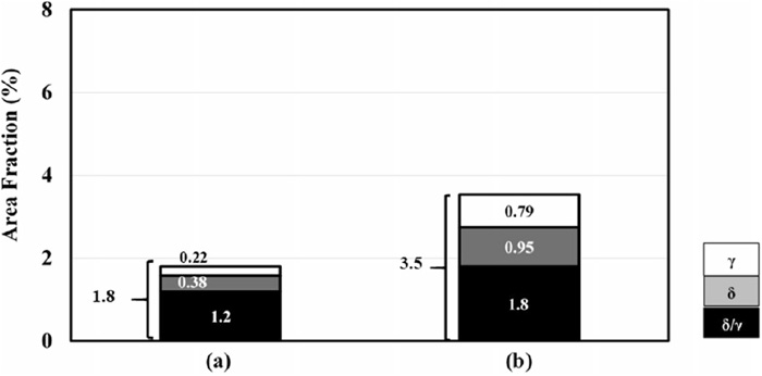
Area fraction of the silver grains in the HMPT images, corresponding to the conditions in Fig. 3. Viewing area and hydrogen-charging time: (a) 1318
μm
2 and 1.5 h, (b) 2164
μm
2 and 24 h, respectively.
Fullsize Image
