Abstract
This review discusses trends in mode of breech delivery in Japan. Recently, primary elective cesarean delivery rates for singleton breech pregnancies have markedly increased due to medical counseling and maternal requests. However, breech extraction skills should be preserved and passed on to future generations of obstetricians. Vaginal breech delivery may be considered if well-trained and full-time medical staff with experience performing breech deliveries are available and comprehensive informed consent is obtained. As specialists of obstetrics and gynecology, it may be necessary to acquire rudimentary techniques for vaginal breech delivery in order to perform fair and objective informed consent procedures regarding the mode of breech delivery.
Recent status of breech delivery
In Japan, primary elective cesarean delivery rates for singleton breech pregnancies have markedly increased due to medical counseling and maternal requests associated with the American College of Obstetricians and Gynecologists (ACOG) recommendation in 2001,1,2,3,4,5,6) which recommended planned cesarean delivery for term singleton breech. The recommendation was made based on the observation by Hannah et al.6) that perinatal and neonatal complication rates were significantly lower in the planned cesarean group relative to the planned vaginal delivery group. Moreover, in the United States, the number of vaginal breech deliveries was deemed too low to teach breech extraction procedures using a ‘hands on’ approach.7) In our institute (one of the major perinatal centers in Tokyo, Japan), the primary elective cesarean delivery rate for breech presentation based on informed consent increased from 59.0% (36/61 cases of singleton breech term deliveries) in 2000 to 76.8% (53/69 cases) in 2005 (P<0.05 using χ2 with Fisher exact test).3) In 2012, Kubonoya et al.4) reviewed cases of breech presentation in obstetrical facilities that are members of the Tokyo Operation group from 1981 to 2010. The rate of breech presentation was 2.4% (64,528/2,672,409 women in labor). The rate of cesarean section for breech presentation in Japan increased progressively from 1981 to 2010 (41.9% in 1981, 70.4% in 1990, 86.7% in 2000, and 94.4% in 2010, P<0.01), and this was accompanied by a progressive decrease in mortality rates for infants with breech presentation during the same period (1.9% in 1981, 0.47% in 1990, 0.39% in 2000, and 0.21% in 2010, P<0.01). In 2008–2010, the mortality rate for infants with vaginal breech was higher than that with cesarean delivery (3.4% vs. 0.07%, P<0.01).4)
ACOG recommendations beyond 2006 have indicated that the decision regarding the mode of delivery should consider patients’ wishes and the experience of the provider,8,9) because some follow-up studies revealed no differences in maternal or neonatal outcomes between planned cesarean and vaginal delivery groups.10,11) However, there has been no significant revival of vaginal breech delivery in Japan, possibility because the techniques for breech delivery are less familiar to today’s young obstetricians. Thus, it may be important to teach the skills needed for breech delivery, although these techniques may only be required occasionally.
In the Guidelines for Obstetrical Practice in Japan (2017 edn),12,13) vaginal delivery in women without a knee or foot presentation, an estimated fetal weight of <2,500 g, a gestational week <37, or presumed cephalic-pelvic disproportion can be considered with the availability of well-trained and full-time medical staff with experience performing breech deliveries after discussing both the risks and benefits of vaginal and cesarean deliveries with the patient and obtaining informed consent.
Correction of breech presentation during pregnancy
To date, various low-invasive attempts such as moxibustion and knee-chest postural management have been introduced to correct term breech presentation.14,15,16,17,18,19) When combined with knee-chest postural management, moxibustion has been suggested to reduce the number of non-cephalic presentations at term. However, there remains a need for well-designed randomized controlled trials that evaluate the effectiveness of moxibustion to correct breech presentation and report on clinically relevant outcomes as well as the safety of the intervention.14) Moxibustion, a form of traditional Chinese medicine, involves the burning of the herb moxa over acupuncture points. The technique has been reported in Japan to convert breech presentation to cephalic presentation. While the mechanism of action of moxibustion is not entirely clear, it has been suggested to stimulate fetal movements and make conversion more likely with increased contractility in response to adrenocortical stimulation.16) Knee-chest postural management for breech presentation at term has not been found to be an effective enough form of care to be offered routinely to women with breech presentation at term.17,18) In addition, Marumo et al.19) observed a negative effect of knee-chest postural management for breech presentation beyond 28 weeks’ gestation. In their study, the rate of breech presentation at term decreased with the discontinuation of knee-chest postural management (2.14% vs. 2.72%, P<0.05). According to the authors, the presence of uterine contractions induced by knee-chest positioning may inhibit fetal self-rotation.
For the safe performance of external cephalic conversion, the Guidelines for Obstetrical Practice in Japan (2017 edn) require confirmation of the following three conditions: (1) emergency cesarean section is available, (2) no previous cesarean delivery, and (3) fetus is mature.12,13) The success rate of external cephalic conversion in Japan is 59–69%.20,21,22) The administration of neuraxial analgesia and/or tocolytic agents significantly increased the success rate of external cephalic conversion and, as a result, the incidence of vaginal delivery as well. Based on a previous study from the United States, the rate of cesarean delivery for dystocia increased after a successful trial of external cephalic conversion.23,24) However, according to a different study, the rate of cesarean delivery and neonatal outcomes after a successful trial of external cephalic conversion were similar to those of natural cephalic presentation.20)
Breech extraction
1. Conditions for breech extraction
Planned vaginal delivery is reasonable for select women with a term singleton breech fetus.25,26,27,28,29,30,31,32,33) In Japan’s current perinatal care system,34) planned induction of labor may also be reasonable. To avoid fetal traction and/or maternal complications such as postpartum urinary retention, the bladder must be empty during the second stage of labor.25,26,27,28,29,30,31,32,33) Assisted extraction of a breech fetus should be started after complete dilation of the uterine os, and the lower abdomen or inferior angle of the scapula of the fetus should be delivered spontaneously. After the above requirements are met, physicians can consider vaginal delivery in women without signs of fetal growth restriction or macrosomia, low estimated fetal weight (especially less than 2,300 g), feto-pelvic disproportion, fetal anomaly incompatible with vaginal delivery, insufficient expulsive force resulting from weak pain, rigidity of the soft birth canal, anomalies of the birth canal, low-lying placenta, hyperextension of the fetal head, presence of a mass obstructing the birth canal, and uterine malformation. In cases of single foot presentation with posterior leg advances, there is a risk that the anterior buttock will be caught on the maternal pubic bone.25,26) In cases of a funnel-shaped pelvis, inadequate labor pain is frequent in cases of simple breech presentation.25,26)
2. Appropriate labor management for breech presentation
The favorable progression of labor has been suggested to be the best indicator of adequate fetal-pelvic proportions for a safe labor, as opposed to findings of radiologic pelvimetry.27,28) However, this does not rule out the clinical utility of radiologic pelvimetry for deciding on the mode of breech delivery.25,31) Parity has been suggested to be associated with the increased success rate of vaginal breech delivery.35,36) Continuous fetal heart monitoring is recommended during the first and second stages of labor.12,13) The ruling out of a prolapsed cord by immediate vaginal examination is required when spontaneous and artificial membranes rupture. Labor pains can be augmented with oxytocin in order to avoid weak pain irrespective of maternal fatigue. In the absence of adequate progression of labor, cesarean section should be considered.
3. Delivery techniques: Partial breech extraction
The term ‘partial’ in the context of breech extraction means extraction of the lower (or the remaining) part of the body only after the infant has been delivered as far as the umbilicus by spontaneous uterine and abdominal forces. A deep unilateral episiotomy should be performed, particularly for primiparous women.
To decide on the appropriate timing to start breech delivery, the physician must feel and confirm sufficient uterine forces for expulsion by pushing in the descending fetal breech with the palm of the dominant hand (Sakaki method).25) This method will facilitate delivery of the fetal head.
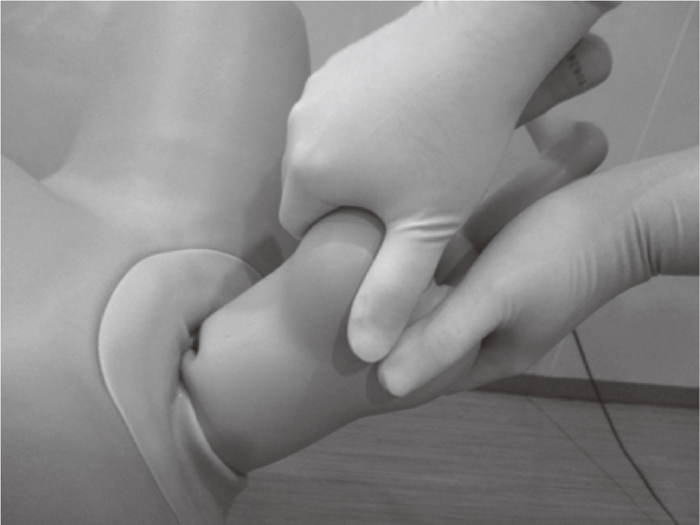
The uterine fundal pressure maneuver during delivery promotes safe breech delivery. If the maternal uterine force is sufficient with or without the maternal fundal pressure maneuver, spontaneous or assisted breech delivery, such as the Bracht method, should be planned.37) In cases of insufficient maternal uterine force for spontaneous breech delivery, ‘transverse figure 8 breech delivery’ (TF8 maneuver: San-ikukai Hospital maneuver first reported by Dr. Takeoka) has been widely recommended in Japan for reducing the incidence of nuchal arm.25,31,32,33) For example, after delivery of the fetal breech as far as the umbilicus, spontaneous or assisted delivery can be expected if the fetal hip rises spontaneously. However, if the fetal hip does not rise spontaneously, then partial breech extraction should be initiated quickly as follows: (1) Thumbs are placed over the sacrum and each index finger wraps over the top of the fetal iliac crest with or without 1 or 2 pieces of gauze to prevent slippage. (2) The fetal body is raised as far as necessary to deliver the inferior angle of the scapula (Figure 1). (3) The fetal body is pulled while twisting 2 or 3 times as in drawing the transverse number 8 in a direction whereby the fetal back always comes to the maternal ventral side until the upper limbs are delivered (Figure 2). Traction towards the lower side is performed with a strong force while upward-lifting is performed gently. This method could be considered one of Mikulitz-Rasicki’s modifications.25,31,32,33)

The TF8 maneuver appears to be the easiest method for delivering the upper limbs while shortening the compression time of the umbilical cord.25) However, in cases of involuntary delivery of fetal upper limbs, such as nuchal arm, the classical method of arm freeing should be performed (Figure 3). The posterior arm must be delivered first. For this, the lower half of the fetal body is raised up and over the maternal groin. The physician’s fingers are inserted under the posterior shoulder and aligned with the humerus.25,30,38) During the method, the uterine fundal pressure maneuver should be interrupted for smooth insertion of the finger. Although the method may reliably deliver the upper limbs of the fetus, more skill is demanded of the physician relative to that needed for other methods such as the TF8 maneuver in order to ensure performance and avoiding neonatal injury.25)

The fetal head should be delivered by Mauriceau-Veit-Smelli’s or Wigand-Martin-Winclel-Veit-Smelli’s maneuver with the assistance of suprapubic pressure to prevent cervical spine injury to the fetus.25,30,37) The fetal head on a flexed position should be extracted in the correct direction.
4. Perinatal outcomes of vaginal breech delivery with TL8 maneuver
Unfortunately, there are only a few detailed records regarding complications of vaginal breech delivery, such as neonatal injury. However, the TF8 maneuver allows for the smooth delivery of the upper limbs without incarceration.31,32) In 1978, Takeoka32) reported improved perinatal outcomes of vaginal breech delivery with the TF8 maneuver. Neonatal mortality and injury rates of vaginal breech delivery at San-ikukai Hospital in 1933–1934 were 12.4% (128/1,031) and 0.8% (8/1,031), respectively, while those in 1946–1962 decreased to 4.5% (60/1,475, P<0.01) and 0.4% (5/1,475, P=0.16), respectively, due to introduction of the TF8 maneuver. In 1972, the neonatal mortality rate of vaginal breech delivery decreased to 1.5% (2/130, P<0.01), independently of the mode of breech delivery.
In 1991–1992, Suzuki and Takeishi attempted vaginal delivery in 40 cases of breech presentation after ruling out of fetal-pelvic disproportion by radiologic pelvimetry at Japanese Red Cross Katsushika Maternity Hospital (unpublished data). The rates of nulliparous women and cases other than frank breech presentation were 73% (29/40) and 60% (24/40), respectively. Of these, 35 cases (88%) were delivered by the TF8 maneuver and 2 cases (5%) by the Bracht method, and 3 cases (8%) required the classical method of arm freeing. The reasons why the 3 cases required the classical method of arm freeing were: inadequate fetal descent complicated by a non-reassuring fetal status associated with a funnel-shaped pelvis for frank breech presentation (2 cases) and oligohydramnios (1 case). Umbilical cord prolapse was recognized in 1 case (3%), but none of the cases required emergent cesarean section. In 5 cases (13%), the fetal head was delivered spontaneously. Wigand-Martin-Winclel-Veit-Smelli’s maneuver was required in the remaining 35 cases (88%). There were no cases of severe neonatal asphyxia.
Conclusions
In reality, breech vaginal delivery is no longer considered the first choice. Nowadays, obstetricians may not have sufficient opportunities to experience vaginal breech delivery in order to evaluate its safety or effectiveness. However, breech extraction is a maneuver that should be passed down to future generations of obstetricians. Vaginal breech delivery can be performed when well-trained and full-time medical staff with experience performing breech deliveries are available, with accompanying informed consent considerations.12,13) As specialists of obstetrics and gynecology, acquiring rudimentary techniques for vaginal breech delivery may be useful and provide more breech delivery options to expecting mothers.
References
- 1. Sato A, Fujimori T, Yamada H, Sugahara N. Breech presentation (In Japanese). Obstetrical and Gynecological Practice. 2005; 54: 1781–1790.
- 2. Suzuki S. Trends in mode of delivery of breech presentation over a 5-year period. J Perinatol. 2001; 27: 464–467.
- 3. Suzuki S, Nakata M. Factors associated with the recent increasing cesarean delivery rate at a Japanese perinatal center. ISRN Obstet Gynecol. 2013; 2013: 863282.
- 4. Kubonoya K, Furukawa S, Machida T, et al. A 30-year case study based on 64,528 breech presentations; Changes in delivery mode and infant mortality. Int J Gynecol Obstet. 2012; 119: S796.
- 5. ACOG Committee Opinion No. 265. Mode of term singleton breech delivery. Obstet Gynecol. 2001; 98: 1189–1190.
- 6. Hannah ME, Hannah WJ, Hewson SA, Hodnett ED, Saigal S, Willan AR. Planned caesarean section versus planned vaginal birth for breech presentation at term: a randomised multicentre trial. Term Breech Trial Collaborative Group. Lancet. 2000; 356: 1375–1383.
- 7. Lavin JP Jr, Eaton J, Hopkins M. Teaching vaginal breech delivery and external cephalic version. A survey of faculty attitudes. J Reprod Med. 2000; 45: 808–812.
- 8. ACOG Committee Opinion No. 340: Mode of term singleton breech delivery. Obstet Gynecol. 2006; 108: 235–237.
- 9. ACOG Committee Opinion No. 745: Mode of Term Singleton Breech Delivery. Obstet Gynecol. 2018; 132: e60-e63.
- 10. Whyte H, Hannah ME, Saigal S, et al. Term Breech Trial Collaborative Group. Outcomes of children at 2 years after planned cesarean birth versus planned vaginal birth for breech presentation at term: the International Randomized Term Breech Trial. Am J Obstet Gynecol. 2004; 191: 864–871.
- 11. Hannah ME, Whyte H, Hannah WJ, et al. Term Breech Trial Collaborative Group. Maternal outcomes at 2 years after planned cesarean section versus planned vaginal birth for breech presentation at term: the international randomized Term Breech Trial. Am J Obstet Gynecol. 2004; 191: 917–927.
- 12. Japan Society of Obstetrics and Gynecology (JSOG) and Japan Association of Obstetricians and Gynecologists (JAOG). Guidelines for obstetrical practice in Japan 2017 edition. http://www.jsog.or.jp/activity/pdf/gl_sanka_2017.pdf (September 22, 2018).
- 13. Minakami H, Maeda T, Fujii T, et al. Guidelines for obstetrical practice in Japan: Japan Society of Obstetrics and Gynecology (JSOG) and Japan Association of Obstetricians and Gynecologists (JAOG) 2014 edition. J Obstet Gynaecol Res. 2014; 40: 1469–1499.
- 14. Coyle ME, Smith CA, Peat B. Cephalic version by moxibustion for breech presentation. Cochrane Database Syst Rev. 2012; 5: CD003928.
- 15. Schlaeger JM, Stoffel CL, Bussell JL, et al. Moxibustion for Cephalic Version of Breech Presentation. J Midwifery Womens Health. 2018; 63: 309–322.
- 16. Ewies A, Olah K. Moxibustion in breech version--a descriptive review. Acupunct Med. 2002; 20: 26–29.
- 17. Hofmeyr GJ, Kulier R. Cephalic version by postural management for breech presentation. Cochrane Database Syst Rev. 2012; 10: CD000051.
- 18. Smith C, Crowther C, Wilkinson C, Pridmore B, Robinson J. Knee-chest postural management for breech at term: a randomized controlled trial. Birth. 1999; 26: 71–75.
- 19. Marumo G, Morita Y, Uchida S, et al. Clinical usefulness of Knee-chest postural management for breech presentation (In Japanese). Obstetrical and Gynecological Therapy. 2010; 100: 99–103.
- 20. Matsuzaki S, Shimoya K, Murata Y. Cesarean delivery after successful external cephalic version of breech presentation at term. Int J Gynaecol Obstet. 2006; 93: 248–249.
- 21. Inohaya A, Sato Y, Moriuchi K, Ri Y, Kin K. External cephalic version in our institute (In Japanese). J Jpn Soc Perin Neon Med. 2018; 54: 606.
- 22. Hayashi S, Ono H, Yamashita A, et al. External cephalic version in our institute (In Japanese). Advances in Obstetrics and Gynecology. 2018; 70: 237.
- 23. Hutton EK, Hofmeyr GJ, Dowswell T. External cephalic version for breech presentation before term. Cochrane Database Syst Rev. 2015; 7: CD000084.
- 24. Vézina Y, Bujold E, Varin J, Marquette GP, Boucher M. Cesarean delivery after successful external cephalic version of breech presentation at term: a comparative study. Am J Obstet Gynecol. 2004; 190: 763–768.
- 25. Takeishi Y. Breech extraction (In Japanese). Self-publishing. 1985.
- 26. Kaneko K. Dystocia in breech delivery (In Japanese). Obstetrical and Gynecological Practice. 1990; 39: 1215–1219.
- 27. Kotaska A, Menticoglou S, Gagnon R; MATERNAL FETAL MEDICINE COMMITTEE. Vaginal delivery of breech presentation. J Obstet Gynaecol Can. 2009; 1: 557–566.
- 28. Kotaska A, Menticoglou S, Gagnon R, et al. Society of Obstetricians and Gynaecologists of Canada. SOGC clinical practice guideline: Vaginal delivery of breech presentation: no. 226, June 2009. Int J Gynaecol Obstet. 2009; 107: 169–176.
- 29. Kasamori S. Extraction in the breech presentation (In Japanese). In: Kuji N, Obata T, eds. Obstetric surgery. Tokyo; Kanehara Shuppan, 1961, 303–351.
- 30. Araki T. Breech presentation (In Japanese). In: The latest obstetrics- abnormal pregnant women 21th ed. Tokyo; Bunko-do, 2008, 310–325.
- 31. Takeoka H. Transverse figure 8 breech delivery (In Japanese). Obstetrical and Gynecological Therapy. 1976; 32: 244–249.
- 32. Takeoka H. Transverse figure 8 breech delivery (In Japanese). Obstetrical and Gynecological Therapy. 1978; 36: 435–441.
- 33. Takeoka H. Transverse figure 8 breech delivery (In Japanese). Obstetrical and Gynecological Therapy. 1981; 42: 417–424.
- 34. Japan Association of Obstetricians and gynecologists. Lack of obstetricians and gynecologists. JAOG Information. 2014: 1–8. http://www.jaog.or.jp/wp/wp-content/ (September 22, 2018).
- 35. Doyle NM, Riggs JW, Ramin SM, Sosa MA, Gilstrap LC 3rd. Outcomes of term vaginal breech delivery. Am J Perinatol. 2005; 22: 325–328.
- 36. Diro M, Puangsricharern A, Royer L, O’Sullivan MJ, Burkett G. Singleton term breech deliveries in nulliparous and multiparous women: a 5-year experience at the University of Miami/Jackson Memorial Hospital. Am J Obstet Gynecol. 1999; 181: 247–252.
- 37. Jotkowitz MW, Picton FC. An appraisal of an anatomically and physiologically correct method of breech delivery: the Bracht manoeuvre. Aust N Z J Obstet Gynaecol. 1970; 10: 151–159.
- 38. Cunningham FG, Leveno KJ, Bloom SL, et al. Breech delivery. In: Cunningham FG, Leveno KJ, Bloom SL, Hauth JC, Dashe JS, Hoffman BL, Casey BM, Dponf GY. eds. Williams Obstetrics. 25th ed. New York: McGraw Hill, 2018; 539–552.
