Abstract
A 72-year-old man visited our hospital with weight loss. CT examination showed right pleural effusion, ascites and mural thickening of the distal ileum. Examination of the ascitic fluid revealed increase in the level of adenosine deaminase associated with an increase in the number of lymphocytes ; there were no malignant cells. PCR assay of the ascitic fluid, and smear examination of the
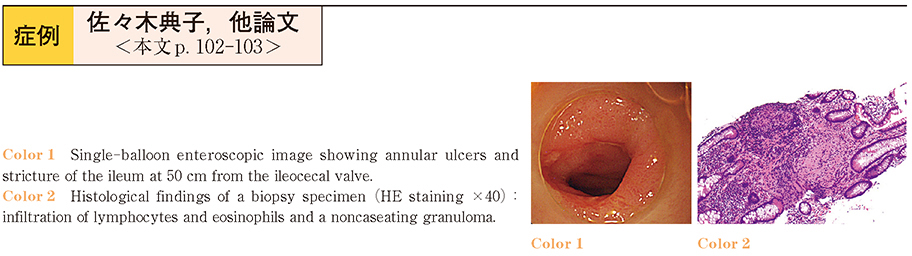
sputum and feces for acid-fast bacilli were all negative, however, the T-spot test was positive. Capsule enteroscopy revealed ulcers and stenosis of the ileum. Single-balloon enteroscopy showed annular ulcers and stricture of the ileum at 50 cm from the ileocecal valve. Histological examination of biopsy specimens showed infiltration of lymphocytes and eosinophils and noncaseating granulomas. Based on these results, we diagnosed the patient as having intestinal tuberculosis. Anti-tuberculous therapy was started with isoniazid, rifampicin, pyrazinamide and ethambutol and the ascites disappeared in 2 months.
Intestinal tuberculosis is often difficult to diagnose, because there are no specific symptoms and the positivity rate for tubercle bacilli in fluids such as ascites and feces is not so high. In our case, single-balloon enteroscopy was useful for the diagnosis.