Abstract
A 49-year-old woman was found to have a tumor of the pancreatic head at a medical checkup. Abdominal ultrasonography revealed a mass measuring 4 cm in diameter in the pancreatic head. Abdominal CT also demonstrated a cystic tumor with a solid tumor component at the pancreatic head. EUS revealed a low-echoic tumor without clear continuity with the muscularis propria. Blood flow was abundant in the margin of the tumor. ERCP revealed compression of the pancreatic head and distal bile duct by the tumor.
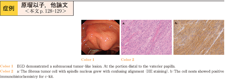
Under the tentative diagnosis of P-NET, pancreatoduodenectomy was performed. Histologically, the tumor showed spindle cells arranged in whorls in a storiform pattern, and immunohistochemically, the tumor was positive for c-kit, which clinched the diagnosis of gastrointestinal stromal tumor (GIST) of the duodenum. Obtaining tissue samples from GISTs is difficult, particularly from tumors located in the distal duodenum. However, it seems essential to obtain samples for a definitive diagnosis of submucosal tumors, such as by EUS-FNA.