Abstract
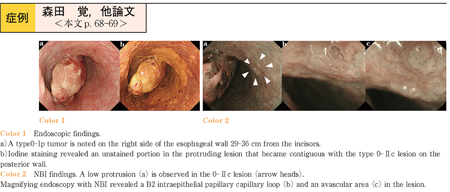
We report a case of so-called carcinosarcoma of the esophagus in which we could examine the correlations between the endoscopic examination and pathological examination findings. A 70-year-old male was referred to us for the investigation of anemia that had been detected during a periodic medical check-up. Barium swallow revealed a protruding tumor on the right side of the esophageal wall in the middle to lower thoracic portion of the esophagus. Upper gastrointestinal endoscopy showed a Type 0-Ip tumor at 29-36 cm from the incisors. Taking these results into consideration, we suspected the tumor as a carcinosarcoma of the esophagus. We performed subtotal esophagectomy with lymph node dissection. Histopathological examination showed that the greater part of the protruding tumor was composed of spindle cells with a sarcoma-like appearance. However, the base was composed of squamous cell carcinoma. Pathologically, there was a transitional zone between the two elements. On immunochemistry, the sarcoma-like spindle cells showed positive staining for vimentin with a few parts of the cells showing slightly positive staining for AE1/AE3 ; on the other hand, the SCC component showed negative staining results for vimentin, S-100, and desmin. Therefore, we diagnosed the tumor as a carcinosarcoma pT2, ly0, v0, n0, Stage II according to the Guideline for Clinical and Pathological Studies on Carcinoma of the esophagus.