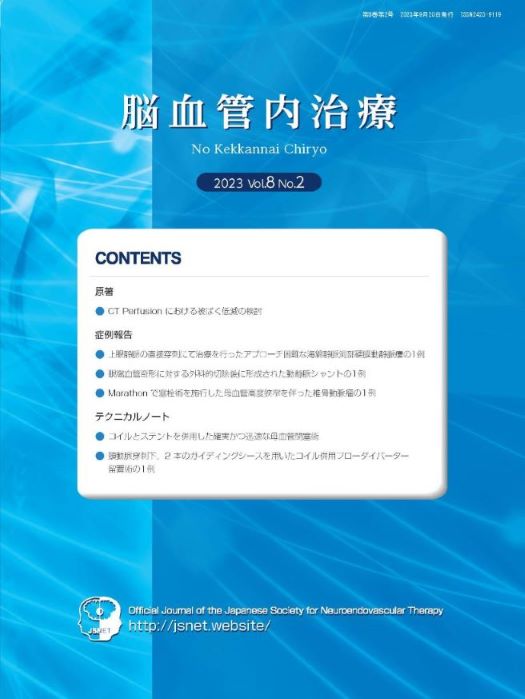
No Kekkannai Chiryo
Online ISSN : 2424-1709
Print ISSN : 2423-9119
ISSN-L : 2423-9119
Volume 8, Issue 2
Displaying 1-6 of 6 articles from this issue
Original Article
  • Fumitaka SATO, Nobuaki TANABE, Takeshi MORISHITA, Takashi SAKAGUCHI, O ...
    2023Volume 8Issue 2 Pages 49-54
    Published: 2023
    Released on J-STAGE: September 20, 2023
    Advance online publication: July 05, 2023
    JOURNAL OPEN ACCESS

    Objective: CT perfusion image (CTP) scans to quantitatively evaluate cerebral blood flow is a useful procedure because of its convenience and rapidity. However, CTP has a disadvantage of increased irradiative exposure several times. We usually conduct CTP scans according to a protocol of maker recommendation. Therefore, we examined retrospectively whether the reduction in scan times could lead to the reduction in exposure keeping the radiological findings.

    Methods: CTP scans were performed for patients with cerebral infarction. The data obtained from CTP according to a protocol of maker recommendation (early phase 16 scans+delayed phase 4 scans) were compared to the CTP data thinning out scan times (early phase 8 scans). Using Bayesian inference, both data were analyzed by visual and quantitative evaluations.

    As for the visual evaluation, these data were evaluated by 4 doctors by using the paired comparison method. As for the quantitative evaluation, each parameter was analyzed from the installed ROI value between the affected and non-affected cerebral cortex.

    Results: As for visual evaluation, no significant differences in all parameters were found between the two groups. In the quantitative evaluation also, there was no significant differences.

    Conclusion: The reduction in scan times in the CTP imaging did not affect the quality of data obtained from CTP, with decreasing exposure dose.

    Download PDF (3332K)
Case Reports
  • Masaru KIYOMOTO, Eishi SATO, Shinya SUEMATSU, Jun NAKAUCHI
    2023Volume 8Issue 2 Pages 55-62
    Published: 2023
    Released on J-STAGE: September 20, 2023
    Advance online publication: May 12, 2023
    JOURNAL OPEN ACCESS

    Objective: Herein, we report a case of cavernous sinus dural arteriovenous fistula (CSDAVF) treated with transvenous embolization and surgical cannulation of the superior ophthalmic vein (SOV).

    Case Presentation: A 61-year-old man with a 4-month history of progressive orbital congestion, exophthalmos, achromatopsia, and high intraocular pressure in the right eye was diagnosed with right CSDAVF using angiography. We attempted embolization via the inferior petrosal sinus and facial vein twice. However, both attempts failed as focally thrombosed, narrowed, and tortuous vein prevented insertion of the microcatheter into the cavernous sinus. Surgical exposure of the SOV allowed its direct cannulation via the orbital septum. The fistula was embolized through the SOV with platinum coils without complication, improving patient’s symptoms.

    Conclusion: Surgical cannulation of the SOV via an opened orbital septum to resolve CSDAVF is a safe and effective approach when conventional transvenous access fails.

    Download PDF (12846K)
  • Hirotsugu NOGAWA, Katsuhiro MIZUTANI, Takenori AKIYAMA, Taichi SAYANAG ...
    2023Volume 8Issue 2 Pages 63-67
    Published: 2023
    Released on J-STAGE: September 20, 2023
    Advance online publication: June 07, 2023
    JOURNAL OPEN ACCESS

    Objective: We report a case of arteriovenous shunt with multilobular venous pouch around the supraorbital fissure, which has not been reported in the literature.

    Case Presentation: A 51-year-old female with a history of craniotomy with a right frontotemporal approach for the surgical resection of a orbital vascular malformation in childhood. She presented with right eye pain, protrusion, and conjunctival hyperemia of the right eyeball. Angiography revealed multilocular venous cavities near the supraorbital fissure from the sphenoid ridge to the middle skull base, with a relatively localized arteriovenous shunt on the venous wall. The shunt blood flow was supplied by the branches of the external carotid artery and drained into the superior ophthalmic vein. The venous cavity where the arteriovenous shunt was formed was also continuous with the cavernous sinus, but it appeared to be an arteriovenous shunt secondary to venous malformation, as the venous structure was multilocular, rather than a single vein. Selective transvenous embolization was performed, and the arteriovenous shunt disappeared and the eye symptoms disappeared.

    Conclusion: After surgery for orbital vascular malformations, an arteriovenous shunt may form in the remaining venous wall. Transvenous embolization may be an effective treatment.

    Download PDF (2638K)
  • Jun ISOZAKI, Osamu MASUO, Yoshiaki TETSUO, Kana TAKASE
    2023Volume 8Issue 2 Pages 68-73
    Published: 2023
    Released on J-STAGE: September 20, 2023
    Advance online publication: July 14, 2023
    JOURNAL OPEN ACCESS

    Objective: We report a case of coil embolization using a Marathon microcatheter for unruptured vertebral artery aneurysm with parent artery tight stenosis.

    Case Presentation: A 59-year-old man was incidentally detected with right vertebral artery (VA) aneurysm of 10 mm in maximum diameter at the junction of the right VA and posterior inferior cerebellar artery (PICA). In addition, the VA beyond the VA–PICA junction came to an end, and the VA at the just proximal neck was tightly stenosed. He had suffered from sudden neck pain one year ago, and the MRI performed four years ago showed the patency of VA without aneurysmal formation. Taking his history and angiographical findings into consideration, this aneurysm could be caused by vertebral dissection. Coil embolization for this aneurysm was planned. After a SL-10 microcatheter was navigated into the aneurysm, the angiography revealed no visualization of the aneurysm and PICA because of the passage of the SL-10 beyond the tight stenosis of the proximal VA. After changing the microcatheter to Marathon, flow stagnation could be resolved and coil embolization was completed. The patient was discharged without any complications 3 days after the treatment.

    Conclusion: While there have been some previous reports referred to aneurysmal coil embolization using a Marathon catheter, this report may be the first case that coil embolization using a Marathon was performed for the aneurysm located at the cerebral main trunk accompanied with tight stenosis. We think that a Marathon microcatheter may be one of the useful options for this situation.

    Download PDF (2457K)
Technical Notes
  • Namito KIDA, Naoki FUKUI, Daichi YAMASAKI, Reina MICHIUE, Fumihiro HAM ...
    2023Volume 8Issue 2 Pages 74-80
    Published: 2023
    Released on J-STAGE: September 20, 2023
    Advance online publication: May 18, 2023
    JOURNAL OPEN ACCESS

    Objective: Endovascular parent artery occlusion (PAO) is a treatment option for complex cerebral aneurysms with the attendant risk of ischemic complications. In this article, we performed a novel PAO technique using both coils and a stent (PACS method) achieving targeted occlusion of parent artery with reduced risk of ischemic complications.

    Case Presentations: We performed PAO for 3 patients with cerebral aneurysms harboring critical arterial branches adjacent to the occlusion site. First, we placed an open-cell stent at the targeted site. Then, a few initial coils were readily anchored and stabilized by the struts of the stent, resulting in targeted embolization with preservation of critical branches in a short time. Patients’ outcomes were favorable without ischemic complications.

    Conclusion: The PACS method is useful in conjunction with PAO to avoid critical branch-perforator ischemia or when anchoring is challenging with coils alone.

    Download PDF (5008K)
  • Kento ASAKURA, Nobuyuki FUKUI, Hirotoshi IMAMURA, Masaomi KOYANAGI, Ma ...
    2023Volume 8Issue 2 Pages 81-87
    Published: 2023
    Released on J-STAGE: September 20, 2023
    Advance online publication: June 13, 2023
    JOURNAL OPEN ACCESS

    Objective: Carotid artery puncture is considered for endovascular treatment of cerebral aneurysm (CA) after surgical treatment of the thoracic aorta or if vascular tortuosity is strong. Here, we report a case of an unruptured giant CA treated by a flow-diverter stent with coils using two guiding sheaths after exposure and direct puncture of the carotid artery, resulting in a favorable outcome.

    Case Presentation: The patient was a 83-year-old man. A giant CA with 31 mm long diameter was detected in the left internal carotid artery’s cavernous sinus on head magnetic resonance imaging. We performed embolization of the CA using guiding sheaths, taking into consideration the small diameter of the puncture hole and other factors.

    Conclusion: Flow-diverter implantation using a guiding sheath under direct carotid artery puncture was effective in treating unruptured cerebral CA after surgical treatment of the thoracic aorta.

    Download PDF (7984K)
feedback
Top