Commentary and Perspective
Detection of singularity in immunity and cancer by novel imaging techniques
2020 Volume 17 Pages 98-99

Details
2020 Volume 17 Pages 98-99
There exist critical moments, such as the Big Bang, where something is created from nothing, or potential moments in the future when artificial intelligence outperforms human intelligence. These moments are called singularities. Singularity events have a major feature where a small number of rare events would become the trigger to cause drastic and irreversible changes throughout the manybody complex system. Even in biological phenomena, it is known that a relatively small number of cells that we call “singularity cell” can become the core to trigger drastic change of the entire multi-cellular system, the mechanisms by which these phenomena occur are largely unknown. In our research project, to look deeply into singularity cells, we are developing the trans-scale-scope AMATERAS (A Multi-scale/modal Analytical Tool for Every Rare Activity in Singularity), which achieves imaging with ultra-wide field of view, high spatial resolution, high speed, long term, and corresponding big data analysis [1]. This will enable us to be at the cutting edge of new scientific fields, thereby we not only uncover the underlying mechanism for generation of singularity cells as well as their biological functions, but also create a novel scientific view point by which we focus on unique outlier rather than averaged majority. Figure 1 describes such concept of our research project.
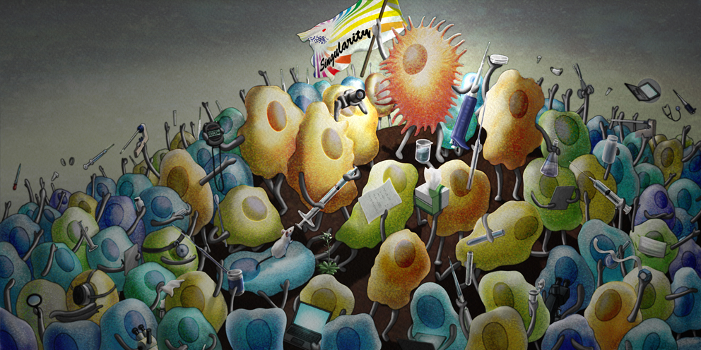
A main visual of Singularity Biology, showing a minor and unique cell becomes a core to induce drastic and irreversible changes throughout the multicellular system.
For this issue, we have a symposium at the 58th Annual Meeting of the Biophysical Society of Japan held in September 2020 inviting seven speakers. In this symposium, we aim to discuss singularities in highly ordered biological system and the way to detect very rare events which act as singularity. It is cosponsored by Scientific Research on Innovative Areas “Singularity Biology”. In the first four talks, we try to input of examples of biological events focusing on acquired immunity, peripheral sensing and the development of cancer.
Dr. Shunsuke Chikuma (Keio University) will summarize the concept of adaptive immunity, in which a few lymphocytes that recognize foreign antigens are activated, expand, and provide life-long protection against pathogens. In particular, he will present recent progress on the mechanism of immune-aging, an age associated defect on immune system, characterized by immunodeficiency and inflammatory phenotypes. In short, the expression and function of TRIM28, a chromatin organizer important in gene silencing, was found to decrease on aged immune cells, which caused unwanted immune reaction, immune-deficiency and exacerbation of autoimmune reaction [2]. The results show so called “epigenome aging” occur in leucocytes, ultimately leading to immune-aging phenotype frequently found in old individuals.
Dr. Shinichiro Sawa (Kyushu University) is an expert in organogenesis, in particular, development of lymph nodes, where immune reaction is initiated. He will present a talk asking a fundamental question “Which cells initiate lymph node formation?” proposing a new model about stepwise activation of only three types of cells; lymphatic endothelial cells, hematopoietic lymphoid tissue inducer cells and mesenchymal cell, that present in lymph node anlagen and interact each other to start organizing lymph nodes.
Dr. Takaharu Okada (RIKEN) has been extensively working on live imaging of the skin barrier system, which is an interface between self and outer environment. He will talk about multi-dimensional imaging of the epidermis, showing how sensory nerves are protected by keratinocytes despite constant turnover of the epidermis in the normal physiological condition, and how this process is disturbed in pathologic conditions [3].
Dr. Shunsuke Kon (Tokyo University of Science) works on physiological reactions upon emergence of cancer cells in normal tissue, trying to elucidate events occurring between normal tissues and cancer cells. Cancer is a mutated “self” that emerges from normal epithelia. He will precisely analyze interactions occurring between normal stromal cells and emerging cancer cells, understanding how obvious cancer cells, as a singularity, induce transition from normal to transformed tissues.
The second part of symposium focuses on the latest imaging technologies that enable the detection of very rare events in the whole body.
Dr. Miya Ishihara (National Defense Medical College) introduces the photoacoustic imaging technique, which utilizes both light and ultrasound. So far, she successfully developed the photoacoustic imaging system, which enables the imaging of the endogenous hemoglobin or exogenous chemical probe in biosample [4]. For examples, the photoacoustic imaging of the morphological change of the human peripheral vascular network induced by surrounding temperature change, the accumulation of the tumor-targeting probe in mouse tumor, and the chromoprotein-expressing cells has been achieved. In this symposium, she will talk about the photoacoustic imaging technique for visualizing the singularity phenomena in biosamples, which is now on-going.
Dr. Toshitada Yoshihara (Gunma University) is developing visualization of hypoxia in tissues by using phosphoresonance lifetime imaging to understand pathophysiology of various hypoxia-related diseases related to hypoxia cancer [5]. He will introduce a method to visualize oxygen tension in the body with spatial resolution at a cellular level using a cell-penetrating phosphorescent probe based on cationic iridium complex and a confocal phosphorescence lifetime imaging microscope (PLIM) to assess hypoxia in tumor. He will also introduce absolute oxygen tensions in cells and capillary vessels in tissues.
Dr. Kenjiro Hanaoka (The University of Tokyo) discusses the development of the ratiometric near-infrared (NIR) fluorescent probe for pH measurements in the whole body [6]. The pH in tissues is strictly regulated and differences of pH are deeply related to key biological events such as tumor. Ratiometric fluorescence imaging is effective for determination of precise pH values and cancelation of the fluorescence differences due to the biodistribution of the fluorescent probe. He will introduce the developed NIR ratiometric fluorescent probe for pH and the whole-body fluorescence imaging of extracellular pH of tumors with the probe.
In conclusion, the symposium includes talks specialized in singularity biology.
We acknowledge the Ministry of Education, Culture, Sports, Science and Technology (MEXT) “Grant-in-Aid for Scientific Research on Innovative Areas” “Singularity biology” (No. 18H05408).