Images in Cardiovascular Medicine
Unique Atrial Tachycardia From the Right Inferior Pulmonary Vein Mimicking Multiple Focal Patterns in the Bilateral Atria and Aorta
2021 年 3 巻 8 号 p. 472-473

詳細
2021 年 3 巻 8 号 p. 472-473
We experienced a rare case of an atrial tachycardia (AT) from the right inferior pulmonary vein (RIPV) presenting with multiple foci in the right atrium (RA), left atrium (LA), and aortic coronary cusp. A 71-year-old man with arrhythmogenic right ventricular cardiomyopathy was referred to Shinshu University Hospital for catheter ablation of a refractory AT. An ablation catheter (ThermoCool SmartTouch; Biosense Webster, Irvine, CA, USA) was used under the guidance of a 3-dimensional mapping system (CARTO 3 version 7; Biosense Webster). We mapped the RA, LA, and aorta, and obtained a focal activation pattern in each chamber (Figure, Left). Because radiofrequency catheter ablation (RFCA) of the earliest site in the RA did not affect the AT, we then mapped the RIPV and found earlier fragmented potentials (Figure, Right). The AT was terminated and became non-inducible by RFCA at the RIPV, but the exact mechanism responsible for the AT remained unclear.
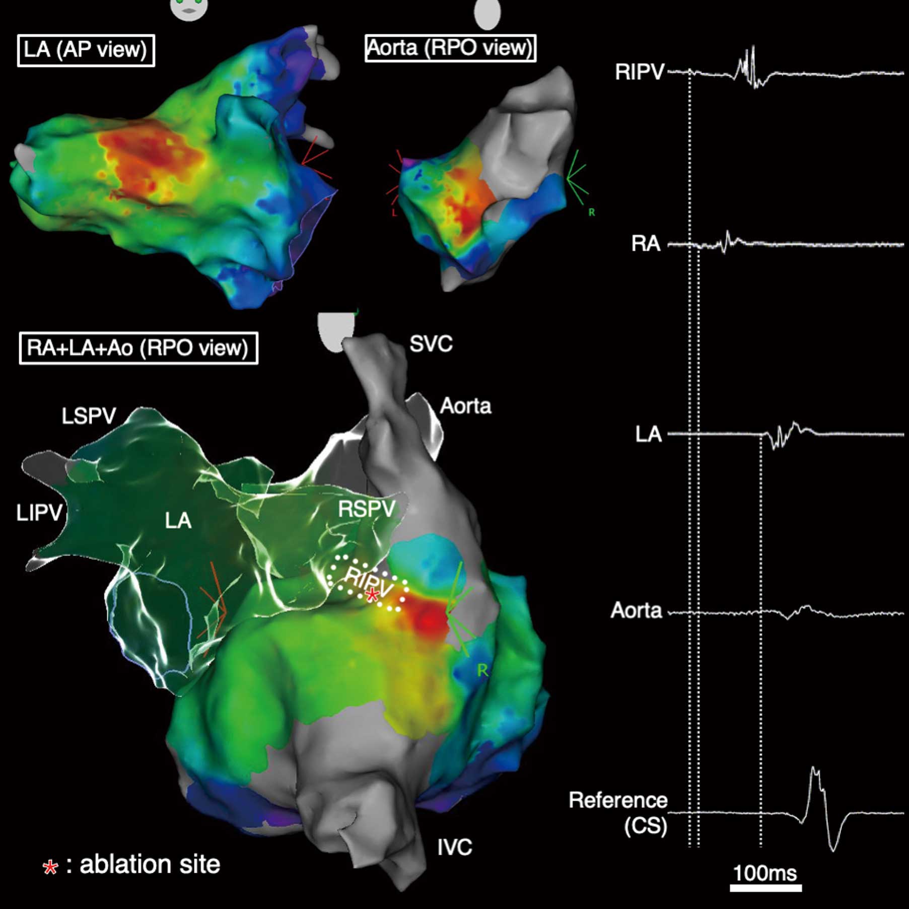
(Left) CARTO activation maps of the atrial tachycardia. (Right) The earliest local electrogram in the left atrium (LA), aorta, right atrium (RA), and right inferior pulmonary vein (RIPV). AP, anteroposterior; CS, coronary sinus; IVC, inferior vena cava; LIPV, left inferior pulmonary vein; LSPV, left superior pulmonary vein; RPO, right posterior oblique; RSPV, right superior pulmonary vein; SVC, superior vena cava.
Our patient had a unique AT presenting with multiple focal activation patterns in the RA, LA, and aorta, whereas the true focus was located in the RIPV. Epicardial preferential pathways adjacent to the thoracic veins have been reported, especially between the right pulmonary veins and RA.1 The epicardial intercaval connections, which are difficult to identify using high-density mapping systems, may mislead one as to the mechanism underlying complex ATs.
K.K. is a member of Circulation Reports’ Editorial Team. The remaining authors have no conflicts of interest to disclose.
This study was approved by the Institutional Review Board of Shinshu University (3856).