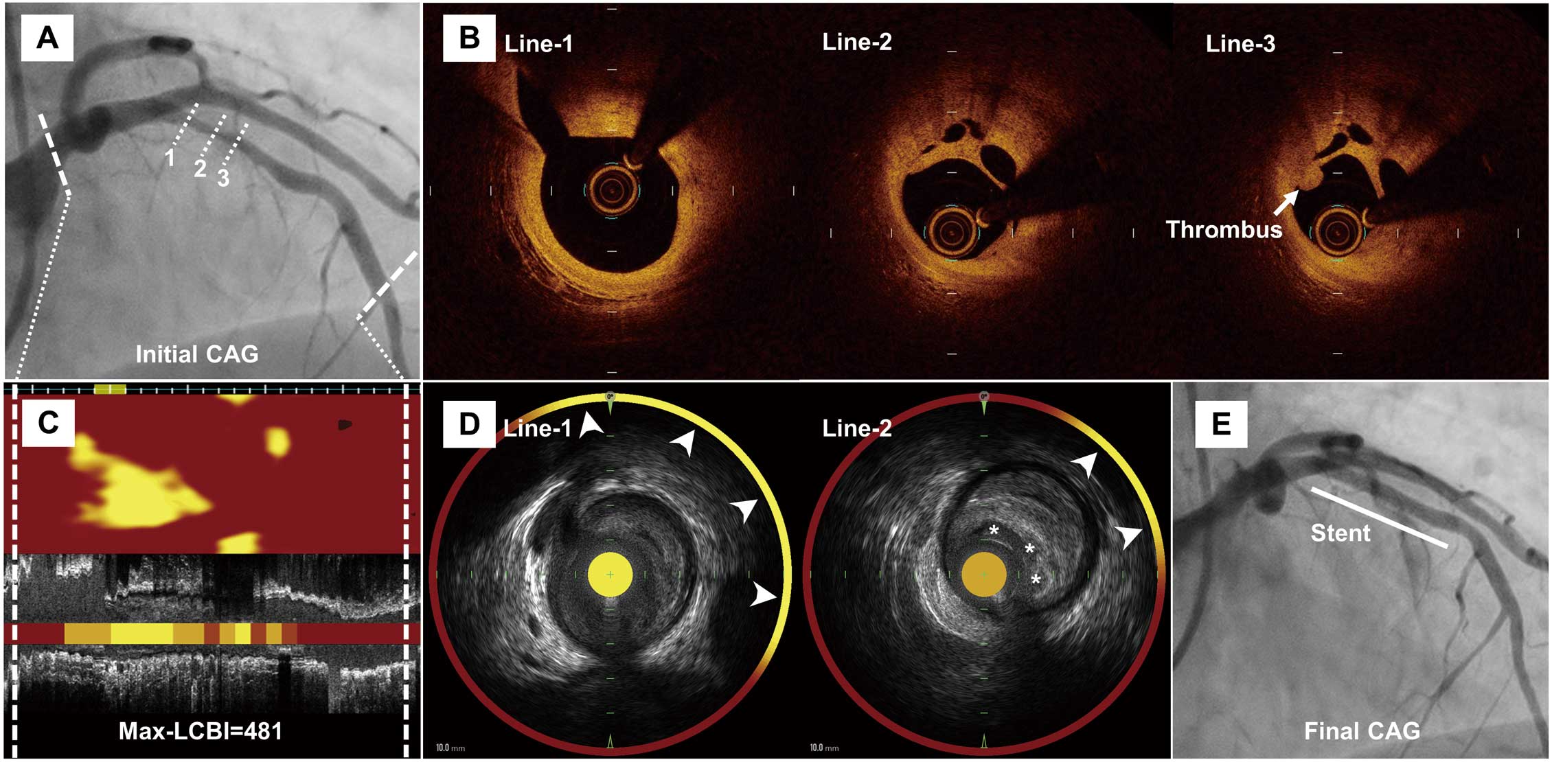
A 50-year-old male current smoker with dyslipidemia presented to Miyazaki Medical Association Hospital with anterior non-ST-elevation myocardial infarction (NSTEMI). The patient complained of frequent chest pain at rest, which had started 3 weeks before admission. Electrocardiography showed T-wave inversions in precordial leads, and echocardiography revealed a wall motion abnormality in the left anterior descending coronary artery (LAD) territory. Coronary angiography identified severe stenosis in the proximal LAD (Figure A). Optical coherence tomography (OCT) showed multiple channels with a ‘lotus root-like’ appearance and mural thrombus on the atheroma (Figure B; Supplementary Movie). Near-infrared spectroscopy (NIRS) and intravascular ultrasound also demonstrated multiple cavities and extensive yellow signals with a maximum lipid core burden index (LCBI) of 481 in a 4-mm segment (Figure C,D), suggesting the presence of a lipid-rich plaque. A drug-eluting stent was successfully implanted at the lesion (Figure E).
This case clearly shows a lotus root-like appearance detected with both NIRS and OCT in a patient with NSTEMI. OCT provides a high-resolution image around the surface of the vessel. Conversely, NIRS can detect lipid components even in deep areas and behind thrombi. The lotus root-like appearance is considered to be caused by the spontaneous recanalization of a blood vessel with a thrombus.1
Several studies have reported a significant association between NIRS-derived LCBI and plaque instability.2
This case demonstrates that a blood vessel segment with a thrombus on a lipid-rich plaque can recanalize spontaneously with the formation of multichannel lumens.
Disclosures
K.K. is a member of
Circulation Reports’ Editorial Team. The remaining authors have no conflicts of interest to declare.
Supplementary Files
Supplementary Movie.
Preprocedural optical coherence tomography.
Please find supplementary file(s);
http://dx.doi.org/10.1253/circrep.CR-21-0127
References
- 1.
Kang SJ, Nakano M, Virmani R, Song HG, Ahn JM, Kim WJ, et al. OCT findings in patients with recanalization of organized thrombi in coronary arteries. JACC Cardiovasc Imaging 2012; 5: 725–732.
- 2.
Kataoka Y, Puri R, Andrews J, Honda S, Nishihira K, Asaumi Y, et al. In vivo visualization of lipid coronary atheroma with intravascular near-infrared spectroscopy. Expert Rev Cardiovasc Ther 2017; 15: 775–785.
