Images in Cardiovascular Medicine
Different Positions of the Impella Device Between the Axillary and Femoral Approaches
2022 年 4 巻 4 号 p. 183-184

詳細
2022 年 4 巻 4 号 p. 183-184
Hemolysis is less frequent following surgical implantation of the Impella 5.0 (Abiomed Japan, Tokyo, Japan) through the axillary artery (AxA) than percutaneous implantation of the Impella CP through the femoral artery (FA);1 however, little is known about the underlying mechanism.
Using the HEARTROID system (JMC, Yokohama, Japan), a training system with a 3-dimensional printed heart model, we took computed tomographic images of the Impella CP inserted through the AxA and FA, and superimposed those images on 3-dimensional constructions (Supplementary Movie). The anterior-posterior view revealed that the Impella from the AxA was located on the lesser curvature side (Figure A). The left anterior oblique 45° view revealed that the Impella from the FA was located on the greater curvature side (Figure B). The left and right oblique 45° views confirmed that the Impella from the AxA was located closer to the left ventricular (LV) apex than that from the FA (Figure B,C). The cranial oblique 90° views demonstrated that the Impella from the AxA was located anteriorly and more coaxially to the LV apex (Figure D), which may suggest that the Impella from the AxA is anatomically favorable for LV suction and subsequent hemolysis.
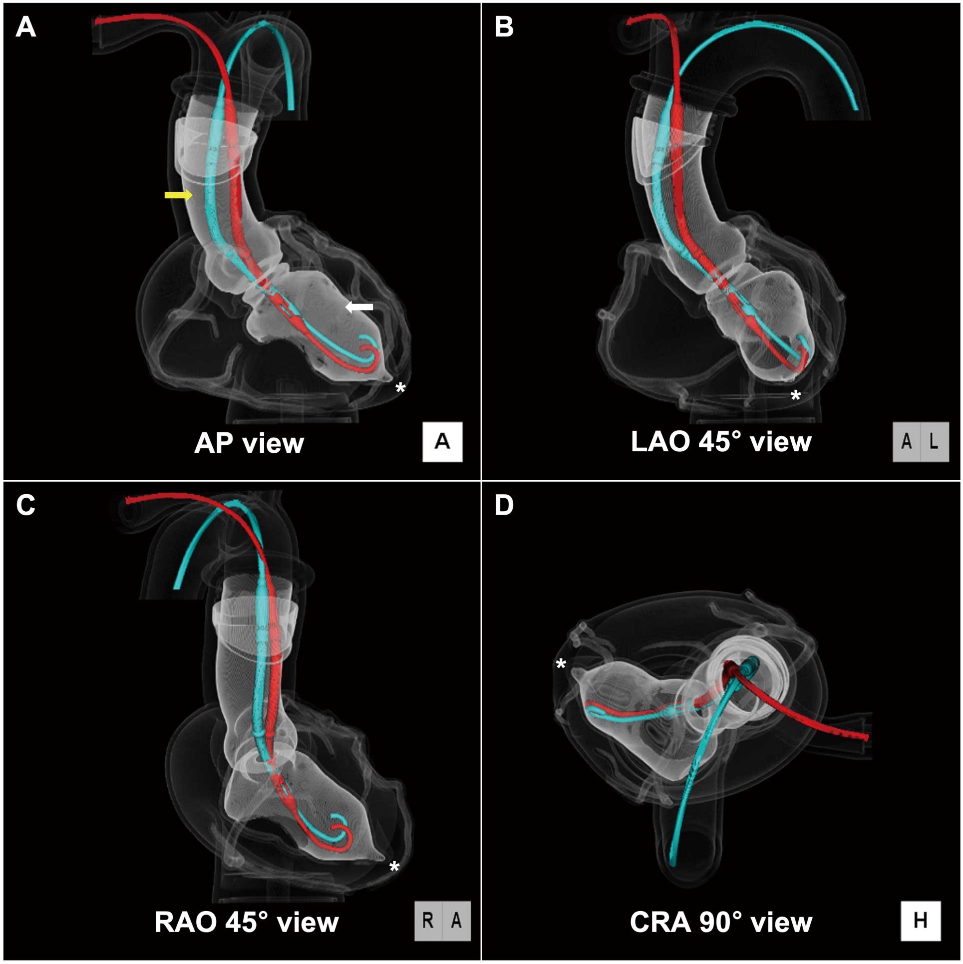
(A) Anterior-posterior (AP), (B) left anterior oblique (LAO) 45°, (C) right anterior oblique (LAO) 45°, and (D) cranial oblique (CRA) 90° views. The red and light blue lines indicate the Impella CP inserted through the axillary and femoral arteries, respectively. The white and yellow arrows indicate the left ventricular and ascending aorta, respectively. The asterisks indicate the left ventricular apex.
The authors are grateful to Mr. Yukihiro Enchi and Mr. John Martin for the 3-dimensional computed tomographic imaging and grammatical assistance, respectively.
None of the authors have any conflicts of interest to declare.
Supplementary Movie. The superimposed 3-dimensional rotational computed tomographic images of the HEARTROID with the impella CP inserted through the axillary and femoral arteries.
Please find supplementary file(s);
http://dx.doi.org/10.1253/circrep.CR-21-0164