Images in Cardiovascular Medicine
Pneumopericardium Resulting After Pericardiocentesis
2023 年 5 巻 4 号 p. 164-165

詳細
2023 年 5 巻 4 号 p. 164-165
A 93-year-old woman with localized scleroderma and on oral prednisolone (5 mg/day) was referred to our hospital because of dyspnea and pericardial effusion. Because the patient presented with precardiac tamponade, an apical-approach echo-guided pericardiocentesis was performed.1 After the puncture, the patient reported relieved symptoms, and no hemodynamic instability was observed. However, the initial chest X-ray after the puncture showed a further enlarged cardiac silhouette (Figure A,B), and chest computed tomography revealed air in the pericardial sac (Figure C,D). A localized pneumothorax with pleural adhesions was found around the puncture site. Pneumopericardium occurred because of direct pleura-pericardial communication (Figure E). With pericardial drainage and an increased dosage of oral prednisolone (20 mg/day), the amount of pericardial drainage gradually decreased. After 7 days, the tube was removed, and the patient was discharged with no pneumopericardium or pneumothorax (Figure F). The catheter was dorsal to the heart, and the air in the pericardial sac remained upward, which could have caused the air to be spontaneously absorbed, in addition to being drained.2 In this case, pneumopericardium could be treated like pericardial effusion. The abundance and, in particular, the speed of constitution of pneumopericardium are the main determinants of clinical severity and will guide the therapeutic strategy. In cases of tension pneumopericardium, definitive management generally involves a pericardial window procedure.
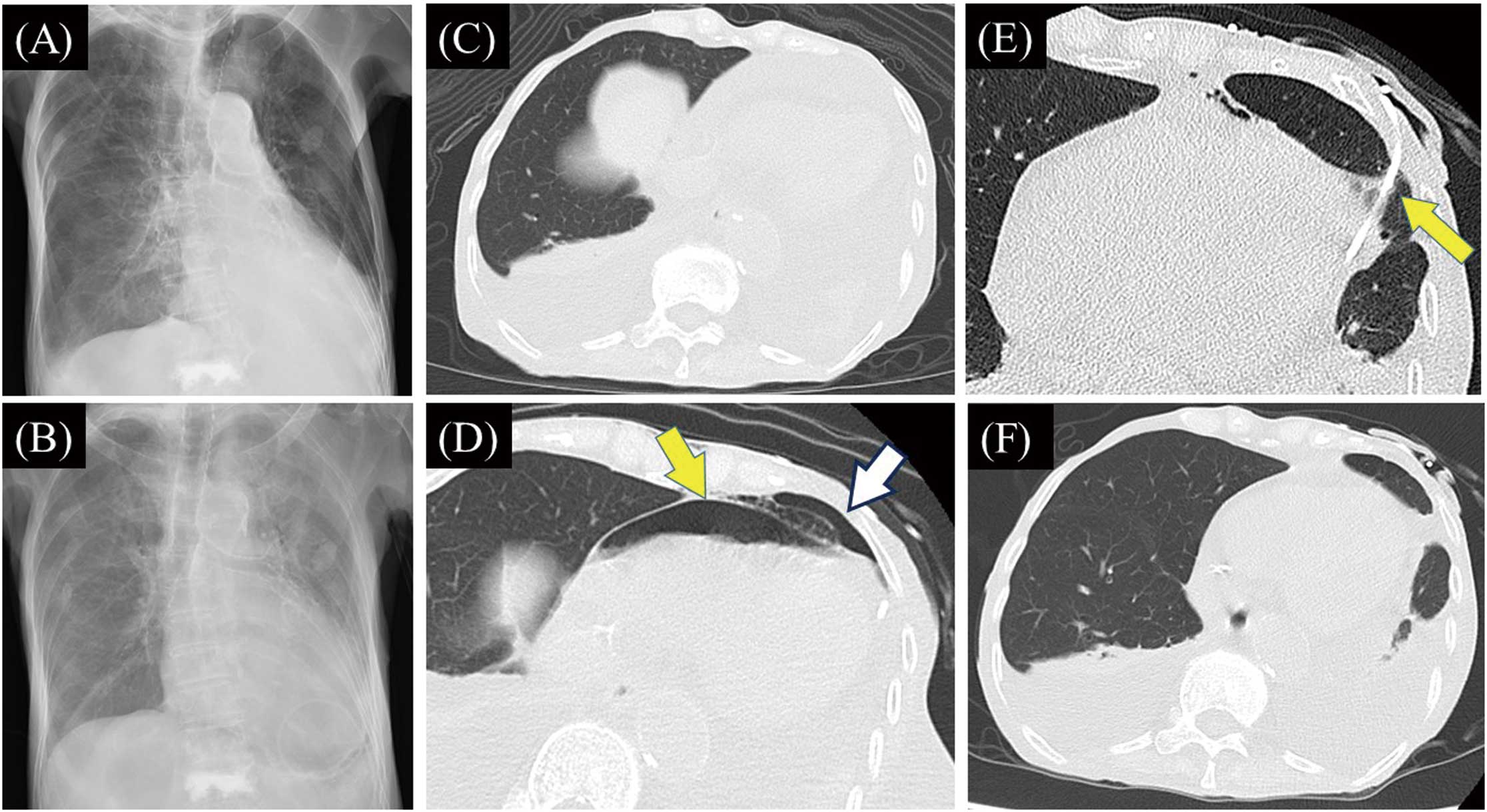
(A,B) Chest X-rays. After the pericardiocentesis, a further enlarged cardiac silhouette (B) was seen than before the procedure (A). (C,D) Chest computed tomography revealed air in the pericardial sac and pneumothorax around the puncture site (D), which was not seen on admission (C). (E) The catheter had penetrated the patient’s lung tissue (arrow). (F) Pneumopericardium and pneumothorax had disappeared 7 days after the pericardiocentesis.
K.T. is a member of Circulation Reports’ Editorial Team.
This study was approved by Shimane University Faculty of Medicine (No. 6174).