Images in Cardiovascular Medicine
Unusual Case of Adult-Onset Congestive Heart Failure Due to Long-Lasting Cardiac Volume Overload Caused by Spinal Epidural Arteriovenous Fistulas
2023 年 5 巻 4 号 p. 162-163

詳細
2023 年 5 巻 4 号 p. 162-163
A 72-year-old woman without significant cardiovascular risk was admitted to our hospital due to worsening dyspnea over several months. Echocardiography showed an ejection fraction (EF) of 35%, left ventricular (LV) diastolic/systolic dimension of 65/54 mm, moderate mitral regurgitation due to tethering, and tricuspid regurgitation with pulmonary hypertension. The inferior vena cava was dilated. Further examination revealed a fine continuous murmur at the back, around the Th10 area. Once the symptoms of congestive heart failure improved with the administration of diuretics, a coronary angiogram and right heart catheterization were performed. The patient’s coronary artery was normal. The cardiac index was high (5.5 L/min/m2) without cardiac shunt. We suspected an extracardiac shunt, and thoracic computed tomography was performed, revealing abnormally dilated intercostal and azygos veins, suggestive of a high-flow arteriovenous shunt (Figure A). An intercostal artery (IA) angiogram demonstrated fistulas at the venous pouches (VP) at the Th10–11 levels. These drained into the epidural venous plexus, the Th9–11 intercostal veins, and finally into the azygos vein. The major feeders of the Th10 VP were the spinal branches of the right Th10 IA (Figure B). The Th11 VP was mainly supplied by the left Th10/11 (Figure C) and Th12 IA spinal branches (Figure D). Transvenous embolization was performed using coils and 25% n-butyl 2 cyanoacrylate via the transazygos approach (Figure E–G). The patient’s symptoms improved, with the LV diastolic dimension decreasing from 65 to 52 mm, EF improving from 35% to 60%, and B-type natriuretic peptide concentrations decreasing from 1,492 to 157 pg/mL.
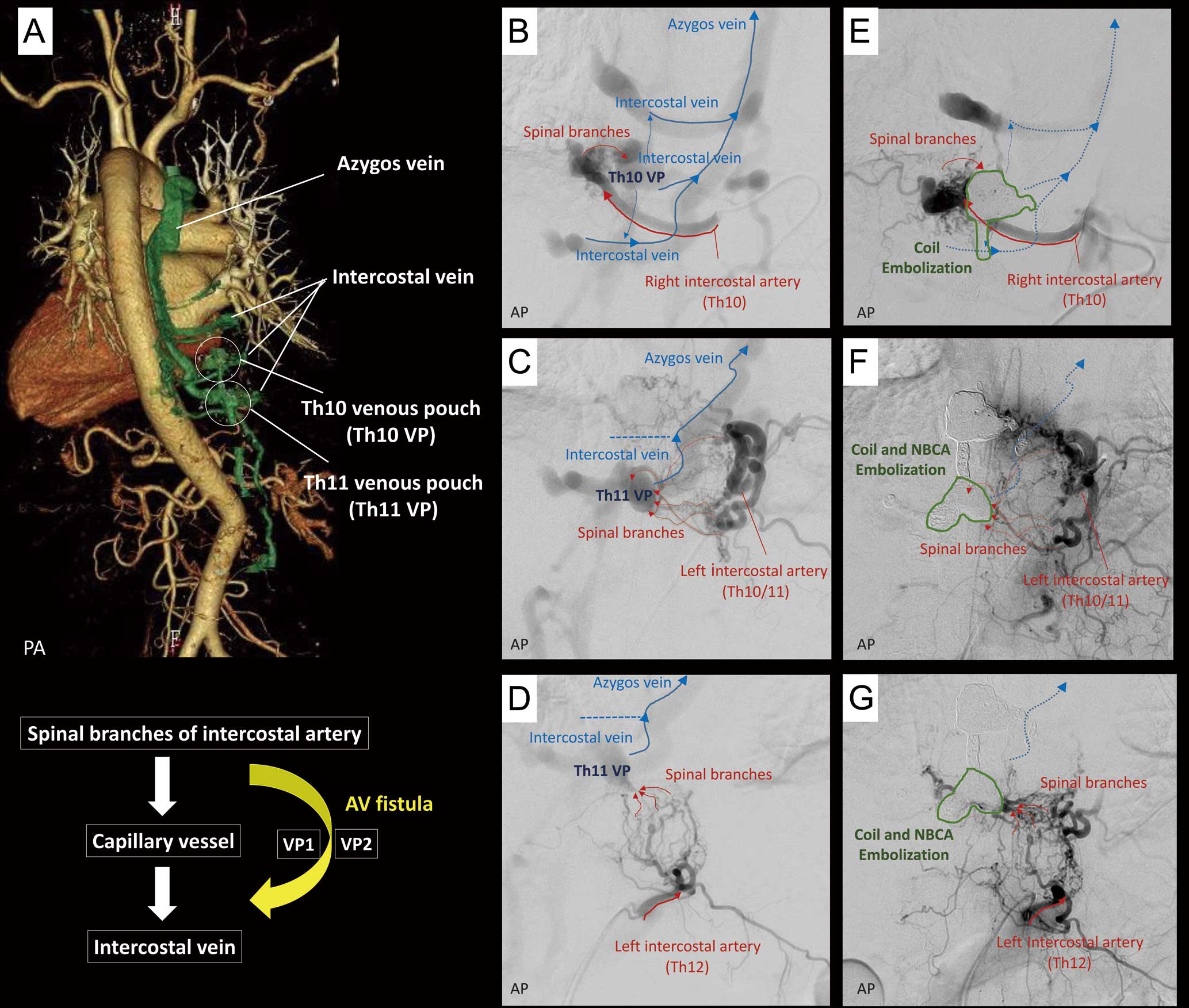
(A) Thoracic computed tomography. (B–D) Intercostal artery angiogram. (E–G) Angiogram after transvenous embolization. Dotted arrows indicate the previously opacified intercostal veins before embolization. AP, antero-posterior view; AV, arteriovenous; NBCA, n-butyl 2 cyanoacrylate; PA, postero-anterior view; VP, venous pouch.
We encountered an unusual case of congestive heart failure due to cardiac volume overload caused by spinal epidural arteriovenous fistulas without typical symptoms, such as radiculopathy or myelopathy.
The authors declare that there are no conflicts of interest.