Caseous Calcification of the Mitral Annulus With Multiple Embolic Strokes
論文ID: CR-24-0188
この記事には本公開記事があります。

詳細
論文ID: CR-24-0188
An 80-year-old woman with history of chronic heart failure and hypertension presented with fever and conscious disturbance. Brain magnetic resonance imaging (MRI) showed multiple embolic strokes. Transthoracic and transesophageal echocardiography revealed no obvious vegetation and all blood cultures were negative. Significant calcification in the posterior mitral annular region with an underlying echolucent area without acoustic shadow and a semilunar echolucent mass located in posterior region was noted in parasternal long-axis and short-axis view (Figure A,B), respectively, indicating a diagnosis of caseous calcification of the mitral annulus (CCMA).1 Computed tomography (CT; Figure C) and cardiac MRI (Figure D) were performed, and both findings was compatible with the diagnosis of CCMA. A brain CT revealed multiple new small calcium deposits in the brain parenchyma, which was unusual for physiologic/age-related intracranial calcification (Figure E), suggesting the correlation between the debris of CCMA and an embolic stroke.2 Three days after admission, the patient was found drowsy and a brain MRI showed multiple new minor embolic strokes. Surgical intervention was considered but withheld due to high risk of morbidity and mortality. Supportive care was maintained and the patient’s clinical course was uneventful thereafter. She was discharged without neurological sequelae.
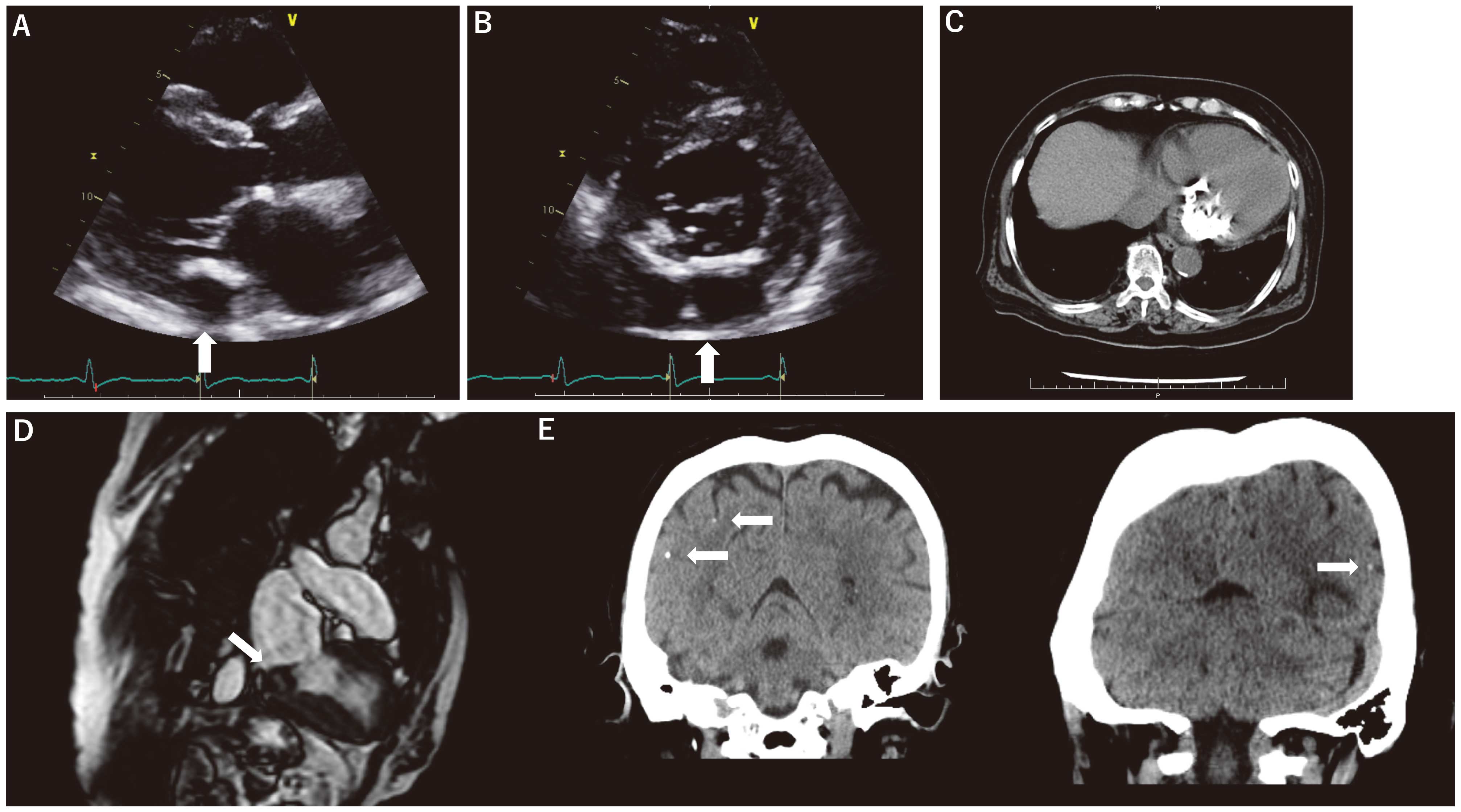
(A,B) Echolucent area (arrow) without acoustic shadow found using transthoracic echocardiography. (C) Computed tomography (CT) revealed significant calcification around the mitral annulus, most remarkable in the posterior annulus. (D) Posterior annulus calcification with central liquification (arrow) in steady-state free-precession cine magnetic resonance imaging. (E) Small calcium deposits (arrow) scattered in brain parenchyma in CT.
CCMA is known as a rare variant of mitral annular calcification, which is usually asymptomatic and found incidentally using cardiac imaging. Mitral valve dysfunction and systemic embolism can be a complication of CCMA, and therefore requires careful attention. To our knowledge, this is the first report to clearly demonstrate caseous material in brain CT.
None.
Not applicable.