Abstract
Objectives: Andrographis paniculata (A. paniculata) is a widely used herb that has potential medical properties. Andrographolide (Andro) is the major component of A. paniculata. We evaluated the anti-tumor activity of Andro using leukemic cell line cells.
Methods: Leukemic cell lines U937, HL60 or H929 cells were cultured in the presence or absence of Andro and compared with the effects of Ara-C or vincristine. The anti-tumor activity was assessed by morphological observations of the cells, DNA fragmentation, MTT assay, Annexin V positive rate, caspase-3/7 activity, and cell cycle analysis.
Results: After addition of Andro, the morphology of cells changed to characteristic shapes with apoptotic bodies. Furthermore, the Annexin V positive rate and caspase-3/7 activities were increased compared with untreated cells. The G1 phase of cell cycle was also similarly increased compared with cells treated with Ara-C.
Conclusions: Our results show that Andro has an anti-tumor activity against leukemic cell lines, very possibly by inducing apoptosis.
Introduction
Medicinal plants and plant-derived drugs are potential sources of alternative medicine used to treat various health conditions.1 Plant-derived natural products have served as important drugs in the field of cancer chemotherapy. The plant-derived anticancer drugs vincristine (VCR) and vinblastine have brought invaluable contributions in modern medicine, and function by inhibiting the separation of cell chromosomes.2–4
Andrographis paniculata (A. paniculata), a plant found in China, India and Thailand, is a famous and widely used medicinal herb plant worldwide. Andrographolide (Andro) is the major component of A. paniculata and is mainly concentrated in leaves.1 Andro has been reported to exhibit therapeutic effects in a variety of diseases.5 This plant is considered as an important source of phytomedicine to treat a wide range of diseases, such as respiratory infection, fever, bacterial dysentery and diarrhea.5
Previous studies showed that Andro inhibits proliferation and induces apoptosis in leukemia cell line cells.6 Furthermore, Andro has been shown to exhibit anti-inflammatory activity, through inhibition of lipopolysaccharide (LPS)-induced TNF-α and interleukin-6 (IL-6).7 Recently, the extract of A. paniculata is being used as a nutritional supplement to prevent inflammatory disease in Taiwan.8 In solid tumors, many studies have reported anti-tumor activities of Andro,9–12 but few have examined its effects in leukemic cells. Hence, in this study, we evaluated the anti-tumor activities of Andro on human leukemic cell line cells. Cytosine β-D-arabinofuranoside (Ara-C) is an anti-cancer chemotherapy drug for leukemia, including acute myelogenous leukemia. We focused on the anti-tumor mechanism of Andro compared with cytosine β-D-arabinofuranoside (Ara-C) or VCR as a potential chemotherapy drug for leukemia.
Materials and Methods
Materials
Andrographolide (Tokyo Chemical Industry, Tokyo, Japan) was dissolved at 10 mM in ethanol and used at 10–50 μM. Ara-C from SIGMA-ALDRICH (MO, USA) was dissolved at 10 mg/mL in phosphate-buffered saline (PBS; 150 mM NaCl, 10 mM phosphate-buffer, pH 7.2) and used at 1 μg/mL. Ara-C inhibits the synthesis of DNA. VCR was obtained from SIGMA-ALDRICH, dissolved at 1 mg/mL in PBS, and used at 0.1 μg/mL. VCR binds to tubulin and inhibits microtubule assembly. Ara-C or VCR treatment was used at the concentration to induce about 50% apoptosis with reference to previous experiments.13,14
Cell culture
U937 cells (human monocytic leukemia cell line; EC85011440), HL60 cells (human promyelocytic leukemia cell line; EC98070106) and NCI-H929 cells (human IgA-producing plasma cell line; EC95050415) were obtained from DS PHARMA BIOMEDICAL (Osaka, Japan) and grown in RPMI 1640 medium (Sigma-Aldrich) supplemented with 10% fetal bovine serum (FBS; Equitech-Bio Inc., Kerrville, USA), 100 U/mL of penicillin, and 100 μg/mL of streptomycin (GIBCO, Carlsbad, USA) at 37°C with 5% CO2.
Morphological observation and DNA fragmentation
Cells were cultured in the presence or absence of Andro, Ara-C or VCR for 24 h. Cells were then deposited on glass slides by the cytospin method at 40×g for 5 min. The glass slides were fixed with Wright solution and stained with Giemsa solution for observing morphological changes.
For the DNA fragmentation assay, cells (1×10⁶ cells) cultured in the presence or absence of Andro, Ara-C or VCR were collected by centrifugation at 300×g for 5 min and washed once with PBS. The cell pellet was suspended in 100 μL of cell lysis buffer (10 mM Tris-HCl buffer, pH 7.4 containing 10 mM EDTA and 0.5% Triton X-100), kept at 4°C for 10 min, and the cell lysate was centrifuged at 16,000×g rpm for 20 min. The supernatants were incubated with RNase A (0.4 mg/mL; Sigma-Aldrich) at 37°C for 60 min, and then with proteinase K (0.4 mg/mL; Sigma-Aldrich) at 37°C for 60 min. Samples were then incubated with 20 μL of 5 M NaCl and 120 μL of isopropyl alcohol at –30°C overnight. Precipitate was then collected by centrifugation at 16,000×g for 15 min and washed by 70% ethanol twice. The samples were allowed to stand for 5 min in a clean bench to dry, and DNA samples thus obtained were dissolved in TE buffer (10 mM Tris-HCl, pH 7.4 and 1 mM EDTA, pH 8.0), and subjected to 2% agarose gel electrophoresis at 100 V for 45 min. DNA was stained with 0.1 mg/L ethidium bromide solution.
MTT assay
MTT (3-(4,5-dimethylthiazol-2-yl)-2,5-diphenyltetrazolium bromide) assay was performed using a MTT cell proliferation assay kit (Cayman Chemical Company, Ann Arbor, USA). Cells incubated in the presence or absence of Andro, Ara-C or VCR were seeded at a density of 2×10⁴ cells/well in 100 μL of culture medium in a 96-well plate (Becton and Dickinson) and incubated at 37°C for 24 h. Next, 10 μL of MTT reagent was added to each well, and after mixing gently, the cells were incubated at 37°C for 4 h in a CO2 incubator. After removing 85 μL of supernatant, 100 μL of lysis buffer was added and mixed in the cell solution. Optical density was measured (550 nm) using a microplate reader (BIO-RAD, Benchmark, Hercules, USA).
Quantification of apoptosis by Annexin V
Detection of apoptosis was performed using The MuseTM Annexin V and Dead Cell Assay Kit (Merck Millipore Corporation, Darmstadt, Germany) and Muse Cell Analyzer (Merck Millipore Corporation), according to the manufacturer’s protocols. Cells incubated in the presence or absence of Andro, Ara-C or VCR for 24 h were collected by centrifugation (300×g at 4°C for 5 min), suspended in 100 μL of RPMI 1640 medium, and incubated with 100 μL of Annexin V reagent at room temperature for 20 min. Cells were measured by the Muse Cell Analyzer.
Measurement of caspase-3/7 activities
Cells incubated in the presence or absence of Andro, Ara-C or VCR were seeded for 24 h at a concentration of 2×10⁵ cells/mL in a 24-well plate dish (Falcon). Cells were collected by centrifugation (300×g at 4°C for 5 min), and suspended in 50 μl of RPMI 1640 medium. Cells were incubated with 5 μl of caspase-3/7 working solution for 30 min at room temperature. Finally, caspase 7-amino actinomycin D (7-AAD) working solution (2 μL of 7-AAD stock solution, 148 μ1 of 1×Assay Buffer BA) was added, and measured by the Muse Cell Analyzer.
Cell cycle analysis
Cells (1×10⁶ cells in 3 mL) incubated in the presence or absence of Andro, Ara-C or VCR for 24 h were collected by centrifugation (300×g at room temperature for 5 min), resuspended in 30 μL of PBS and fixed by 200 μL of 80% ethanol (final 70% ethanol concentration) for over 3 h at –20°C. After fixation, cell pellets obtained by centrifugation (300×g, 5 min) were suspended in 500 μL of PBS, and incubated with 200 μL of Muse Cell Cycle Reagents in the dark for 30 min and measured by the Muse Cell Analyzer.
Statistical analysis
Data were analyzed using Excel software and the student’s t-test was used to assess statistical significance between treatments. Results are expressed as mean±SD of three independent experiments. P<0.05 was considered as statistically significant.
Results
Morphological changes and detection of nuclear DNA fragmentation
Chromatin structure and nuclear fragmentation were observed by Wright-Giemsa staining in U937 cells incubated with Ara-C (1.0 μg/mL) or Andro (50 μM) for 24 h. The morphological changes by Andro resembled the changes of DNA fragmentation induced by Ara-C treatment (Figure 1). The appearance of DNA fragmentation was observed by agarose gel electrophoresis in U937, HL60 and H929 cells treated with Ara-C (1.0 μg/mL), VCR (0.1 μg/mL) or Andro (50 μM) (Figure 2).
Inhibition of cell proliferation by MTT assay
We performed MTT assay to measure the inhibition rate of cell proliferation and viability of the cells incubated with Ara-C, VCR or Andro for 24 h. As shown in Figure 3, the proliferation of all cells was inhibited by the treatment with Ara-C, VCR or Andro for 24 h. Inhibition rates of Andro (50 μM for 24 h) on cell proliferation in each leukemic cell line were 85.5% in U937 cells, 76.3% in HL60 cells and 86.5% in H929 cells compared with untreated cells. These rates were higher than those of Ara-C (1.0 μg/mL) and VCR (0.1 μg /mL) treatment.
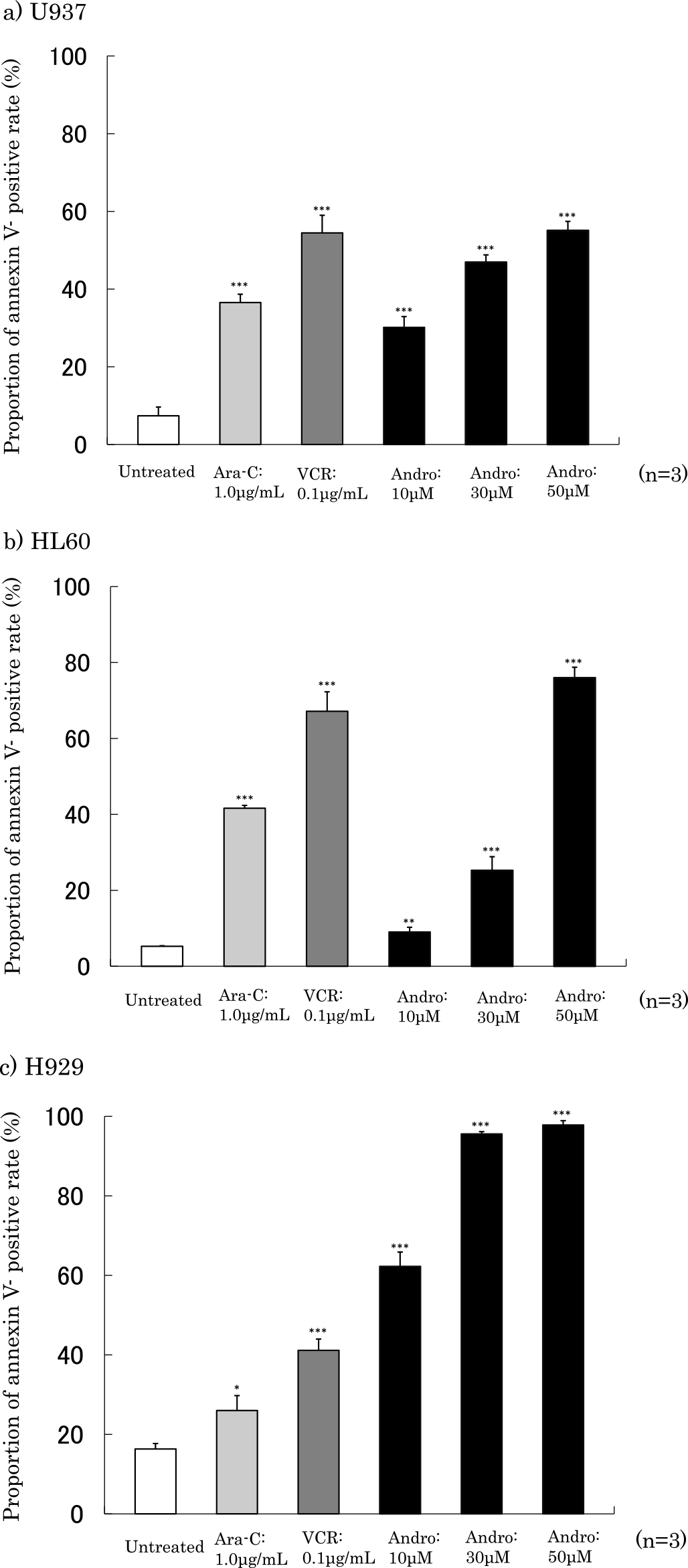
Detection of Annexin V-positive cells
Proportions of annexin V-positive cells after 24 h of treatment with Ara-C, VCR and Andro are shown in Figure 4. The proportions of Annexin V positive cells in cells incubated with Andro (50 μM) were 55.1% in U937 cells, 76.0% in HL60 cells and 97.8% in H929 cells. This rate was increased in a concentration-dependent manner.
Dot plots of Annexin V-positive cells showed a clearly increase after addition of Ara-C, VCR or Andro compared with untreated cells (Figure 5).
Activation of caspase-3/7
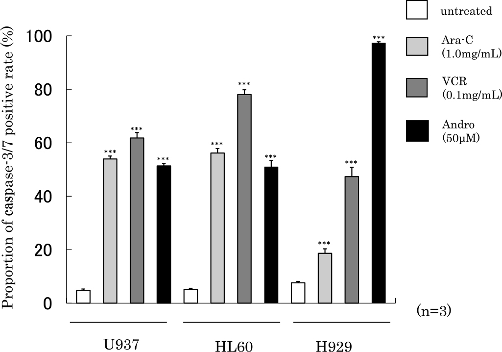
We next examined caspase-3/7 activities after 24 h of treatment with Ara-C, VCR or Andro in U937, HL60 or H929 cells (Figure 6). The proportions of caspase-3/7 positive activities after addition of Andro was 51.4% in U937 cells, 51.0% in HL60 cells and 97.3% in H929 cells. We observed a similar increase after addition of Ara-C or VCR compared with untreated cells.
Analysis of cell cycle
After addition of Andro, the percent of cells in the G1 phase of cell cycle was 50.1% in U937 cells, 42.1% in HL60 cells or 53.8% in H929 cells (Figure 7), indicating an increase in G1 phase cells compared with each untreated cell. As a positive control, we confirmed increase of G1 phase after addition of Ara-C in each leukemic cell line, and increase of G2/M phase after addition of VCR in U937 or H929 cells.
Discussion
A. paniculata grows widely in many Asian countries and is a potential medical plant. The extracts of A. paniculata have been shown to possess many pharmacological effects such as anti-cancer and anti-inflammatory activities.15 The major bioactive component extracted from A. paniculata is Andro, a diterpenoid lactone with three hydroxyls at C-3, C-19 and C-14.16 In this study, we examined the anti-tumor mechanism of Andro, focusing on apoptotic induction by Andro in leukemic cells.
Apoptosis is characterized by a series of typical morphological features, such as shrinkage of the cell, fragmentation into membrane-bound apoptotic bodies. Addition of Andro was shown to change the morphology to that of apoptotic cells, and we also detected DNA fragmentation in treated leukemic cells.
In MTT assay, the proliferation of leukemic cells was significantly inhibited compared with untreated cells after Andro treatment. Annexin V allows for the quantitative of early or late apoptosis. In early apoptosis, these are externalization of phosphatidyl serine to the cell surface. Annexin V is a calcium-dependent phospholipid-binding protein with a high affinity for phosphatidyl serine. In late apoptosis, cleavage and degradation of specific cellular proteins occurs, as well as fragmentation of nuclear chromatin and loss of membrane integrity. 7-AAD is a dead cell marker, and allows late apoptosis. The Annexin V positive rate was shown to increase significantly as compared with untreated cells after Andro treatment in leukemic cells.
Caspases, a family of cysteine proteases, are crucial mediators of apoptosis. Caspases are present as inactive pro-enzymes that are activated by proteolytic cleavage. Caspases have been subclassified by their mechanism of action and are either initiator caspases (caspase-8 and -9) or executioner caspases (caspase-3, -6, and -7). The activation of caspase 3/7 induces DNA fragmentations. The Caspase-3/7 positive rate was shown to increase significantly as compared with untreated cells after Andro treatment in leukemic cells.17–19
The cell cycle is important for chemotherapy, because many chemotherapeutic drugs work on cells that are actively replicating. Ara-C damages DNA when the cell cycle in the S phase (synthesis of DNA). VCR inhibits microtubule assembly by binding tubulin and producing a transient G2/M phase block. The Muse cell cycle kit includes the nuclear DNA intercalating stain propidium iodide (PI). In the cell cycle, G0/G1 phase contain two copies of each chromosome, and the G2/M phase contain four copies of double chromosome. The G1 phase of cell cycle was increased after addition of Andro as same as Ara-C treatment in leukemic cells.
The present results, including the morphological observation of nuclear fragmentation using Wright-Giemsa and the occurrence of DNA fragmentation observed using agarose electrophoresis, inhibition of cell proliferation by MTT assay, increase in annexin V-positive cells, and increase in caspase 3/7 activities by Andro treatment, suggested the occurrence of apoptosis in U937, HL60 and H929 cells upon treatment with Andro. Ara-C and VCR, basic anti-cancer drugs used for leukemic treatment, were used as a positive control.
Many reports have examined the anti-tumor activity of Andro. Andro has been reported to induce apoptosis with cell cycle arrest, and its mechanism occurred via a mitochondria-dependent pathway in human acute promyelocytic leukemic cell line HL-60 cells.20 Andro was reported to induce apoptosis in the T-cell acute lymphoblastic leukemia cell line Jurkat.21
In this study, we observed the highest annexin V positive rate in H929 leukemic cell. H929 is a human multiple myeloma cell established from myeloma (IgA kappa) at relapse. Multiple myeloma is an incurable plasma cell malignancy. In the last decades, significant changes were achieved in the field of multiple myeloma treatment. A cardinal change came with the introduction of so-called novel agents bortezomib, thalidomide and lenalidomide.22 Bortezomib is a proteasome inhibitor proven to be clinically useful for multiple myeloma and shows anti-tumor activity through inhibition of NF-kB pathway.23 Andro, a novel nuclear factor-kB inhibitor, was reported to inhibit the growth of multiple myeloma cells.24 Andro has shown to inhibit cancer stem cell activity in multiple myeloma and to inhibit multiple myeloma cells.25
Our findings showed that Andro exhibits anti-tumor activity in leukemic cells by inducing apoptosis. Although the present study focuses on in vitro analyses, our results suggest that Andro is an interesting pharmacophore with anti-cancer activity, and hence has the potential for being developed as a cancer therapeutic agent.
Conflict of Interest
The authors declare no competing financial interests.
Acknowledgments
This work was supported by Institute of Health and Immunology Science.
References
- 1. Varma A, Padh H, Shrivastava N. Andrographolide: a new plant-derived antineoplastic entity on horizon. Evid Based Complement Alternat Med 2011; 10: 1–9.
- 2. Cragg GM, Newman DJ. Plants as a source of anti-cancer agents. J Ethnopharmacol 2005; 100: 72–79.
- 3. Palem PP, Kuriakose GC, Jayabaskaran C. An endophytic fungus, talaromyces radicus, isolated from catharanthus roseus, produces vincristine and vinblastine, which induce apoptotic cell death. PLoS One 2015; 11: 1–22.
- 4. Rajabalian S. Methanolic extract of Teucrium polium L. potentiates the cytotoxic and apoptotic effects of anticancer drugs of vincristine, vinblastine and doxorubicin against a panel of cancerous cell lines. Exp Oncol 2008; 30: 133–138.
- 5. Hossain MS, Urbi Z, Sule A, Hafizur Rahman KM. Andrographis paniculata (Burm. f.) Wall. ex Nees: a review of ethnobotany, phytochemistry, and pharmacology. Scientific World Journal 2014; 24: 1–50.
- 6. Kumar D, Das B, Sen R, Kundu P, Manna A, Sarkar A, Chowdhury C, Chatterjee M, Das P. Andrographolide analogue induces apoptosis and autophagy mediated cell death in U937 cells by inhibition of PI3K/Akt/mTOR pathway. PLoS One 2015; 5: 1–39.
- 7. Parichatikanond W, Suthisisang C, Dhepakson P, Herunsalee A. Study of anti-inflammatory activities of the pure compounds from Andrographis paniculata (burm.f.) Nees and their effects on gene expression. Int Immunopharmacol 2010; 10: 1361–1373.
- 8. Lu WJ, Lee JJ, Chou DS, Jayakumar T, Fong TH, Hsiao G, Sheu JR. A novel role of andrographolide, an NF-kappa B inhibitor, on inhibition of platelet activation: the pivotal mechanisms of endothelial nitric oxide synthase/cyclic GMP. J Mol Med (Berl) 2011; 89: 1261–1273.
- 9. Mishra SK, Tripathi S, Shukla A, Oh SH, Kim HM. Andrographolide and analogues in cancer prevention. Front Biosci 2015; 7: 256–266.
- 10. Bao GQ, Shen BY, Pan CP, Zhang YJ, Shi MM, Peng CH. Andrographolide causes apoptosis via inactivation of STAT3 and Akt and potentiates antitumor activity of gemcitabine in pancreatic cancer. Toxicol Lett 2013; 222: 23–35.
- 11. Zhang C, Qiu X. Andrographolide radiosensitizes human ovarian cancer SKOV3 xenografts due to an enhanced apoptosis and autophagy. Tumour Biol 2015; 36: 8359–8365.
- 12. Banerjee M, Chattopadhyay S, Choudhuri T, Bera R, Kumar S, Chakraborty B, Mukherjee SK. Cytotoxicity and cell cycle arrest induced by andrographolide lead to programmed cell death of MDA-MB-231 breast cancer cell line. J Biomed Sci 2016; 23: 1–17.
- 13. Akiyama H, Ino T, Tokunaga E, Katsuda I, Ezaki K. A synergistic increase of apoptosis utilizing Fas antigen expression induced by low doses of anticancer drug. Rinsho Byori 2003; 51: 733–739.
- 14. Akiyama H, Endo M, Matsui T, Katsuda I, Emi N, Kawamoto Y, Koike T, Beppu H. Agaritine from Agaricus blazei Murrill induces apoptosis in the leukemic cell line U937. Biochim Biophys Acta 2011; 1810: 519–525.
- 15. Rajagopal S, Kumar RA, Deevi DS, Satyanarayana C, Rajagopalan R. Andrographolide, a potential cancer therapeutic agent isolated from Andrographis paniculata. J Exp Ther Oncol 2003; 3: 147–158.
- 16. Shen YC, Chen CF, Chiou WF. Suppression of rat neutrophil reactive oxygen species production and adhesion by the diterpenoid lactone andrographolide. Planta Med 2000; 66: 314–317.
- 17. Mishra SK, Tripathi S, Shukla A, Oh SH, Kim HM. Andrographolide and analogues in cancer prevention. Front Biosci (Elite Ed) 2015; 7: 255–266.
- 18. Pratheeshkumar P, Sheeja K, Kuttan G. Andrographolide induces apoptosis in B16F-10 melanoma cells by inhibiting NF-κB-mediated bcl-2 activation and modulating p53-induced caspase-3 gene expression. Immunopharmacol Immunotoxicol 2012; 34: 143–151.
- 19. Yang L, Wu D, Luo K, Wu S, Wu P. Andrographolide enhances 5 fluorouracil-induced apoptosis via caspase-8-dependent mitochondrial pathway involving p53 participation in hepatocellular carcinoma (SMMC 7721) cells. Cancer Lett 2009; 276: 180–188.
- 20. Cheung HY, Cheung SH, Li J, Cheung CS, Lai WP, Fong WF, Leung FM. Andrographolide isolated from Andrographis paniculata induces cell cycle arrest and mitochondrial-mediated apoptosis in human leukemic HL-60 cells. Planta Med 2005; 71: 1106–1111.
- 21. Yang T, Yao S, Zhang X, Guo Y. Andrographolide inhibits growth of human T-cell acute lymphoblastic leukemia Jurkat cells by downregulation of PI3K/AKT and upregulation of p38 MAPK pathways. Drug Des Devel Ther 2016; 10: 1389–1397.
- 22. Štork M, Krejčí M, Sandecká V, Král Z, Pour L. Use of new drugs within primary therapy of multiple myeloma. Vnitr Lek 2016; 62: 413–422.
- 23. Montagut C, Rovira A, Albanell J. The proteasome: a novel target for anticancer therapy. Clin Transl Oncol 2006; 8: 313–317.
- 24. Gao H, Wang J. Andrograholide inhibits multiple myeloma cells by inhibiting the TLR/NF-kB signaling pathway. Mol Med Rep 2016; 13: 1827–1832.
- 25. Gunn EJ, Williams JT, Huynh DT, Iannotti MJ, Han C, Barrios FJ, Kendall S, Glackin CA, Colby DA, Kirshner J. The natural products parthenolide and andrographolide exhibit anti-cancer stem cell activity in multiple myeloma. Leuk Lymphoma 2011; 52: 1085–1097.