Abstract
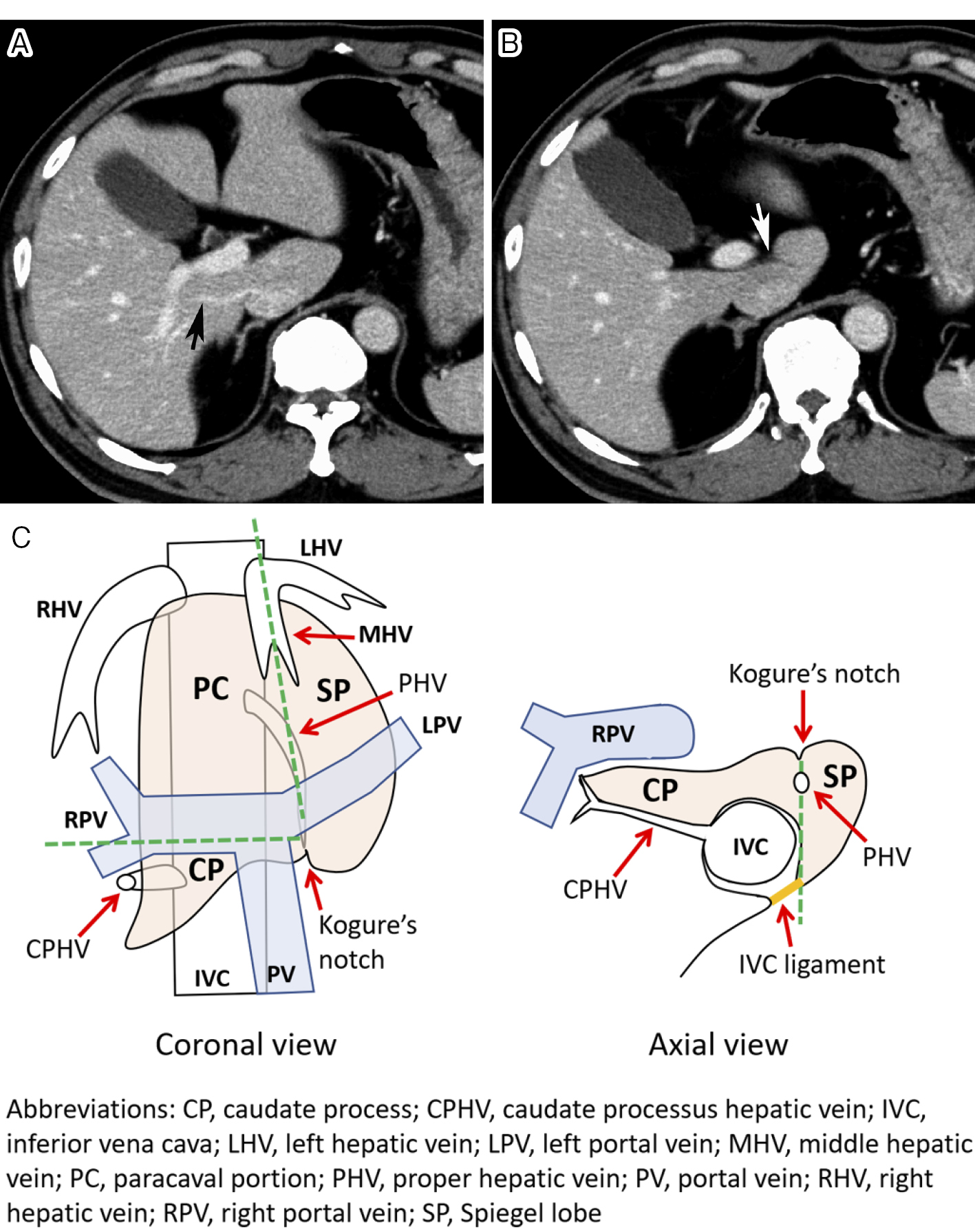
The caudate lobe is located between the bilateral hepatic lobes and is divided into three subsegments: the Spiegel lobe, paracaval portion, and caudate process. The caudate artery arises from various sites of the bilateral hepatic arteries as an independent branch, common trunk, or arcade. Extrahepatic arteries can enter the caudate lobe mainly by the right inferior phrenic artery. The caudate artery also supplies the main bile duct and posterior aspect of segment IV. Although catheterization into the caudate artery is occasionally difficult because of its small size and sharp angulation, selective embolization of a tumor feeder is a significant prognostic factor in patients with hepatocellular carcinoma originating there. Therefore, we should recognize the peculiarity of its vascular anatomy and should be familiar with catheterization and embolization techniques.