Japanese Journal of Medical Technology
Online ISSN : 2188-5346
Print ISSN : 0915-8669
ISSN-L : 0915-8669
Part 3
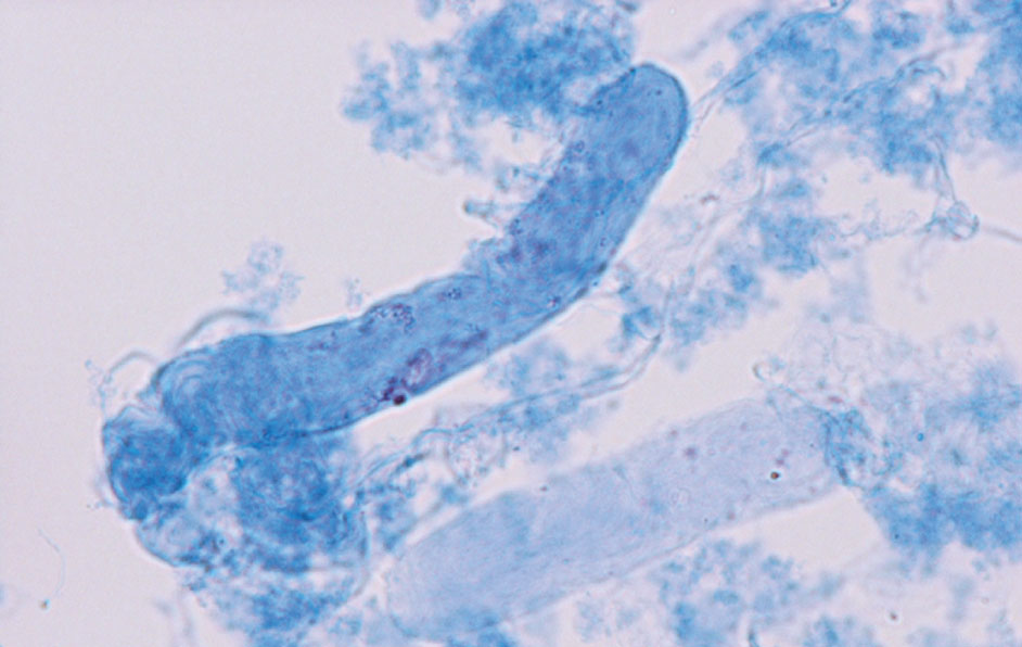
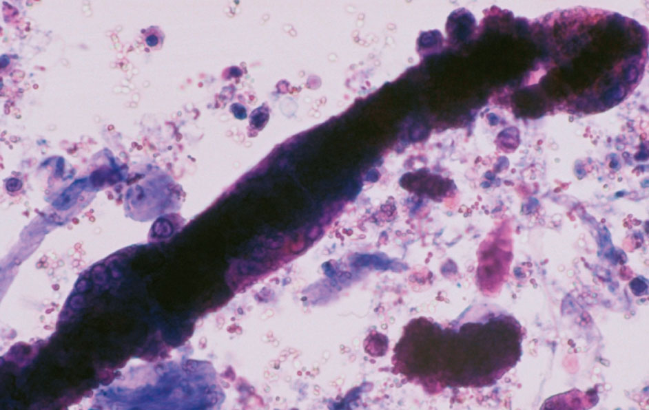
Atlas of Urinary Sediment: IV Casts
Japanese Association of Medical Technologists; Editorial Committee of the Special Issue: Urinary Sediment
Author information
JOURNAL FREE ACCESS FULL-TEXT HTML

2017 Volume 66 Issue J-STAGE-1 Pages 123-141

Details
Abstract

尿沈渣検査は,尿中に出現する成分を尿の遠心操作にて得られた沈殿物を観察する検査である。尿沈渣の標本作成における操作が単純であるにもかかわらず,尿沈渣に出現する成分は多種多様であるため,鑑別が非常に複雑である。その要因としては,尿沈渣に出現する尿中有形成分が,ひとつの成分においても様々な形態で存在することがある。たとえばシュウ酸カルシウム結晶では正八面体型とビスケット型,コマ型などが存在し,尿細管上皮細胞に至っては基本型,特殊型と細胞形態のバリエーションが多岐にわたる。このように尿沈渣検査では成分を正しく鑑別するための知識と技術が必要である。この部では,尿沈渣に出現する基本的な尿中有形成分を鑑別する知識を習得するために,最も基本となる成分の写真について「尿沈渣検査法2010」の尿沈渣アトラスを引用(一部改編)し掲載する。また「*」でマークした写真は,尿沈渣成分の新たな情報として追加したものである.この尿沈渣アトラスを利用し,各成分の特徴を捉えることをしっかりと身につけ,今後遭遇するであろう鑑別困難な成分に対しても対処できるよう,基礎知識を学習することを目的とする。

IV  円柱類   Casts
Figure 3.211 

硝子円柱 40× 無染色

Hyaline cast 40× No staining

基質内には,有形成分がみられない。基質構造は薄くみえるため,観察には光源が明るすぎると見落としやすいので注意を要する。

There are no visible components in the matrix. The matrix structures appear thin. Care must be taken during observation because the structure is easily overlooked if the light source is too bright.

Figure 3.212 

腎組織像 20× HE染色

Renal histology 20× HE staining

ネフローゼ症候群症例の腎生検組織像。尿細管腔内にエオジンに好染する硝子円柱がみられる。間質の線維化,炎症細胞の浸潤はみられない。

Renal biopsy tissue image of a nephrotic syndrome case. There are hyaline casts well-stained with eosin in the renal tubular lumens. Interstitial fibrosis and inflammatory cell infiltration are not observed.

Figure 3.213 

硝子円柱 40× 無染色

Hyaline cast 40× No staining

一端が細く類円柱状であるが,明瞭な硝子基質であるため,硝子円柱に分類する。

This is a cylindroid with one thin end. Because it has a clear hyaline matrix, it is classified as a hyaline cast.

Figure 3.214 

硝子円柱 40× S染色

Hyaline cast 40× S staining

基質の一部が,屈曲状を呈する硝子円柱である。

This is a hyaline cast exhibiting a bent shape in a part of the matrix.

Figure 3.215 

硝子円柱 40× S染色

Hyaline casts 40× S staining

染色性が異なる2個の硝子円柱がみられる。基質の蛋白濃度の差や濃縮力の差によって染色の濃淡が異なったものと考えられる。

There are two hyaline casts that exhibit different stainability. The differing shade of staining appears to be due to the difference in the concentration of protein in the matrix and the concentrating power.

Figure 3.216 

硝子円柱,幅広円柱 40× 無染色

Hyaline cast, broad cast 40× No staining

円柱の幅が60 μm以上の硝子円柱である。成分の分類では,硝子円柱と幅広円柱の両成分として報告する。

This is a hyaline cast in which the width is wider than 60 μm. In the classification of components, it is reported as both a hyaline cast and a broad cast.

Figure 3.217 

上皮円柱 40× S染色

Epithelial cast 40× S staining

基質内の尿細管上皮細胞の細胞質は赤紫色に,核は青色に染め出されている。

The cytoplasm of renal tubular epithelial cells in the matrix are stained reddish purple, and the nuclei are stained blue.

Figure 3.218 

腎組織像 40× HE染色

Renal histology 40× HE staining

巣状糸球体硬化症症例の腎生検組織像。尿細管腔内に尿細管上皮細胞を封入した上皮円柱がみられる。間質にリンパ球を主体とした炎症細胞の浸潤がみられる。

Renal biopsy tissue image of focal a glomerular sclerosis case. Epithelial casts encapsulating renal tubular epithelial cells are found in the tubular cavities. Infiltration of inflammatory cells, which are mainly composed of lymphocytes, are observed in the interstitium.

Figure 3.219 

上皮円柱 40× 無染色

Epithelial cast 40× No staining

基質内に尿細管上皮細胞が隙間なく封入された上皮円柱である。よく見ると細胞境界が見える。

This is an epithelial cast that encloses renal tubular epithelial cells without gaps in its matrix. Looking closely, cell boundaries are visible.

Figure 3.220 

上皮円柱 40× S染色

Epithelial cast 40× S staining

Figure 3.219と同様の円柱のS染色像である。核は不明瞭だが,尿細管上皮細胞である。

This is an S-stained image of a cast similar to Figure 3.219. Although the nucleus is unclear, these are renal tubular epithelial cells.

Figure 3.221 

上皮円柱 40× 無染色

Epithelial cast 40× No staining

基質内に3個以上の角柱型や角錐台型の尿細管上皮細胞がみられる上皮円柱である。

An epithelial cast which has more than three renal tubular epithelial cells exhibiting prism and pyramid shapes in the matrix.

Figure 3.222 

上皮円柱 40× 無染色

Epithelial cast 40× No staining

円形・類円形型の尿細管上皮細胞が封入された上皮円柱である。無染色では白血球と鑑別が必要となり,大小不同で細胞質が厚いことから鑑別される。

This is an epithelial cast encapsulating renal tubular epithelial cells of circular or near-circular shapes. When unstained, differentiation from leukocytes is necessary. They are identified as tubular epithelial cells based on the thick cytoplasm and irregular sizes.

Figure 3.223 

上皮円柱 40× 無染色

Epithelial cast 40× No staining

洋梨・紡錘型の尿細管上皮細胞が3個以上封入された上皮円柱である。

This is an epithelial cast that encapsulates more than three renal tubular epithelial cells exhibiting pear/spindle shapes.

Figure 3.224 

上皮円柱 40× S染色

Epithelial cast 40× S staining

Figure 3.223と同様の円柱のS染色像で洋梨・紡錘型の尿細管上皮細胞が確認できる。

Pear/spindle-shaped renal tubular epithelial cells can be confirmed with the S-stained image of the same type of cast as that shown in Figure 3.223.

Figure 3.225 

上皮円柱,幅広円柱 40× 無染色

Epithelial cast, broad cast 40× No staining

基質内に円形・類円形型の尿細管上皮細胞が封入されている。大食細胞との鑑別が必要になるが,細胞質の辺縁が明瞭で核が膨化状であることから鑑別可能である。

In the matrix, circular- and near-circular-shaped renal tubular epithelial cells are enclosed. Differentiation from macrophages is necessary. That is possible because epithelial cells have clear cytoplasm margins and swollen nuclei.

Figure 3.226 

上皮円柱 40× S染色

Epithelial cast 40× S staining

Figure 3.225と同様の円形・類円形型の尿細管上皮細胞が円柱内にみられる。

Circular- and near-circular-shaped tubular epithelial cells, similar to cells shown in Figure 3.225, are found in the cast.

Figure 3.227 

上皮円柱,幅広円柱 40× 無染色

Epithelial cast, broad cast 40× No staining

円柱の辺縁に尿細管上皮細胞(矢印)が3個以上付着または封入された上皮円柱であり,円柱の幅が60 μm以上あるので幅広円柱としても報告する。

This is an epithelial cast on which three or more tubular epithelial cells (arrows) are attached or enclosed on the margin, and it is also reported as a broad cast due to a width of 60 μm or more.

Figure 3.228 

上皮円柱 40× S染色

Epithelial cast 40× S staining

3個の尿細管上皮細胞が円柱に付着または封入されている上皮円柱である。

Three tubular epithelial cells are attached on or enclosed in the epithelial cast.

Figure 3.229 

上皮円柱 40× 無染色

Epithelial cast 40× No staining

ビリルビン色素に着色された尿細管上皮細胞を3個以上封入した上皮円柱である。

An epithelial cast that encloses three or more renal epithelial cells stained with bilirubin pigment.

Figure 3.230 

上皮円柱 40× S染色

Epithelial cast 40× S staining

Figure 3.229と同様の円柱のS染色像である。ビリルビン色素のため本来の染色性とは異なり赤褐色に染色されている。

An S-stained image of a cast similar to Figure 3.229. Due to the bilirubin pigment, the cast is stained reddish brown, unlike the original stainability.

Figure 3.231 

顆粒円柱 40× 無染色

Granular cast 40× No staining

円柱全体に顆粒成分が散在的にあり,全体の1/3以上を占めるため顆粒円柱に分類する。

Granular components are scattered throughout the cast and occupy more than one-third of the region. Thus, it is classified as a granular cast.

Figure 3.232 

顆粒円柱 40× S染色

Granular cast 40× S staining

円柱の一端に顆粒成分が集中してみられ,1/3以上を占めるため顆粒円柱に分類する。

Granular components are concentrated on one end of the cast and occupy at least one-third of it. Thus, it is classified as a granular cast.

Figure 3.233 

顆粒円柱 40× 無染色

Granular cast 40× No staining

やや粗大な顆粒成分で構成された顆粒円柱である。

A granular cast composed of somewhat coarse granule components.

Figure 3.234 

顆粒円柱 40× S染色

Granular cast 40× S staining

S染色で赤紫色に染色された顆粒成分で構成された顆粒円柱である。

A granular cast composed of granular components stained reddish purple by S staining.

Figure 3.235 

顆粒円柱,幅広円柱 40× 無染色

Granular cast, broad cast 40× No staining

全体に微細な顆粒で構成された顆粒円柱である。幅広円柱もあわせて報告する。

A granular cast composed entirely of fine granules. This is also reported as a broad cast.

Figure 3.236 

顆粒円柱,幅広円柱 40× S染色

Granular cast, broad cast 40× S staining

S染色で青紫色に染色された顆粒成分で構成された顆粒円柱である。幅広円柱もあわせて報告する。

A granular cast composed of granular components stained bluish purple by S staining. This is also reported as a broad cast.

Figure 3.237 

ろう様円柱 40× 無染色

Waxy cast 40× No staining

硝子円柱に比べ基質に厚み,光沢があり,均一無構造で輪郭は明瞭である。一部に切れ込みもみられるろう様円柱である。

Compared to a hyaline cast, the matrix is thick and glossy, has uniformly no structure and has a clear outline. This is a waxy cast with a few cuts.

Figure 3.238 

ろう様円柱 40× S染色

Waxy cast 40× S staining

赤紫色に染色され厚みがあり光沢がみられるろう様円柱である。4~6箇所に切れ込みがみられる。

A waxy cast stained reddish purple and is thick and glossy. Four to six notches are seen.

Figure 3.239 

ろう様円柱 40× 無染色

Waxy cast 40× No staining

屈曲,蛇行の形状を呈するろう様円柱である。

Waxy cast exhibiting a bending and meandering shape.

Figure 3.240 

ろう様円柱,幅広円柱 40× 無染色

Waxy cast, broad cast 40× No staining

均一無構造のろう様円柱である。同時に幅広円柱である。

A uniformly unstructured waxy cast. This is also a broad cast.

Figure 3.241 

ろう様円柱 40× 無染色

Waxy cast 40× No staining

大小のイクラ状を呈したろう様円柱である。

Waxy cast shaped like an irregularly-sized salmon roe.

Figure 3.242 

ろう様円柱 40× S染色

Waxy cast 40× S staining

S染色で青紫に染色されているイクラ状のろう様円柱である。硝子円柱に封入されていることから,下部ネフロンで再形成された円柱と推定される。

This is a salmon roe-shaped waxy cast stained bluish purple via S staining. Because it is enclosed in a hyaline cast, it is presumed to be a cast reconstituted in the lower nephron.

Figure 3.243 

ろう様円柱 40× 無染色

Waxy cast 40× No staining

非常に小さいイクラ状を呈したろう様円柱である。数珠玉がつながったようにもみえる(矢印) 。

A waxy cast with a very small salmon roe shape. It looks like connected beads (arrow).

Figure 3.244 

ろう様円柱 40× S染色

Waxy cast 40× S staining

S染色で赤紫に染色されている非常に小さいイクラ状を呈したろう様円柱である。

Waxy cast with a very small salmon roe shape stained reddish purple with S stain.

Figure 3.245 

ろう様円柱 40× 無染色

Waxy cast 40× No staining

輪郭明瞭で角張った形状を示すろう様円柱である。繊維と紛らわしいので誤判定しないように注意する。

A waxy cast with a clear contour and angular shape. Care must be taken not to erroneously identify it as it can be confused with fibers.

Figure 3.246 

ろう様円柱 40× S染色

Waxy cast 40× S staining

Figure 3.245と同様のろう様円柱で,S染色で赤紫に染色されている。

A waxy cast, similar to that shown in Figure 3.245, stained reddish purple with S stain.

Figure 3.247 

脂肪円柱 40× 無染色

Fatty cast 40× No staining

硝子基質の中に光沢のある黒色や黄色調の脂肪顆粒が3個以上封入されている。脂肪顆粒を3個以上封入するものは脂肪円柱に分類する。

Three or more glossy black and yellowish fatty granules are enclosed in the hyaline matrix. Those containing three or more fatty granules are classified as fatty casts.

Figure 3.248 

脂肪円柱 40× S染色

Fatty cast 40× S staining 

青色に染色された硝子基質内に光沢のある脂肪顆粒が3個以上封入されている。 脂肪成分は染色されない。

Three or more glossy fatty granules are enclosed in a hyaline matrix stained blue. The fat components are not stained.

Figure 3.249 

脂肪円柱 40× 無染色

Fatty cast 40× No staining

光沢のある黄色調の脂肪顆粒を全体に満たした卵円形脂肪体を2個封入している。卵円形脂肪体を1個以上含めば脂肪円柱である。

Two oval fat bodies filled with glossy, yellowish fatty granules are enclosed in the fatty cast. If it contains one or more oval fat bodies, it is considered to be a fatty cast.

Figure 3.250 

脂肪円柱 40× 無染色

Fatty cast 40× No staining

硝子基質の中に光沢のある大小の脂肪顆粒が多数封入されている脂肪円柱である。円柱の中に方形板状のコレステロール結晶も封入されているが,1個なのでこれは脂肪円柱とコレステロール結晶として報告する。

A fatty cast filled with a large number of glossy, various-sized fatty granules in the hyaline matrix. A rectangular plate-like cholesterol crystal is also enclosed in the cast, but there is only one cholesterol crystal, so this specimen is reported as a fatty cast and cholesterol crystal.

Figure 3.251 

脂肪円柱 40× 無染色

Fatty cast 40× No staining

基質内に大小の脂肪顆粒が充満した脂肪円柱である。

A fatty cast filled with various sizes of fatty granules in the matrix.

Figure 3.252 

脂肪円柱 40× 偏光顕微鏡像

Fatty cast 40× Polarizing microscope image

Figure 3.251の偏光顕微鏡像である。脂肪顆粒はMaltese crossと呼ばれる重屈折性偏光像を示しており,コレステロールエステルまたはリン脂質と推測される。

A polarizing microscope image of the same fatty cast shown in Figure 3.251. Fatty granules show a birefringence polarized image called a Maltese cross and appear to be cholesterol esters or phospholipids.

Figure 3.253 

脂肪円柱 40× S染色

Fatty cast 40× S staining 

卵円形脂肪体,脂肪顆粒を封入した脂肪円柱である。S染色では脂肪顆粒は染まらないが,卵円形脂肪体の細胞質が赤紫色を示している。

A fatty cast with enclosed oval fat bodies and fatty granules. With S staining, fatty granules are not stained, but the cytoplasm of the oval fat bodies is stained reddish purple.

Figure 3.254 

脂肪円柱 40×  Sudan III染色

Fatty cast 40× Sudan III staining

脂肪顆粒は橙赤色に染色されている。

With Sudan III staining, the fatty granules are stained orange-red.

Figure 3.255 

脂肪円柱 40× 無染色

Fatty cast 40× No staining

基質内の脂肪顆粒を含有した細胞は,辺縁構造が不明瞭なことから大食細胞由来の卵円形脂肪体が考えられる。

Cells containing lipid granules in the matrix appear to be oval fat bodies derived from macrophage cells because the marginal structure is unclear.

Figure 3.256 

脂肪円柱,白血球円柱 40× S染色

Fatty cast, WBC cast 40× S staining

基質内の卵円形脂肪体は,細胞質辺縁構造が不明瞭なことから大食細胞由来が考えられる。また,基質内に2個の好中球と1個の単球がみられる。

Oval fat bodies in the matrix are derived from macrophage cells because the cytoplasmic marginal structure is unclear. The matrix contains two neutrophils and one monocyte.

Figure 3.257 

赤血球円柱 40× S染色

RBC cast 40× S staining

基質内の赤血球がS染色で赤色調を呈している。

The red blood cells in the substrate exhibit a red tone on S staining.

Figure 3.258 

腎組織像 40× HE染色

Kidney histology 40× HE staining

間質性腎炎症例の腎生検組織像。尿細管腔内に赤血球を封入し一部顆粒化した赤血球円柱がみられる。間質にリンパ球を主体とした炎症細胞の浸潤がみられる。

Renal biopsy tissue image of an interstitial nephritis case. A red blood cell cast that the tubular cavity filled with red blood cells and is partially granulated is seen. Infiltration of inflammatory cells mainly composed of lymphocytes is observed in the interstitium.

Figure 3.259 

赤血球円柱 40× 無染色

RBC cast 40× No staining

円柱内に非溶血の赤血球を多数封入した赤血球円柱である。

A red blood cell cast in which a large number of non-hemolytic red blood cells are enclosed.

Figure 3.260 

赤血球円柱 40× S染色

RBC cast 40× S staining

基質内の一部は顆粒状にみえる部分もあるが,広範囲で赤血球の輪郭が確認できるので赤血球円柱として報告する。

Part of the inside of the substrate appears to be granular, but the contours of the red blood cells are confirmed in a wide area. Therefore, it is reported as a red blood cell cast.

Figure 3.261 

赤血球円柱 40× 無染色

RBC cast 40× No staining

円柱内に非溶血の赤血球を多数封入した赤血球円柱である。

A red blood cell cast in which a large number of non-hemolytic red blood cells are enclosed.

Figure 3.262 

赤血球円柱 40× 無染色

RBC cast 40× No staining

赤血球が隙間なく封入されている赤血球円柱である。

A red blood cell cast in which the red blood cells are enclosed without any gaps.

Figure 3.263 

白血球円柱 40× S染色

WBC cast 40× S staining

円柱に封入された細胞は大きさが10 μm程度で多形核を示すことから,白血球(好中球)である。分葉状の核は青色に染め出されている。

Cells enclosed in the cast are leukocytes (neutrophils) because they are approximately 10 μm and polymorphonuclear. The segmented nucleus is stained blue.

Figure 3.264 

腎組織像 20× HE染色

Kidney histology 20× HE staining

尿細管腔内にみられた白血球円柱である。

A white blood cell cast in the renal tubule cavity.

Figure 3.265 

白血球円柱 40× 無染色

WBC cast 40× No staining

約20個の白血球を封入した白血球円柱である。円柱内に封入された多形核を示す白血球(好中球)がみられる。

A white blood cell cast enclosing approximately 20 white blood cells. Polymorphonuclear leukocytes (neutrophils) are enclosed in the cast.

Figure 3.266 

白血球円柱 40× S染色

WBC cast 40× S staining

円柱内に封入された多形核を示す細胞は白血球(好中球)であり,白血球円柱である。

Polymorphonuclear cells enclosed in the cast are leukocytes (neutrophils), and this cast is a white blood cell cast.

Figure 3.267* 

白血球円柱 40× S染色

WBC cast 40× S staining

円柱内にはN/C比大の単核のリンパ球を主体とした白血球がみられる。細胞質の染色性は尿細管上皮細胞に比べ淡染である。

Leukocytes mainly composed of mononuclear lymphocytes with a large N/C ratio are found in the cast. Cytoplasmic staining is paler than for renal tubular epithelial cells.

Figure 3.268 

白血球円柱 40× S染色

WBC cast 40× S staining

円柱内には好中球多形核球と単球およびリンパ球がみられる。

Polymorphonuclear neutrophils, monocytes, and lymphocytes are visible in the cast.

Figure 3.269 

白血球円柱 40× 無染色

WBC cast 40× No staining

基質内の単核の白血球は単球で,褐色から黒色調の顆粒をもつ白血球は好酸球(矢印)である。

Mononuclear leucocytes in the substrate are monocytes, and leukocytes with brown-to-blackish granules are eosinophils (arrows).

Figure 3.270 

白血球円柱 40× Hansel染色

WBC cast 40× Hansel staining

基質内の好酸球はHansel染色で赤色調に染色されている。

Eosinophils in the substrate are stained red with Hansel staining.

Figure 3.271 

白血球円柱 40× S染色

WBC cast  40× S staining

矢印の白血球は好酸球で,残りの多くは単球である。好酸球の顆粒はS染色では染色されない。

Leukocytes indicated by the arrows are eosinophils, and the majority of the remaining cells are monocytes. Eosinophil granules are not stained with S staining.

Figure 3.272 

白血球円柱 40× 無染色

WBC cast 40× No staining

基質内の白血球は好中球より大きく,灰白色調で細胞質は淡く綿菓子状を示しており,単球と判定される。

Leukocytes in the substrate are larger than the neutrophils and have a grayish color. The cytoplasm of the leukocytes is pale and exhibits a cotton candy shape. The leukocytes are judged to be monocytes.

Figure 3.273 

白血球円柱 40× S染色

WBC cast 40× S staining

基質内中央には細胞質の辺縁構造が不明瞭で,核形に切れ込みがある典型的な単球を確認することができる(矢印)。

In the center of the matrix, there is a typical monocyte, with an unclear marginal cytoplasm structure and a nucleus with a cut (arrow).

Figure 3.274 

白血球円柱 40× S染色

WBC cast 40× S staining

尿細管上皮細胞に類似するが,細胞質辺縁構造は不明瞭であることから単球である。

Although they appear similar to renal tubular epithelial cells, they are monocytes because the cytoplasmic marginal structures are unclear.

Figure 3.275 

白血球円柱 40× PB染色

WBC cast 40× PB staining

黒色の成分は好中球で,赤色の成分はリンパ球や崩壊の強い好中球である。PB染色では好中球も赤色を示すことがあり注意する。

The black components are neutrophils. The red components are lymphocytes or severely disintegrated neutrophils. Be aware that neutrophils may also be stained red with PB staining.

Figure 3.276 

空胞変性円柱 40× 無染色

Vacuolar denatured cast 40× No staining

円柱全体が大小の空胞で占められている円柱で空胞変性円柱に分類する。糖尿病性腎症などに多くみられる。

The cast is classified as a vacuolar denatured cast that is occupied by vacuoles of different sizes. Many of these are found in cases of diabetic nephropathy.

Figure 3.277 

空胞変性円柱 40× S染色

Vacuolar denatured cast 40× S staining

赤紫色に染色された空胞変性円柱である。空胞変性円柱は赤紫色のろう様基質であることが多い。

A vacuolar denatured cast stained reddish purple. Vacuolar denatured casts often have reddish purple waxy substrates.

Figure 3.278 

空胞変性円柱 40× S染色

Vacuolar denatured cast 40× S staining

青紫色に染色された空胞変性円柱である。

A vacuolar denatured cast stained bluish purple.

Figure 3.279 

空胞変性円柱 40× S染色

Vacuolar denatured cast 40× S staining

大きな空胞によって円柱の幅が一部広くなっているようにみえる。空胞変性円柱である。

The cast is partially widened by large vacuoles. It is a vacuolar denatured cast.

Figure 3.280 

塩類・結晶円柱 40× 無染色

Salt/crystal cast 40× No staining

リン酸塩を1/3以上封入した円柱であり,塩類・結晶円柱に分類する。

A cast enclosing at least one-third phosphates. It is classified as a salt/crystal cast.

Figure 3.281 

塩類・結晶円柱 40× 無染色

Salt/crystal cast 40× No staining

小形ビスケット状のシュウ酸カルシウム結晶を3個以上封入した円柱であり,塩類・結晶円柱に分類する。赤血球に似ているので注意が必要である。

A cast enclosing three or more small biscuit-like calcium oxalate crystals. It is classified as a salt/crystal cast. Care must be taken as these cells resemble red blood cells.

Figure 3.282 

塩類・結晶円柱 40× 偏光顕微鏡像

Salt/crystal cast 40× Polarizing microscopic image

Figure 3.281の塩類・結晶円柱の偏光顕微鏡像である。結晶類は偏光顕微鏡で確認が容易である。

A polarizing microscopic image of the salt/crystal cast shown in Figure 3.281. Crystals are easily identified with a polarizing microscope.

Figure 3.283 

塩類・結晶円柱 40× S染色

Salt/crystal cast 40× S staining

シュウ酸カルシウム結晶が円柱内に封入されているがS染色では染色されない。

Calcium oxalate crystals are enclosed in the cast. However, they are not stained with S staining.

Figure 3.284 

塩類・結晶円柱 40× 無染色

Salt/crystal cast 40× No staining

橙色で大小不同の尿酸塩が円柱内に封入された塩類・結晶円柱である。赤血球と類似しているので鑑別に注意が必要である。

Urate crystals in orange and various sizes are enclosed in the salt/crystal cast. Attention is required during differentiation as the crystals resemble red blood cells.

Figure 3.285 

大食細胞円柱,脂肪円柱 40× S染色

Macrophage cast, fatty cast 40× S staining

基質内の細胞は細胞質辺縁が不明瞭であることから大食細胞と考えられる。左の細胞(矢印)は大食細胞由来の卵円形脂肪体であることから大食細胞円柱,脂肪円柱である。

The cells in the matrix are considered to be macrophages because they have an unclear cytoplasm margin. The cell on the left (arrow) is an oval fat body from a macrophage. Therefore, this cast is a macrophage cast and a fatty cast.

Figure 3.286 

腎組織像 20× HE染色

Kidney histology 20× HE staining

尿細管腔内にみられた大食細胞である。

These are macrophages observed in a renal tubular cavity.

Figure 3.287 

大食細胞円柱 40× 無染色

Macrophage cast 40× No staining

細胞質辺縁が綿菓子状で不明瞭な灰白色調の大食細胞が3個以上封入されているので大食細胞円柱に分類する。

This cast encloses at least three macrophages in which cytoplasmic margins that are unclear and cotton candy-shaped with a light gray color. Therefore, this is categorized as a macrophage cast.

Figure 3.288 

大食細胞円柱 40× S染色

Macrophage cast 40× S staining

基質内の大食細胞はS染色で細胞質が赤紫色に染色されている。細胞質辺縁は不明瞭である。

The cytoplasm of the macrophages in the matrix is stained reddish purple with S staining. The margins of the cytoplasm are unclear.

Figure 3.289 

腎組織像 20× HE染色

Kidney histology 20× HE staining

一部の尿細管腔は,尿細管上皮細胞が脱落して大食細胞に置き換わっている像である。

Some renal tubular cavities contain macrophages that have been replaced with renal tubular epithelial cells.

Figure 3.290 

腎組織像 40× HE染色

Renal histology 40× HE staining

Figure 3.289の拡大である。大型で多核の大食細胞が確認できる。

A magnified image of Figure 3.289. A large polynuclear macrophage can be identified.

Figure 3.291 

大食細胞円柱,上皮円柱,ろう様円柱 20× S染色

Macrophage cast, epithelial cast, waxy cast

20× S staining

円柱基質の右上,中央,左下に多核の大食細胞が付着していることが確認できる。

Macrophages are attached to the matrix of the cast on the upper right, center, and lower left.

Figure 3.292 

大食細胞円柱,上皮円柱,ろう様円柱 40× S染色

Macrophage cast, epithelial cast, waxy cast

40× S staining

Figure 3.291の拡大である。大型で多核の大食細胞がろう様円柱を巻き込んでいることが確認できる。

A magnified image of Figure 3.291. A large polynuclear macrophage is engulfing a waxy cast.

Figure 3.293 

赤血球円柱,上皮円柱 40× 無染色

RBC cast, epithelial cast 40× No staining

ヘモグロビン色調の赤血球が凝集し,周囲には,特殊型の尿細管上皮細胞が付着している。

Red blood cells with hemoglobin tone aggregate in the cast, and renal tubular epithelial cells with special shapes are attached around the cast.

Figure 3.294 

赤血球円柱,上皮円柱,幅広円柱 40× 無染色

RBC cast, epithelial cast, broad cast 40× No staining

赤血球円柱に類円形の尿細管上皮細胞がみられる。幅が60 μm以上あるため幅広円柱としても報告する。

Near circular-shaped renal tubular epithelial cells are found in a red blood cell cast. It is also reported as a broad cast because its width is at least 60 μm.

Figure 3.295 

上皮円柱,顆粒円柱 40× 無染色

Epithelial cast, granular cast 40× No staining

微細な顆粒円柱の中に尿細管上皮細胞(矢印)が3個以上みられるため上皮円柱と顆粒円柱に分類する。

There are at least three renal tubular epithelial cells (arrows) in a fine granular cast. This is classified as an epithelial cast and a granular cast.

Figure 3.296 

上皮円柱,顆粒円柱 40× S染色

Epithelial cast, granular cast 40× S staining

顆粒の中に核が青色に染められた尿細管上皮細胞が3個以上みられるため上皮円柱と顆粒円柱に分類する。

In the granule, there are at least three renal tubular epithelial cells in which the nuclei are stained blue. Therefore, the cast is categorized as an epithelial cast and a granular cast.

Figure 3.297 

上皮円柱,顆粒円柱 40× 無染色

Epithelial cast, granular cast 40× No staining

顆粒円柱の側面に類円形の尿細管上皮細胞が付着しているため上皮円柱と顆粒円柱に分類する。

This is categorized as an epithelial cast and a granular cast due to the near-circular-shaped renal tubular epithelial cells being attached to the side surfaces of a granular cast.

Figure 3.298 

上皮円柱,顆粒円柱 40× 無染色

Epithelial cast, granular cast 40× No staining

顆粒円柱に線維細胞型の尿細管上皮細胞が付着しているため上皮円柱と顆粒円柱に分類する。

This is categorized as an epithelial cast and a granular cast because fibrous renal tubular epithelial cells are attached to a granular cast.

Figure 3.299 

上皮円柱,ろう様円柱 40× 無染色

Epithelial cast, waxy cast 40× No staining

ろう様円柱の中に黄色調の尿細管上皮細胞が3個以上みられるため上皮円柱とろう様円柱に分類する。

There are at least three yellowish renal tubular epithelial cells in a waxy cast. This is classified as an epithelial cast and a waxy cast.

Figure 3.300 

上皮円柱,ろう様円柱 40× S染色

Epithelial cast, waxy cast 40× S staining

ろう様円柱の中に核が青く染色されている尿細管上皮細胞が確認できる。

In a waxy cast, renal tubular epithelial cells with their nuclei stained blue can be confirmed.

Figure 3.301 

顆粒円柱,ろう様円柱 40× 無染色

Granular cast, waxy cast 40× No staining

顆粒円柱の中にろう様化した部分(矢印)があるため顆粒円柱とろう様円柱に分類する。

Because there is a waxy region (arrow) in a granular cast, that is categorized as a granular cast and a waxy cast.

Figure 3.302 

上皮円柱,塩類・結晶円柱 40× 無染色

Epithelial cast, salt/crystal cast 40× No staining

尿酸塩を封入した円柱に線維型の尿細管上皮細胞が3個以上付着している(矢印)ため上皮円柱と塩類・結晶円柱に分類する。

Three or more fibrous renal tubular epithelial cells (arrows) are attached to the cast enclosing urate crystals. Thus, it is categorized as an epithelial cast and a salt/crystal cast.

Figure 3.303 

腎組織像 20× HE染色

Kidney histology 20× HE staining

尿細管腔内にみられる円柱は,石灰化成分(矢印)が沈着していることが確認できる。

In the cast in the renal tubular cavity, there are depositions of calcification components (arrows).

Figure 3.304 

上皮円柱,塩類・結晶円柱 40× S染色

Epithelial cast, salt/crystal cast 40× S staining

黄褐色調の尿酸塩を封入した塩類・結晶円柱に尿細管上皮細胞が3個以上付着しているため上皮円柱と塩類・結晶円柱に分類する。

Because three or more renal tubular epithelial cells are attached to a salt/crystal cast enclosing yellowish brown urates, this is classified as an epithelial cast and a salt/crystal cast.

Figure 3.305 

上皮円柱,塩類・結晶円柱 40× 無染色

Epithelial cast, salt/crystal cast 40× No staining

円柱に沿って尿細管上皮細胞(矢印)が付着し,円柱内には薬物と思われる針状の結晶成分もみられるため上皮円柱と塩類・結晶円柱に分類する。

Renal tubular epithelial cells (arrows) adhere to the cast, and needle-like crystalline components thought to be drugs are also found in the cast. Therefore, this is classified as an epithelial cast and a salt/crystal cast.

Figure 3.306 

ヘモジデリン円柱 40× 無染色

Hemosiderin cast 40× No staining

無染色において,ヘモジデリン成分は暗褐色調の顆粒状を示す。

When unstained, Hemosiderin components exhibit dark brownish granular.

Figure 3.307 

ヘモジデリン円柱 40× S染色

Hemosiderin cast 40× S staining

S染色では暗赤褐色の顆粒円柱としてみえ,本来のヘモジデリン色は失われる。持続的な溶血性疾患に出現しやすい。

A hemosiderin cast appears as a granular cast appearing dark reddish brown with S staining. The original color of the hemosiderin is lost. This finding is likely to emerge with hemolytic diseases.

Figure 3.308 

ヘモジデリン円柱 40× Berlin blue染色

Hemosiderin cast 40× Berlin blue staining

Berlin blue染色によって青藍色に染色されている。

This cast is stained blue/indigo with Berlin blue staining.

Figure 3.309 

ミオグロビン円柱 40× 無染色

Myoglobin cast 40× No staining

無染色では赤褐色調の線維束が不規則に重合したろう様円柱のようにみえることがある。免疫学的な証明がなければろう様円柱に分類する。

Without staining, a myoglobin cast may appear as a waxy cast with reddish brown fiber bundles randomly polymerized. If there is no immunological evidence, it is classified as a waxy cast.

Figure 3.310 

ミオグロビン円柱 40× S染色

Myoglobin cast 40× S staining

S染色では濃赤紫色や濃青紫色に染色される。典型的な線維の束状以外にも,細かなイクラ状や顆粒状,ろう様状など種々の形態をとる。

With S staining, a myoglobin cast is stained in deep reddish purple or deep bluish purple. In addition to the typical bundles of fiber, the cast forms various shapes, such as the fine salmon roe, granular, and waxy shapes.

Figure 3.311 

ミオグロビン円柱 40× 免疫組織化学染色(Streptavidin-Biotin法)

Myoglobin cast 40× Immunocytochemical staining

(Streptavidin–Biotin method)

免疫染色でのミオグロビン陽性像である。ミオグロビンは酸性下で凝集沈殿しやすく尿細管腔を閉塞することで急性腎不全を惹起する。

This is a myoglobin-positive image with immunostaining. Myoglobin tends to reveal coagulative precipitation under acidic conditions and causes acute renal failure by occluding the renal tubule lumen.

Figure 3.312 

ミオグロビン円柱 40× 免疫組織化学染色(Streptavidin-Biotin法)

Myoglobin cast 40× Immunocytochemical staining

(Streptavidin–Biotin method)

ろう様円柱と同様の切れ込みが確認できる。ミオグロビン円柱を同定するには免疫化学染色が有用である。

Cuts similar to a waxy cast are confirmed. Immunochemical staining is useful for identifying a myoglobin cast.

Figure 3.313 

Bence Jones蛋白円柱 40× S染色

Bence Jones protein cast 40× S staining

S染色では赤紫色に染色されたイクラ状のろう様円柱としてみえる。

With S staining, it appears as a salmon roe-shaped waxy cast, stained reddish purple.

Figure 3.314 

Bence Jones蛋白円柱 40× 免疫組織化学染色(ポ‍リマー間接法ENVISION法)

Bence Jones protein cast 40× Immunocytochemical

staining (Polymer indirect method, ENVISION method)

免疫染色でL鎖のKappa鎖もしくはLambda鎖を染色することによりBence Jones蛋白円柱として証明される。  

This is proven to be a Bence Jones protein cast by staining of the Kappa or Lambda chain of the L chain with immunostaining.

Figure 3.315 

フィブリン円柱 40× 無染色

Fibrin cast 40× No staining

線維質成分が詰まったフィブリン円柱である。無染色でも円柱基質の繊維質成分が十分確認できる。

A fibrin cast filled with fibrous components. The fibrous components in the cast substrate are easily observed without staining.

Figure 3.316 

フィブリン円柱 40× S染色

Fibrin cast  40× S staining

フィブリン円柱はS染色では染色性が不良であり,線維質構造も確認できる。

Stainability of fibrin casts is poor with S staining. The fibrous structure is identifiable.

Figure 3.317 

フィブリン円柱 40× S染色

Fibrin cast 40× S staining

線維質成分の詰まったフィブリン円柱であり,円柱基質の染色性は不良である。背景の硝子円柱の青い基質に比較してほとんど染まっていない。

This is a fibrin cast filled with fibrous components, and the stainability of the cast substrate is poor. Compared to the blue matrix of the hyaline casts in the background, the matrix of the fibrin cast is scarcely stained.

Figure 3.318 

フィブリン円柱,上皮円柱 40× S染色

Fibrin cast, epithelial cast 40× S staining

線維質成分の詰まったフィブリン円柱であり,円柱基質の染色性は不良である。尿細管上皮細胞との染色態度の差は明瞭である。

This is a fibrin cast filled with fibrous components, and the substrate stainability of the cast is poor. Its staining appearance is different from that of renal tubular epithelial cells.

Figure 3.319 

フィブリン円柱 40× Azan染色

Fibrin cast 40× Azan staining

円柱基質はAzan染色で赤く染色される線維質であり,フィブリンの染色態度に合致する。写真左端の円柱から飛び出た線維自体が赤染している。

The cast substrate is fibrous, stained red with Azan staining, and matches the staining appearance of fibrin. Fibers protruding from the cast at the left end of the image are stained red.

Figure 3.320 

フィブリン円柱 40× Cytokeratin染色

Fibrin cast 40× Cytokeratin staining

円柱基質はCytokeratin染色で染まらず,細胞由来の成分は否定される。

As substrates of the cast are not stained with cytokeratin, the possibility of cell-derived components is ruled out.

 
© 2017 Japanese Association of Medical Technologists
feedback
Top