日本ヒトプロテオーム機構第6回大会
セッションID: S10-2
会議情報
主催: 日本ヒトプロテオーム機構
ヒトアミロイドーシスの分子病態解明に向けて
詳細
主催: 日本ヒトプロテオーム機構
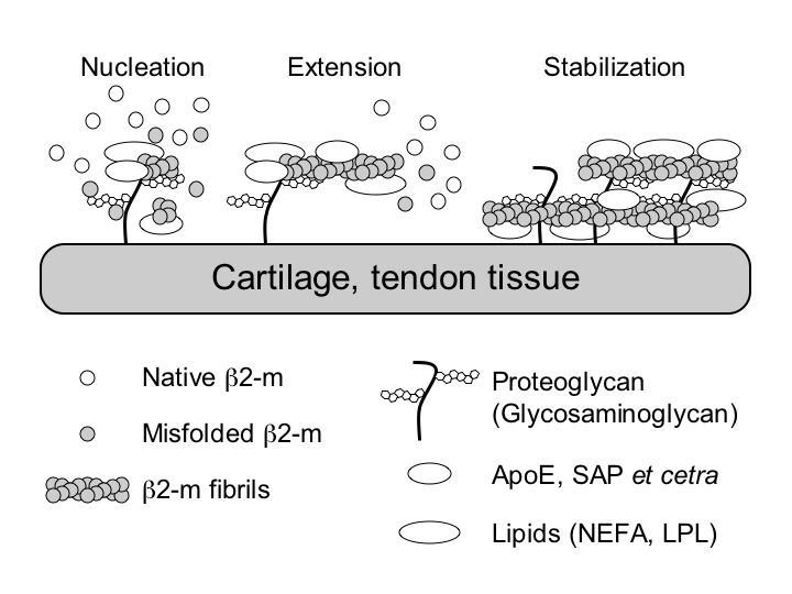
Beta2-microglobulin-related (Ab2M) amyloidosis is a common and serious complication in long-term hemodialysis patients. Intact beta2-microglobulin (b2-m) is a major structural component of amyloid fibrils deposited in the tissue, but the mechanism of the deposition of these amyloid fibrils is not fully understood. Based on a nucleation-dependent polymerization model and by using fluorescence spectroscopy with thioflavin T and electron microscopy, we systematically examined the effects of several classes of biological molecules on the formation and stabilization of Ab2M amyloid fibrils in vitro. Ab2M amyloid deposition takes place predominantly in the cartilaginous and tendinous tissues, suggesting that the specific interaction between b2-m and the extracellular matrix molecules in these tissues, such as type II collagen, glycosaminoglycans (GAGs), and proteoglycans (PGs), causes Ab2M amyloid deposition. We first observed that various types of GAGs and PGs stabilize the Ab2M amyloid fibrils and inhibit their depolymerization at a neutral pH. We next reported that some GAGs, especially heparin, dose-dependently enhanced the 2,2,2-trifluoroethanol-induced fibril extension at a neutral pH. In the mechanism of amyloidogenesis of natively folded proteins such as b2-m and transthyretin, partial unfolding is believed to be prerequisite to its assembly into amyloid fibrils both in vitro and in vivo. We recently found that some non-esterified fatty acids (NEFAs) and lysophospholipids (LPLs) induced not only the extension of Ab2M amyloid fibrils but also the formation of Ab2M amyloid fibrils from the b2-m monomer at a neutral pH, by partially unfolding the compact structure of b2-m to an amyloidogenic conformer, as well as stabilizing the extended fibrils. Based on these observations, we propose a working model for the molecular mechanism of the deposition of Ab2M amyloid fibrils in vivo (Figure).