Abstract
Albite porphyroblasts occur in schists in higher-grade zones of the Sanbagawa metamorphic belt, Japan. In this study of pelitic schists from the Nagatoro area of the belt, various microstructures in albite porphyroblasts were identified and indicate that grain size reduction of the albite porphyroblasts occurred after their formation. Schists with recognizable porphyroblasts contain fine grains of albite aligned in the matrix. In comparison, schists without recognizable albite porphyroblasts contain lenticular aggregations of albite grains in quartz-rich layers that suggest grain size reduction of the original porphyroblasts. These observations suggest that the absence of albite porphyroblasts does not necessarily indicate different pressure, temperature, or chemical conditions but can instead be explained by grain size reduction of albite porphyroblasts during deformation.
INTRODUCTION
Albite porphyroblasts occur widely in schists in higher-grade zones of the Sanbagawa metamorphic belt, Japan. The albite porphyroblasts are referred to as ‘albite spots’ and have been used as a useful index of higher-grade metamorphism, as they are recognizable with the naked eye. These porphyroblasts are thought to have formed during the later stages of or immediately after the peak of Sanbagawa metamorphism (Itaya, 1978; Hosotani, 1980; Otsuki, 1980; Banno and Sakai, 1989; Fushii and Ii, 2001; Okamoto and Toriumi, 2001). The Sanbagawa metamorphic belt is known to have undergone intense deformation during exhumation (Toriumi et al., 1988; Takeshita, 2021). The orientations of trails of inclusions in the albite porphyroblasts are oblique to the matrix schistosity and have been used to unravel the deformation history of the Sanbagawa metamorphic rocks (Takagi and Hara, 1979; Okamoto, 1998). However, the cause of the growth of the albite porphyroblasts and how their shape and size were influenced by host-rock deformation during the exhumation stage remain unclear. Toriumi (1975a) attributed the growth of albite porphyroblasts to collision and coalescence of albite grains. However, the mechanism is not assured to be universal in the Sanbagawa metamorphic belt. A better understanding of the history of the albite porphyroblasts is required to establish their metamorphic and tectonic significance.
Experimental studies have suggested that, compared with quartz, feldspars are more likely to deform by the occurrence of microcracks or recrystallization owing to their limited dislocation activity under low- to medium-temperature conditions (Tullis and Yund, 1985; Shigematsu, 1999). However, it is challenging to apply experimental results to observations of natural rocks because the deformation characteristics of minerals depend on many factors, such as temperature, strain rate, and the presence of water. Relative plasticity (or other deformation mechanisms) of minerals may also play an important role in the deformation of the whole rock. In addition, the relative strength of quartz and feldspar switches places depending on the temperatures under which deformation takes place (Tullis et al., 2000; Passchier and Trouw, 2005).
In this paper, we report on microstructures that indicate that grain size reduction of albite porphyroblasts probably occurred as a result of strong shear deformation of the host schists. Microstructures in the Sanbagawa pelitic schists sampled in the Nagatoro area of the Kanto Mountains reveal that fracturing of the albite porphyroblasts has occurred. Some of the analyzed samples have no recognizable albite porphyroblasts, although their microstructures suggest that larger albite porphyroblasts were present previously. This observation is important, as it constrains the controls on the presence or absence of albite porphyroblasts. The deformation microstructures of albite porphyroblasts constitute new evidence for constraining the conditions and history of deformation of the Sanbagawa metamorphic belt during exhumation.
GEOLOGICAL SETTING
The Sanbagawa metamorphic belt is a metamorphosed accretionary complex exposed in southwestern Japan, mostly along the Japan island arc. The belt has therefore been studied intensively as the product of a typical subduction zone (e.g., Banno and Sakai, 1989; Wallis and Okudaira, 2016). Geochronological studies have shown that Mesozoic trench sediments, accompanied by basaltic rocks and chert, were metamorphosed during the Late Cretaceous (Isozaki and Itaya, 1990; Tsutsumi et al., 2009). The highest metamorphic grade was achieved by rocks that are now exposed in central Shikoku and have been metamorphosed to eclogite-facies conditions (Takasu, 1984; Aoya, 2001; Kouketsu et al., 2014). Metamorphic zonation in central Shikoku is defined by mineral assemblages in pelitic rocks, comprising the chlorite, garnet, albite-biotite, and oligoclase-biotite zones, in order of increasing metamorphic grade (Enami, 1983; Higashino, 1990). In central Shikoku, the highest metamorphic grade rocks are located in the middle of the structural sequence, and the grade decreases both northward and southward. The same structure is observed in the Kanto Mountains. The Sanbagawa metamorphic rocks generally show a gently northward-dipping foliation and subhorizontal east-west mineral lineation generated by ductile deformation during the retrograde metamorphic stage. The crystallographic preferred orientation of quartz has been studied to determine the conditions of deformation that produced the thermal structure of the Sanbagawa metamorhic belt during the exhumation stage (Takeshita, 1996, 2021). It was indicated that the quartz c-axis fabric transition from type I to type II crossed-girdle, which occurs at deformation temperatures around 400 °C, is preserved in the high-grade part of the Sanbagawa metamorphic rocks. The lithology and the metamorphic zonation boundaries are oriented roughly parallel to the foliation.
Pelitic and psammitic schists of the Sanbagawa metamorphic belt are exposed in the Kanto Mountains in Saitama and Gunma prefectures, together with mafic schists and less common quartz schists and serpentinites. Albite porphyroblasts occur in both the mafic and pelitic schists, appearing as white elliptical spots in the former and as black elliptical spots in the latter on account of abundant carbonaceous inclusions. The albite porphyroblasts in the pelitic schists occur mainly in micaceous layers that form the foliation of the pelitic schists.
The metamorphic zonation identified in the Kanto Mountains by Hashimoto et al. (1992) comprises Zone I (quartz + albite + muscovite + chlorite), Zone II (Zone I minerals + garnet), and Zone III (Zone II minerals + biotite), which are equivalent to the abovementioned chlorite, garnet, and albite-biotite zones, respectively, of central Shikoku. The Nagatoro area is located several kilometers south of the highest-grade rocks in the Kanto Mountains. Most schists in the Nagatoro area are in Zone I (chlorite zone), whereas rocks of Zone II (garnet zone) and Zone III (biotite zone) are only locally exposed. The peak temperature in the studied outcrop has been estimated as 423 ± 18 and 409 ± 24 °C using Raman spectroscopy applied to carbonaceous materials (Umeda and Enami, 2014). This estimated metamorphic temperature is slightly lower than the temperature determined for the garnet zone in the Shikoku area (Enami et al., 1994). Hashimoto et al. (1992) argued that the complex spatial variation in peak metamorphic temperatures in the Kanto Mountains was generated by the piling up of thin sheets formed under different thermal conditions, described as a ‘shuffled-cards’ structure. Tagiri et al. (2003) attempted to delineate boundaries between the ‘cards’ on the basis of analysis of carbonaceous materials. The samples used in the present study were obtained from the same exposure as that sampled by Tagiri et al. (2003).
METHODS
Pelitic schists with and without recognizable albite porphyroblasts were sampled from the locality shown in Figure 1. The samples were cut parallel to the stretching lineation of the minerals and normal to the foliation to make XZ thin sections for examination using an optical polarizing microscope. Mapping and semi-quantitative point analyses were performed using energy-dispersive X-ray spectroscopy (EDS) with a JEOL JSM-6010LA (JEOL, Tokyo, Japan) scanning electron microscope (SEM) at Kokushikan University, Tokyo, Japan. The acceleration voltage during analyses was 20.0 kV. Crystallographic orientations of quartz were analyzed using an electron backscattered diffraction (EBSD) system (Oxford Instruments INCA Synergy AZTEC v4.2 HKL-Channel5) attached to an SEM (HITACHI S-3400N Type II) at the Rock and Mineral Laboratory of Nagoya University, Nagoya, Japan. The sample tilt was 70° from the horizontal. Thin sections were polished using colloidal silica to remove surface damage prior to EBSD analysis. Abbreviations for the names of minerals and end-member components follow Whitney and Evans (2010).

SAMPLE DESCRIPTIONS
The studied outcrop is located just downstream of the Takasago bridge (Fig. 1). Schists with and without albite porphyroblasts are juxtaposed at the outcrop (Tagiri et al., 2003). Between these rocks lies a 2-m-thick band consisting of breccias and serpentinites. Both the mafic and pelitic schists occur with and without recognizable albite porphyroblasts. This outcrop was identified as being in the garnet zone by Hashimoto et al. (1992), with garnet occurring in schists with and without recognizable albite porphyroblasts. Eight samples of pelitic schists were obtained from the outcrop as shown in Figure 1: seven samples with albite porphyroblasts, one without. Several more samples, both with and without albite porphyroblasts, were taken from the outcrop and observed, although the sampling point records were not accurate enough to show in Figure 1.
The mineral assemblage of schists from the outcrop consists of quartz + albite + muscovite + chlorite + calcite + garnet + titanite + K-feldspar ± tourmaline ± apatite. No obvious difference in the major mineral assemblage was observed between samples with and without albite porphyroblasts. The foliation is defined mainly by the preferred orientation of muscovite and chlorite. The schists comprise alternating micaceous (muscovite, chlorite, albite, and garnet) and quartz-rich layers. Calcite is observed in both the micaceous and quartz-rich layers. Albite porphyroblasts in thin section are generally elliptical in shape and elongated parallel to the lineation. The porphyroblasts are mostly 0.5-2.0 mm long and 0.2 mm wide. Albite grains in schist samples without albite porphyroblasts are mostly <0.2 mm in length. The spatial distribution of samples with or without albite porphyroblasts is shown in Figure 1. Identification of albite grains was possible with the optical microscope due to their slightly lower refractive index, however it proved difficult to identify all grains, so the task was undertaken by SEM-EDS mapping of Na. Details of the distribution of albite grains are given in the following section. K-feldspar occurs mostly as infilling cracks within albite porphyroblasts (Fig. 2).

Figure 2. Photomicrographs of boudinaged albite porphyroblasts. Sample No. 20181203KS8. (a) Minerals names labelled on a plane-polarized-light photomicrograph. Areas not filled with colors comprise mainly muscovite and chlorite (Ab, albite; Qz, quartz; Kfs, K-feldspar; Grt, garnet; Ms, muscovite; Chl, chlorite). (b) Cross-polarized-light photomicrograph showing K-feldspar infilling microveins formed by boudinage of an albite porphyroblast. Quartz also infills cracks formed in the albite porphyroblast. The crack shapes suggest deformation subsequent to infilling.
In schists with albite porphyroblasts, garnet occurs as inclusions in albite porphyroblasts and as grains in the matrix. Inclusions of garnet grains are euhedral and measure 0.1-0.2 mm in diameter. Garnet grains in the matrix are anhedral with morphologies indicative of dissolution after grain growth. In contrast, smaller euhedral garnet grains with diameters of <0.05 mm occur in schists without albite porphyroblasts and are found only in micaceous layers.
MICROSTRUCTURES OF THE STUDIED SCHISTS
Microstructures in and near albite porphyroblasts
Albite porphyroblasts observable to the naked eye occur mainly in micaceous layers. These porphyroblasts are elongated parallel to the lineation and have lengths of 0.5-2.0 mm. Micas are oriented along the outlines of albite porphyroblasts (Fig. 2). Pressure shadows filled with quartz occur at both ends of the porphyroblasts. The albite porphyroblasts commonly contain cracks infilled by other minerals, and these cracks can be divided into two types. One type of crack is infilled by quartz and/or calcite (QC-crack), and the other type is infilled by K-feldspar (K-crack) (Fig. 2). K-feldspar occurs mostly as infills in K-cracks and is not normally observed in the surrounding host schists. QC- and K-cracks occur in the same thin section. There is no pattern in the spatial distribution of samples containing QC- and K-cracks at outcrop scale (Fig. 1). Finer-grained albites are often aligned in the direction parallel to the lineation (Fig. 3). The fine-grained albites are <0.1 mm in length and <0.05 mm in width and have angular to nearly elliptical elongated morphologies. The grain size variation suggests that the large albite porphyroblasts are original grains and that the alignments of fine-grained albites are produced by grain size reduction of the porphyroblasts. Some of the porphyroblasts show undulose extinction and subgrain boundaries indicating the occurrence of breaking-ups of porphyroblasts (Fig. 3d). Crystallographic preferred orientations of quartz obtained using the EBSD technique show a cleft-girdle fabric of the quartz c-axis with a distinct Y-maximum (Fig. 4a), indicating ductile behavior of quartz-rich layers during the final stage of deformation. Ductile behavior of the host rock enables the finer albite grains to align as the consequence of shear deformation.

Figure 3. Microstructures in pelitic schist sample No. 20191205NY03, in which albite porphyroblasts are visible to the naked eye. Panels (a)-(c) show the same area within the sample. Panel (d) shows the area marked by a red frame in (b). Fine-grained albite grains are aligned parallel to the schistosity (as shown by ellipses depicted by dashed lines) near albite porphyroblasts. (a) Plane-polarized-light photomicrograph. Porphyroblastic albite grains are labelled ‘Ab’. (b) Cross-polarized-light photomicrograph. (c) Chemical map of Na. Contents of Na are defined according to the color bar (bottom left). Albite is the only mineral that contains Na in this map. (d) An albite porphyroblast showing undulose extinction.

Figure 4. Crystallographic preferred orientation of quartz (c-axis) measured using electron backscattered diffraction. (a) Schist with albite porphyroblasts (sample No. 20191205NY03). The quartz c-axis fabric shows a type-I crossed-girdle structure with a slight Y-maximum. (b) Schist without albite porphyroblasts (sample No. 20220725NG). The quartz c-axis fabric shows a type-II crossed-girdle structure.
The pelitic schist without albite porphyroblasts contain albite grains in both the micaceous and quartz-rich layers. The grain size and distribution of albite grains differ in these layer types. Albite grains in the micaceous layers are generally small in size, typically <0.1 mm in length and <0.05 mm in width, and mostly elliptical. No microstructures indicating pressure solution are observed along boundaries between albites and micas. In quartz-rich layers, much larger albite grains occur (∼ 0.2 mm in length), as well as smaller grains about the same size as those in the micaceous layers. Groups of albite grains are found forming lenticular-shaped aggregations in the quartz-rich matrix (Fig. 5), referred to as ‘lenticular aggregations’ herein. Larger albite grains are slightly angular compared to surrounding quartz grains, and tend to be located in the central part of the lenticular aggregations. In some places in Figures 5c and 5d, the outlines of neighboring grains within a single lenticular aggregation show corresponding curves, suggesting that the grains once constituted a single large grain. This example and others imply that the lenticular aggregations were originally larger albite porphyroblasts. No pressure shadows of quartz are observed around the albite grains. Crystallographic preferred orientations of quartz exhibit a type-II crossed-girdle structure of the c-axis (Fig. 4b), indicating ductile deformation of the quartz layers. Ductile deformation of the host quartz layers could have made possible for the fine albite grains, which were produced by breaking up of the porphyroblasts, to scatter and distribute in the observed lenticular aggregations.

Figure 5. Microstructures in pelitic schist sample No. 20220725NG, in which albite porphyroblasts are not visible to the naked eye. Panels (a) and (b) show the same area within the sample. Panels (c) and (d) show the area marked by a red frame in (a). Lenticular aggregations composed of scattered fine-grained albites (with one such aggregation depicted by the dashed-line ellipse) are observed in the quartz-rich layer. (a) Plane-polarized-light photomicrograph. Some of the larger albite grains are labelled ‘Ab’. (b) Chemical map of Na. The content of Na is defined according to the color bar (bottom left). Albite is the only mineral that contains Na in this map. (c) Plane-polarized-light photomicrograph of part of the lenticular aggregation of albite shown in (a). Albite grains are barely distinguishable by their slightly angular shape and smaller refractive index, although it is difficult to identify all. (d) Cross-polarized-light photomicrograph of the area shown in (c). Albite grains are depicted with blue dotted lines.
GARNET CHEMISTRY
Inclusions of garnet in albite porphyroblasts have spessartine-rich cores, and contents of Mn decrease toward rims, compensated by increased contents of Fe. An abrupt increase in contents of Ca is observed in the middle of growth (Fig. 6a). Anhedral garnets in the matrix of samples with albite porphyroblasts exhibit similar chemical zoning but lack the highest-Fe rims (Fig. 6b). Garnets in the samples without albite porphyroblasts are found only within the micaceous layers. These grains are euhedral and very small, mostly <0.05 mm in diameter. They also have spessartine-rich cores, but decreases in contents of Mn toward rims are compensated by increases in contents of Ca, rather than Fe content (Fig. 6c). This chemical trend is different from that of garnet grains contained in the rocks with albite porphyroblasts.
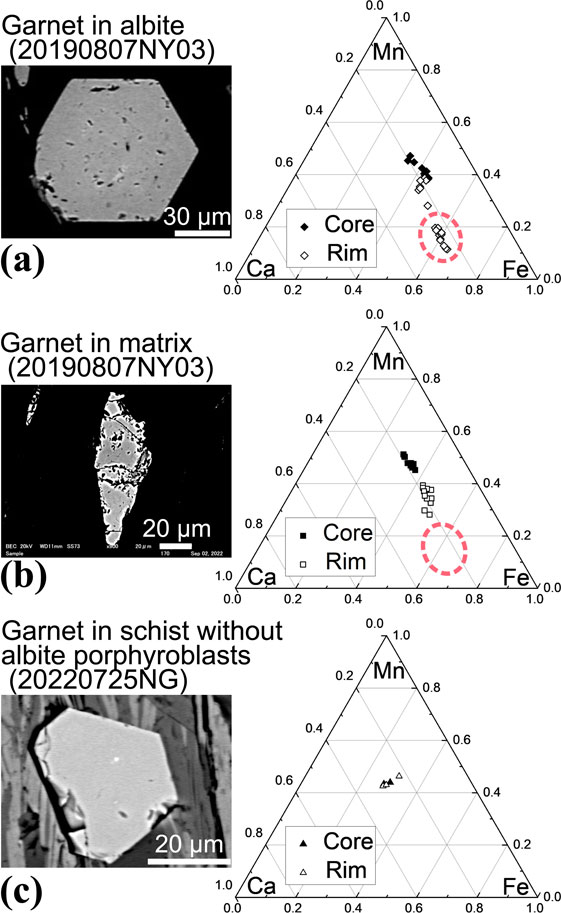
Figure 6. Backscattered electron images and Mn-Fe-Ca ternary diagrams of garnet chemistry. The content of the Mg-endmember in garnet is generally very low (<1 wt%). (a) Garnet inclusion in an albite porphyroblast (sample No. 20190807NY03). The garnet shape is euhedral. The red-dashed-line ellipse shows the chemical composition of the garnet rim. (b) Garnet in the same sample as (a), but not as an inclusion in an albite porphyroblast. Compared with the inclusion-type garnet shown in panel (a), the garnet shape is anhedral, grains are generally smaller, and their chemical trends lack the rim component (shown as the red-dashed-line ellipse lacking data). (c) Garnet in a micaceous layer of schist without albite porphyroblasts. These garnets are euhedral and are very small, mostly less than 50 µm in diameter. The chemical trend from core to rim is different compared with (a) or (b).
DISCUSSION
Grain size reduction of albite porphyroblasts
The grain sizes and microstructures of albite grains investigated in this study strongly indicate that the albite porphyroblasts decreased in size during post-peak deformation of the Sanbagawa metamorphic belt through two different mechanisms. One is fracturing and breaking-up of porphyroblasts into fine grained albite less than 0.2 mm in diameter. Microstructures indicating this mechanism are observed in schists both with and without visible albite porphyroblasts. Another is boudinaging of the porphyroblasts observed in samples with visible porphyroblasts (Fig. 2). Schists with visible porphyroblasts contain not only large grains but also numerous fine-grained albites aligned in the matrix. Some of the alignment is associated with the porphyroblasts like tails. This distribution cannot be explained by the growth or segregation of albite grains during metamorphism. In schists without albite porphyroblasts, lenticular aggregations of albite grains are observed within quartz-rich layers. It is possible that the microstructure shows the process of formation of the albite porphyroblasts through dynamic segregation and coalescence of finer grains as argued by Toriumi (1975b). However, segregation seems less plausible since the direction of extension of the lenticular aggregation is oblique to the metamorphic layering. It seems more straightforward to interpret that these albite grains are on the way of dispersing. Corresponding outlines of neighboring grains also imply that these grains were originally a single grain.
The textural evidence of quartz c-axis fabric suggests the occurrence of ductile deformation in the host schists accomodated at least by quartz. Ductile behavior of the host rock is critical for the interpretation of the microstructures since without that the broken-up pieces of the old albite porphyroblasts could not scatter or align as observed. In schists with albite porphyroblasts, quartz c-axis fabrics show a single-girdle structure with a distinct Y-maximum, indicating deformation in high temperature conditions, namely above 400 °C since it implies the dominant slip system to be prism <a> (Law, 2014). The expected ductile behavior of quartz is concordant with the alignment of the fractured fine grained albite by shear deformation. The fine fractured grains do not surround the original porphyroblasts but are aligned parallel to the lineation in the matrix. Therefore it is unclear which grain their origin is. In schists without albite porphyroblasts, quartz c-axis fabrics show a type-II crossed-girdle structure, which is also interpreted as evidence of ductile deformation at temperatures above 400 °C (e.g., Takeshita, 1996; Law, 2014). The grain size of albites in micaceous layers is comparable to that of dynamically fractured grains in samples with albite porphyroblasts, indicating that the mechanism of grain size reduction was similar. Relatively large grains of albite are present in quartz-rich layers but are not as large as the proper albite porphyroblasts. Smaller albite grains in the quartz-rich layers are similar in size to the finer grains in the sample with albite porphyroblasts. In addition, the occurrence of lenticular aggregations of albite grains in quartz-rich layers suggests that these grains had once been a single large grain. Thus, the original albite grains have been substantially disaggregated. The substantial break-up of the original grains suggests that total strain was larger in schists without (cf. with) albite porphyroblasts.
Pressure shadows of quartz observed in schists with visible porphyroblasts indicate that dissolution-precipitation of quartz also occurred. The original grains in schists with albite porphyroblasts are commonly boudinaged, and cracks are filled by other minerals, such as quartz and calcite or K-feldspar (Fig. 2). The cracks are thin, around 100 µm in width, and the cracked grains mostly keep their shape. The thin cracking seems inconsistent with the finer grains, since the sizes of the resulted grains are much different. It is suggested that the cracks in the remnant original porphyroblasts were formed during a later stage that involved deformation via dissolution-precipitation of quartz at lower temperature conditions. Quartz c-axis fabric of the sample is weak and not very clear (Fig. 4a), presumably due to the mixing of two different deformation mechanism recorded. Some of the surviving albite porphyroblasts show undulose extinction indicating formation of subgrain boundary within the porphyroblasts. The shape of the subgrain boundary is large and angular and resembles the boudinage, not the fracturing into finer grains (Fig. 3d), indicating that the boudinage postdated the fracturing. No microstructures suggesting the bulging recrystallization were found, that were reported from highly deformed albite (Allard et al., 2021). K-feldspar is not contained in the host schists and is found only in cracks of the original porphyroblasts. K-feldspar has been observed in veins of the Sanbagawa schists of the Kanto Mountains (Okamoto et al., 2008). It is therefore reasonable to assume that the cracks infilled with K-feldspar formed as microveins during a later stage of deformation.
Geological implications
Chemical trends observed in garnets indicate a slight difference in metamorphic condition between the schists with and without albite porphyroblasts. Chemical trends and occurrences of garnet observed in this paper are within the variation of garnet hitherto found from the Sanbagawa metamorphic rocks. Bell-shaped chemical profiles of contents of spessartine, which decrease from core to rim, are typical of garnet from the Sanbagawa metamorphic belt (Banno and Sakai, 1989; Higashino, 1990; Enami, 1998). Albite porphyroblasts containing garnet grains are commonly observed in the Sanbagawa metamorphic rocks (Sakai et al., 1985; Inui, 2010). The content of grossular in the garnets usually increases outward in the core, followed by a decrease toward the rim, with a maximum recorded in the middle of garnet growth (Banno and Kurata, 1972; Enami, 1998; Inui and Toriumi, 2002), which is not the case in this study. However, garnets having the maximum grossular content at the rims have been found, although rare (Enami, 1998). The chemical zoning of inclusions of garnet in the albite porphyroblasts observed in this study resembles that of typical garnets from the Sanbagawa metamorphic belt, except at rims, where contents of grossular do not decrease (Fig. 6a). It is inferred that garnets in the albite porphyroblasts formed in response to Sanbagawa metamorphism and that their growth ceased when they became trapped inside albite. Garnet grains in the matrix have irregular shapes, indicating dissolution after growth, but the chemical composition of the remaining parts is similar to that of garnet inclusions. Thus, garnet inclusions in albite porphyroblasts and matrix garnet grew under the same conditions, probably during prograde metamorphism. Garnets in schists without albite porphyroblasts show a different occurrence, and their size and chemical composition also differ. Garnet grains are distinctly small and euhedral in comparison with those in schists with albite porphyroblasts. Garnet only occur in the micaceous layers and does not occur as inclusions in albite, nor in the quartz-rich layers where the lenticular aggregations of albite grains are found. The occurrence implies that the garnet grains were never included in albites and, therefore, that they grew after the formation of the present microstructure. Very small euhedral garnet grains occurring only within micaceous layers have been reported the Nagatoro area (Inui et al., 2020) and also from the Asemigawa district of the Sanbagawa metamorphic belt (Inui, 2010). It seems reasonable to interpret that the schists narrowly reached the garnet-in condition.
In summary, the schists with recognizable albite porphyroblasts show evidence of deformation at high (<400 °C) and low temperatures, causing fracturing of albite porphyroblasts into fine grains and boudinaging, respectively. In contrast, the schists without recognizable albite porphyroblasts only show evidence of deformation at high temperature conditions. The total strain was larger in the latter, indicated by the fact that most of the albite porphyroblasts are fractured into fine grains. Their microstructure indicates that at least some of the lower-grade rocks of the Sanbagawa metamorphic belts underwent strong deformation while the temperature was still around the peak condition. It is also important to note that remnants of albite porphyroblasts are possibly more widespread than currently realized and are yet to be discovered in other areas of the Sanbagawa metamorphic belt.
CONCLUSIONS
This study identified grain size reduction of albite porphyroblasts in Sanbagawa metamorphic rocks from an outcrop in the Nagatoro area, Kanto Mountains, Japan. Lenticular aggregations of albite grains occur in pelitic schist without albite porphyroblasts. Schists with recognizable porphyroblasts also contain aligned fine grains of albite, indicative of grain size reduction. The observations suggest that the absence of albite porphyroblasts is not necessarily due to different P, T, or chemical conditions but can be simply explained by grain size reduction of albite porphyroblasts during deformation.
ACKNOWLEDGMENTS
The authors are grateful to Y. Kouketsu for her handling of the manuscript and T. Takeshita and two other anonymous reviewers for the kind reviews. Their comments and suggestions helped us to bring the scattered discussion together. We thank K. Michibayashi very much for his kind assistance on their EBSD analyses in Nagoya University. We are also grateful to T. Okudaira for his encouragement. Finally, we are always thankful to the members of the lab who supplied and organized their samples for use in this study.
REFERENCES
- Allard, M., Ildefonse, B., Oliot, É. and Barou, F. (2021) Plastic deformation of plagioclase in oceanic gabbro accreted at a slow-spreading ridge (Hole U1473A, Atlantis Bank, Southwest Indian Ridge). Journal of Geophysical Research: Solid Earth, 126, e2021JB021964.
- Aoya, M. (2001) P-T-D Path of Eclogite from the Sambagawa Belt Deduced from Combination of Petrological and Microstructural Analyses. Journal of Petrology, 42, 1225-1248.
- Banno, S. and Kurata, H. (1972) Distribution of Ca in zoned garnet of low-grade pelitic schists. Journal of the Geological Society of Japan, 78, 507-512 (in Japanese with English abstract).
- Banno, S. and Sakai, C. (1989) Geology and metamorphic evolution of the Sanbagawa belt. In Evolution of Metamorphic Belts (Daly, J.S., Cliff, R.A. and Yardley, B.W.D. Eds.). Geological Society, London, Special Publication, 43, 519-532.
- Enami, M. (1983) Petrology of pelitic schists in the oligoclase-biotite zone of the Sanbagawa metamorphic terrain, Japan: phase equilibria in the highest grade zone of a high-pressure intermediate type of metamorphic belt. Journal of Metamorphic Geology, 1, 141-161.
- Enami, M. (1998) Pressure-temperature path of Sanbagawa prograde metamorphism deduced from grossular zoning of garnet. Journal of Metamorphic Geology, 16, 97-106.
- Enami, M., Wallis, S.R. and Banno, Y. (1994) Paragenesis of sodic pyroxene-bearing quartz schists: implications for the P-T history of the Sanbagawa belt. Contributions to Mineralogy and Petrology, 116, 182-198.
- Fushii, K. and Ii, H. (2001) Texture to decipher growth stage of albite-porphyroblasts in pelitic schists from the Sambagawa metamorphic belt. Journal of Mineralogical and Petrological Sciences, 96, 148-153.
- Hashimoto, M., Tagiri, M., Kusakabe, K., Masuda, K. and Yano, T. (1992) Geologic structure formed by tectonic stacking of sliced layers in the Sanbagawa metamorphic terrain, Kodama-Nagatoro area, Kanto Mountains. Journal of the Geological Society of Japan, 98, 953-965 (in Japanese with English abstract).
- Higashino, T. (1990) The higher grade metamorphic zonation of the Sambagawa metamorphic belt in central Shikoku, Japan. Journal of Metamorphic Geology, 8, 413-423.
- Hosotani, H. (1980) Growth stage of albite porphyroblast in pelitic schist of the Sanbagawa metamorphic belt. Journal of Geological Society of Japan, 86, 537-543 (in Japanese with English abstract).
- Inui, M. (2010) Two types of garnet in Sanbagawa pelitic schists along Asemigawa River, central Shikoku. Journal of Mineralogical and Petrological Sciences, 105, 274-279.
- Inui, M. and Toriumi, M. (2002) Prograde pressure-temperature paths in the pelitic schists of the Sambagawa metamorphic belt, SW Japan. Journal of Metamorphic Geology, 20, 563-580.
- Inui, M., Wakai, Y. and Sakuragi, H. (2020) Nucleation and initial growth of garnet in low-grade metamorphic rocks of the Sanbagawa metamorphic belt, Kanto Mountains, Japan. Minerals, 10, 292-305.
- Isozaki, Y. and Itaya, T. (1990) Chronology of Sanbagawa metamorphism. Journal of Metamorphic Geology, 8, 401-411.
- Itaya, T. (1978) Notes on petrography and rock-forming mineralogy (3) Albite porphyroblasts containing retrograde TiO2-minerals from Sanbagawa pelitic schists in central Shikoku, Japan. The Journal of the Japanese Association of Mineralogists, Petrologists and Economic Geologists, 73, 339-345.
- Kouketsu, Y., Enami, M., Mouri, T., Okamura, M. and Sakurai, T. (2014) Composite metamorphic history recorded in garnet porphyroblasts of Sambagawa metasediments in the Besshi region, central Shikoku, Southwest Japan. Island Arc, 23, 263-280.
- Law, R.D. (2014) Deformation thermometry based on quartz c-axis fabrics and recrystallization microstructures: A review. Journal of Structural Geology, 66, 129-161.
- Okamoto, A. and Toriumi, M. (2001) Application of differential thermodynamics (Gibbs’ method) to amphibole zonings in the metabasic system. Contributions to Mineralogy and Petrology, 141, 268-286.
- Okamoto, A., Kikuchi, T. and Tsuchiya, N. (2008) Mineral distribution within polymineralic veins in the Sanbagawa belt, Japan: implications for mass transfer during vein formation. Contributions to Mineralogy and Petrology, 156, 323-336.
- Okamoto, K. (1998) Inclusion-trail geometry of albite porphyroblasts in a folded structure in the Sambagawa Belt, central Shikoku, Japan. Island Arc, 7, 283-294.
- Otsuki, M. (1980) Notes on petrography and rock-forming mineralogy (7) Zonal structure of albite porphyroblast from basic Sanbagawa schists in central Shikoku. The Journal of the Japanese Association of Mineralogists, Petrologists and Economic Geologists, 75, 196-202.
- Passchier, C.W. and Trouw, R.A.J. (2005) Microtectonics, Second Edition. pp.366, Springer.
- Sakai, C., Banno, S., Toriumi, M. and Higashino, T. (1985) Growth history of garnet in pelitic schists of th Sanbagawa metamorphic terrain in central Shikoku. Lithos, 18, 81-95.
- Shigematsu, N. (1999) Dynamic recrystallization in deformed plagioclase during progressive shear deformation. Tectonophysics, 305, 437-452.
- Tagiri, M., Matsumoto, T. and Tanimoto, K. (2003) Reevaluation of the precision of the degree of graphitization, and the boundaries between shuffled cards in the Sanbagawa metamorphic belt, Kanto Mountains, Japan. Japanese Magazine of Minera-logical and Petrological Sciences, 32, 175-184 (in Japanese with English abstract).
- Takagi, K. and Hara, I. (1979) Relationship between growth of albite porphyroblasts and deformation in a Sambagawa schist, Central Shikoku, Japan. Tectonophysics, 58, 113-125.
- Takasu, A. (1984) Prograde and Retrograde Eclogites in the Sambagawa Metamorphic Belt, Besshi District, Japan. Journal of Petrology, 25, 619-643.
- Takeshita, T. (1996) Estimate of the physical conditions for deformation based on c-axis fabric transitions in naturally deformed quartzite. Journal of Geological Society of Japan, 102, 211-222 (in Japanese with English abstract).
- Takeshita, T. (2021) Quartz microstructures from the Sambagawa metamorphic rocks, southwest Japan: indicators of deformation conditions during exhumation. Minerals, 11, 1038.
- Toriumi, M. (1975a) Petrological study of the Sanbagawa metamorphic rocks in the Kanto Mountains, central Japan. Bulletin of the University Museum, University of Tokyo, 9.
- Toriumi, M. (1975b) A dynamic aspect of metamorphic differentiation. Journal of Geological Society of Japan, 81, 505-511 (in Japanese with English abstract).
- Toriumi, M., Masui, M. and Mori, K. (1988) Strain in metacherts as determined from deformed shapes of radiolaria and Fry’s method. Tectonophysics, 145, 157-161.
- Tsutsumi, Y., Miyashita, A., Terada, K. and Hidaka, H. (2009) SHRIMP U-Pb dating of detrital zircons from the Sanbagawa Belt, Kanto Mountains, Japan: need to revise the framework of the belt. Journal of Mineralogical and Petrological Sciences, 104, 12-24.
- Tullis, J. and Yund, R.A. (1985) Dynamic recrystallization of feldspar: A mechanism for ductile shear zone formaion. Geology, 13, 238-241.
- Tullis, J., Stünitz, H., Teyssier, C. and Heilbronner, R. (2000) Deformation microstructures in quartzo-feldspathic rocks. Journal of the Virtual Explorer, 2.
- Umeda, H. and Enami, M. (2014) Testing for robustness on estimation of graphitization degree by Raman spectroscopy. Journal of Mineralogical and Petrological Sciences, 109, 279-285.
- Wallis, S.R. and Okudaira, T. (2016) Paired metamorphic belts of SW Japan: the geology of the Sanbagawa and Ryoke metamophic belts and the Median Tectonic Line. In The Geology of Japan (Moreno, T., Wallis, S., Kojima, T. and Gibbons, W. Eds.). Geological Society, London, 101-124.
- Whitney, D.L. and Evans, B.W. (2010) Abbreviations for names of rock-forming minerals. American Mineralogist, 95, 185-187.
