Abstract
Pyriofenone demonstrates outstanding efficacy in controlling powdery mildew. We investigated the impact of pyriofenone on the infection processes and cytological features of Blumeria graminis f. sp. tritici on wheat leaves. The preventive application of pyriofenone before inoculation did not inhibit conidial germination but effectively suppressed both appressorial and haustorial formation. Notably, haustorial formation was effectively inhibited, resulting in the complete suppression of successive lesion development and sporulation. Curative application of pyriofenone after inoculation also inhibited lesion expansion and sporulation. Furthermore, it had considerable impact on the morphogenesis of powdery mildew fungus. We observed multi-formed secondary appressoria, shrunken or bifurcated hyphae, abnormal conidiophores, and clubbed conidia-like structures. Subsequently, we employed a histochemical approach to analyze the localization of essential components for the polar growth of fungal hyphae. Pyriofenone induced mislocalization of the actin cytoskeleton, β-glucan and cytoplasmic vesicles, although it did not affect tubulin orientation.
Introduction
Fungi causing powdery mildew can target numerous economically important crop species, infecting the leaves, stems, flowers, and fruits of nearly 10,000 angiosperms species.1,2) To manage powdery mildew, fungicides are frequently applied throughout each crop’s growing season. Generally, the causative agents of powdery mildew are prone to mutations and the development of fungicide resistance due to their rapid conidial production during their short period of alternation of generation.3,4) This has led to the emergence of strains displaying increased tolerance to existing powdery mildew fungicides in recent years, significantly diminishing the effectiveness of these active compounds. Therefore, to combat powdery mildew and prevent the proliferation of fungicide-resistant strains, rotation spray program that includes fungicides with different modes of action is employed.5–7)
Pyriofenone, (5-chloro-2-methoxy-4-methyl-3-pyridyl) (4,5,6-trimethoxy-o-tolyl)methanone (code names: IKF-309, Property®, PROLIVO®, Kusabi®, Unicicut®, CROSSOUT®, and CASSINI®) (Supplemental Fig. S1), is a new fungicide developed by Ishihara Sangyo Kaisha, Ltd.8,9) It exhibits exceptional efficacy in controlling powdery mildew in several economically significant crops. Official tests, conducted by the Japan Plant Protection Association (JPPA) since 2008, have demonstrated the effectiveness of pyriofenone against powdery mildew diseases on wheat, cucumber, strawberry, and eggplant.8) In a previous report, we provided details regarding the fungicidal spectrum and biological properties of pyriofenone.9) Pyriofenone possesses features such as favorable residual activity, rainfastness, curative properties, translaminar activity, and vapor activity, all contributing to its excellent performances under field conditions. Furthermore, it does not exhibit cross-resistance with existing powdery mildew fungicides.8,9) Therefore, pyriofenone is considered a potent tool for rotational spray programs and holds significant utility as an agricultural agent.
Wheat powdery mildew, caused by Blumeria graminis f. sp. tritici, stands as one of the most significant diseases affecting wheat in the field. B. graminis is an obligate pathogen, exclusively parasitizing living plants.1,2) In this study, we delineated the effects of pyriofenone on the infection processes and cytological characteristics of B. graminis on wheat leaves. Based on the results obtained, we delved into the mode of action of pyriofenone against powdery mildew fungi.
Materials and methods
1. Chemicals and formulationsPyriofenone 180 g/L suspension concentrate (Property®180SC) or 300 g/L suspension concentrate was employed in this study.
2. Plant diseaseWheat powdery mildew fungus, Blumeria graminis f. sp. tritici, was cultivated on wheat seedlings (cv. Norin 61) under fluorescent lights at 20°C in a thermostatic chamber. The wheat seedlings were inoculated by applying B. graminis f. sp. tritici conidia to the leaves and subsequently incubated at 20°C. Conidia formed on the leaves 7–10 days after inoculation were used for the successive experiment.
3. Effect of pyriofenone on infection processes of Blumeria graminis on wheat leaves3.1. Application of chemicalsPyriofenone was dissolved in water containing 0.2% (v/v) of the surfactant SHINDINE®. The first leaves of wheat (cv. Norin 61), positioned horizontally, were treated with the test solutions using a handgun sprayer (20 mL/0.2 m2, equivalent to 1000 L/ha water volume). To evaluate the preventive effect of pyriofenone on conidial germination, formation of appressorium and haustorium, mycelial growth, and sporulation, we applied test solutions before inoculation at the same day of inoculation (preventive condition). Conversely, to evaluate the curative effect of pyriofenone on mycelial growth and sporulation, test solutions were applied 4 days after inoculation (curative condition).
3.2. Inoculation of wheat powdery mildewThe wheat seedlings leaves were inoculated by dusting them with B. graminis conidia. Simultaneously, the leaves were positioned horizontally to ensure uniform inoculation. Next, the seedlings were placed in a thermostatic chamber at 20°C under fluorescent lighting for incubation.
3.3. Evaluation of infection processesEvaluations were conducted using a fluorescence microscope following staining with a 0.01% water solution of Fluorescent Brightener 28 (Sigma-Aldrich Japan G. K., Tokyo, Japan), except for the assessment of haustorial formation. Conidial germination and appressorial formation were observed at 1 and 2 days post-inoculation, respectively. The percentages of conidial germination and appressorial formation were calculated. The assessment of haustorial formation, mycelial growth and sporulation under preventive conditions was conducted 7 days after inoculation. For haustorial formation evaluation, the inoculated leaves were decolorized using a compound liquid of ethanol and acetic acid (3 : 1) and then were stained with a 0.5% cotton-blue solution. To assess haustorial formation, we visually compared the treated leaves with the untreated ones under microscope. Mycelial growth and sporulation areas in each field of view were visually observed and rated on a scale of 1 to 10, then averaged across 10 fields of view. The number of scale was equivalent to 10% (scale 1) to 100% (scale 10) of mycelial growth and sporulation areas.
The evaluation of mycelial growth and sporulation of the curative condition was conducted 7 days after inoculation. Mycelial growth and sporulation areas within a field of view were visually assessed across 10 fields of view, rated on a scale of 1 to 10, and then averaged. Lesion expansion values were calculated by subtracting the initial value at the time of pyriofenone application (4 days after inoculation) from the final value (7 days after inoculation). No sporulation was observed 4 days after inoculation even in the untreated control.
4. Effect of pyriofenone on the fungal morphogenesis of Blumeria graminis on wheat leavesThe effect of pyriofenone on the fungal morphogenesis of B. graminis on wheat leaves was observed using scanning electron microscope (SEM) and fluorescence microscope following the curative application of pyriofenone. Pyriofenone applications were performed 3 or 4 days after inoculation, and the treated seedlings were subsequently incubated at 20°C for an additional 3 or 4 days. For SEM observations, leaf segments were rapidly frozen using liquid nitrogen. These frozen samples were then mounted on the chilled stage of a scanning electron microscope (HITACHI S-3200N) and observed under low-vacuum conditions. In the case of fluorescence microscopy, Fluorescent Brightener 28 and Hoechst 33342 (Sigma) were employed as stains to highlight the septa in fungal cell walls and the nuclei, respectively.
5. Cytological techniquesFluorescent Brightener 28 was employed for staining the β-glucan in fungal cell walls. A 0.01% solution of Fluorescent Brightener 28 was prepared in water with 0.05% Tween 20 (Sigma). Subsequently, this solutions was applied to leaf segments (10 mm×5 mm) prior to fluorescence microscopy, using an excitation wavelength of 330–385 nm and an emission wavelength of 420 nm.
For nuclei staining, Hoechst 33342 was utilized. Leaf segments were immersed in a 1 µg/mL Hoechst 33342 solution in a 25 mM phosphate buffer (PB, pH 6.8) for 10 min. After rinsing in PB, fluorescence was observed using a fluorescent microscope with an excitation wavelength of 346 nm and an emission wavelength of 400–460 nm.
To visualize cytoplasmic vesicles, we employed FM4-64 (Invitrogen™, ThermoFisher Scientific, Tokyo, Japan). Leaf segments were submerged for 5 min in a 20 mM 4-(2-hydroxyethyl)-1-piperazineethanesulfonic acid (HEPES) buffer (pH 7.2) containing 10 µM FM4-64. The samples were subsequently washed with HEPES buffer and directly examined using a fluorescent microscope with an excitation wavelength of 530–550 nm and an emission wavelength of 573 nm.
Actin and microtubules were visualized through indirect immunofluorescence microscopy using anti-actin and anti-tubulin antibodies, respectively. Leaf segments were fixed in a fixation buffer (containing 4% formaldehyde, 25 mM piperazine-1,4-bis(2-ethanesulfonic acid) (PIPES), 2 mM ethylene glycol tetraacetic acid, 2 mM MgCl2, 250 µM m-maleimidobenzoyl-N-hydroxysuccinimide ester, 0.5 mg/mL Tween 20; pH 6.8) at 25°C for 40 min. Next, the leaf segments were rinsed successively in 25 mM PIPES (pH 6.8) and 25 mM PB (pH 6.5) before a final rinse in 25 mM PB (pH 6.5). Subsequently, they were transferred to a solution containing 50 mg/mL Glucanex (Sigma), 50 mg/mL Driselase (Sigma), and 10 mg/mL bovine serum albumin (BSA) dissolved in 25 mM PB (pH 6.5) at 25°C for 15 min. After another rinse in 25 mM PB (pH 6.5), they were treated with 5 g/L Triton X-100 in 25 mM PB (pH 6.8) for 10 min. The specimens were washed with 25 mM PB (pH 6.8), followed by TB buffer (comprising 50 mM Tris-HCl and 150 mM NaCl at pH 7.6). After rinsing, the segments were incubated with a monoclonal mouse anti-actin antibody (anti-β-actin [AC-15]; Sigma) for actin localization or an anti-tubulin antibody (anti-α-tubulin [clone DM1A]; Sigma) for tubulin localization, diluted at a 1 : 200 ratio in TB-BSA buffer (50 mM Tris-HCl at pH 7.6, 150 mM NaCl, and 10 mg/mL BSA) at 25°C for 60 min. Vacuum infiltration was repeated three times for 1 min to promote infiltration. The specimens were washed twice in TB-T buffer (50 mM Tris-HCl at pH 7.6, 150 mM NaCl, 0.25% Tween 20) before being incubated in goat anti-mouse IgG Alexa Fluor 488 (anti-mouse IgG, F [ab′] 2 Fragment; Invitrogen™, ThermoFisher Scientific), diluted at a 1 : 200 ratio in TB-BSA buffer at 25°C for 1.5 hr. Vacuum infiltration was repeated as described above. Finally, the leaf segments were rinsed with TB-T buffer and TB buffer (pH 7.6). The specimens were mounted in TB buffer (pH 7.6) on glass slides and examined using fluorescence microscopy with an excitation wavelength of 470–495 nm and an emission wavelength of 510–550 nm.
Results
1. Effect of pyriofenone on the infection processes of Blumeria graminis on wheat leaves1.1. Effect on conidial germinationPyriofenone did not inhibit the germination of conidia, even at a high concentration of 100 µg/mL. The germination rates of conidia were not considerably different from those of the untreated control (Table 1).
Table 1. Effect of pyriofenone on conidial germination, appressorial formation, haustorial formation, lesion expansion and sporulation of
Blumeria graminis| Application dose (µg/mL) | % Germinationa) | EC50 (µg/mL) | % Appressorial formationa) | EC50 (µg/mL) | Haustorial formation | % Lesion areaa) | EC50 (µg/mL) | % Sporulation areaa) | EC50 (µg/mL) |
|---|
| 100 | 64.8±5.0 | >100 | 26.4±2.0** | 9.7 | − | 0±0** | 0.1 | 0±0** | 0.1 |
| 12.5 | 71.7±9.8 | 36.6±2.0* | − | 0±0** | 0±0** |
| 1.6 | 71.6±3.1 | 41.7±3.0* | − | 0±0** | 0±0** |
| 0.4 | 71.2±4.9 | 41.6±1.7* | + | 1.6±2.6** | 0±0** |
| 0.1 | 68.8±9.1 | 41.8±8.9* | ++ | 47.0±19.2** | 56.5±28.0** |
| 0 | 75.2±6.1 | 67.8±9.9 | ++ | 74.0±16.0 | 94.0±5.5 |
Conidial germination and appressorial formation were evaluated at 1day and 2 days after inoculation, respectively. Haustorial formation, lesion expansion and sporulation were evaluated after 7 days after inoculation. Mycelial growth and sporulation areas in each field of view were visually observed and rated on a scale of 1 to 10, then averaged across 10 fields of view. a) Shows significant difference to untreated control (* p<0.005 ** p<0.001 Tukey HSD).
While the inhibitory effect of pyriofenone on appressorial formation was observed across all tested concentrations ranging from 0.1 to 100 µg/mL, the level of inhibition remained insufficient. Even at a concentration of 100 µg/mL of pyriofenone application, 26.4% of conidia were observed to form appressoria (Table 1).
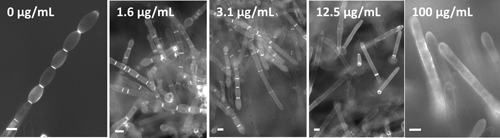
1.3. Effect on haustorial formationPyriofenone strongly inhibited haustorial formation. No haustorial formation was observed at a concentration of 1.6 µg/mL, and only a few were observed at a concentration of 0.4 µg/mL. However, there was no difference in haustorial formation between the untreated control and the 0.1 µg/mL concentration (Table 1).
1.4. Effect on mycelial growth and lesion expansionPyriofenone exhibited notable inhibition of mycelial growth when applied preventively. Complete inhibition of mycelial growth occurred at a concentration of 1.6–100 µg/mL, with nearly 90% control observed at the 0.4 µg/mL concentration (Table 1). When pyriofenone was applied curatively 4 days after inoculation, lesion expansion was inhibited at concentrations exceeding 1.6 µg/mL. However, even at higher concentrations of pyriofenone, complete inhibition of lesion expansion was not achieved (Table 2).
Table 2. Effect of pyriofenone on lesion expansion and sporulation of
Blumeria graminis| Application dose (µg/mL) | % Lesion areaa) | EC50 (µg/mL) | % Sporulation areaa) | EC50 (µg/mL) |
|---|
| 100 | 4.0±7.1** | 1.9 | 0.0±0.0** | 0.6 |
| 12.5 | 10.7±11.7** | 0.1±0.2** |
| 1.6 | 11.2±13.1** | 6.5±6.7** |
| 0.4 | 39.2±15.0 | 62.0±20.4* |
| 0.1 | 41.5±11.9 | 70.5±15.4 |
| 0 | 46.5±9.4 | 77.0±12.2 |
Lesion expansion and sporulation were evaluated at 4 days and 7 days after inoculation. Mycelial growth and sporulation areas within a field of view were visually assessed across 10 fields of view, rated on a scale of 1 to 10, and then averaged. Lesion expansion values were calculated by subtracting the initial value at the time of pyriofenone application (4 days after inoculation) from the final value (7 days after inoculation). No sporulation was observed 4 days after inoculation. a) Shows significant difference to untreated control (* p<0.005 ** p<0.001 Tukey HSD).
Pyriofenone exhibited potent inhibition of sporulation when applied both preventively and curatively. When pyriofenone was applied preventively, sporulation was nearly eradicated at concentrations of 0.4–100 µg/mL (Table 1). Similarly, with the curative application performed 4 days after inoculation, sporulation was seldom observed at concentrations of 1.6–100 µg/mL (Table 2).
Based on these results, we could summarize the effect of pyriofenone on the infection processes of wheat powdery fungus. The preventive application of pyriofenone before inoculation did not inhibit conidial germination. However, it inhibited the formation of appressoria and haustoria. Notably, haustorial formation was effectively inhibited, resulting in the complete prevention of successive lesion development and sporulation. Additionally, the curative application of pyriofenone after inoculation also effectively inhibited lesion expansion and sporulation.
2. Effect of pyriofenone on fungal morphogenesis of Blumeria graminis on wheat leavesSince the curative application of pyriofenone following inoculation effectively inhibited lesion expansion and sporulation, fungal morphology on wheat leaves was examined using both Chilled SEM and a fluorescence microscope subsequent to the curative application of pyriofenone.
Pyriofenone applications at a concentrations of 6 µg/mL were conducted 3 days after inoculation. The treated seedlings were subsequently incubated at 20°C for an additional 4 days. On untreated leaves, B. graminis exhibited smooth growth of cylindrical hyphae (Fig. 1-A). In contrast, pyriofenone treatment led to the collapse of mycelium and the formation of shrunken hyphae (Fig. 1-B). Occasionally, cytoplasm-like structures were observed at the hyphal tips (data not shown). In the untreated control group, a pair of secondary appressoria was formed (Fig. 1-A); however, on pyriofenone-treated leaves, numerous secondary appressoria were formed closely together (Fig. 1-C).
The hyphal cell wall was stained with Fluorescent Brightener 28 and observed using a fluorescent microscope. This fluorescent dye specifically stains β-glucan in the cell wall. In the untreated control group, we observed smooth growth of cylindrical hyphae (Fig. 2-A). However, in the group treated with fungicide, the hyphae exhibited abnormally swelling and shrinkage (Fig. 2-B), and hyphal tips were sometimes bifurcated (Fig. 2-C). At the swollen hyphal tips, we observed hall-like areas with low fluorescence (data not shown). Furthermore, Fluorescent Brightener 28 staining revealed the formation of numerous secondary appressoria (Fig. 2-E) in contrast to the simple secondary appressoria observed in the untreated control (Fig. 2-D).
Pyriofenone applications, with concentrations ranging from 1.6 to 100 µg/mL, were conducted 4 days after inoculation. The treated seedlings were subsequently incubated at 20°C for an additional 3 days. While the untreated control displayed normal septum formation and chains of conidia, abnormal septation occurred in a concentration-dependent manner with pyriofenone, leading to the formation of clubbed conidia-like structures devoid of septa at the 100 µg/mL concentration (Fig. 3). Furthermore, when these aberrant structures were employed as inoculum on new wheat leaves, no lesions were observed, indicating a loss of pathogenicity in these abnormal structures (data not shown).
Pyriofenone applications at a concentration of 6 µg/mL were conducted 3 days after inoculation. The treated seedlings were subsequently incubated at 20°C for an additional 5 days. The cell walls and septa were stained with Fluorescent Brightener 28, while the nuclei were stained with Hoechst 33342. In the untreated control group, normal cell division occurred, leading to the formation of conidial chains containing a single nucleus clearly delimited by septa (Fig. 4-A). Conversely, in the pyriofeone-treated samples, abnormal septation occurred, resulting in the presence of two or multiple nuclei in the conidiophore mother cell (arrows in Fig. 4-B) or clubbed conidia-like structures (arrows in Fig. 4-C).
3. Histochemical analysis of the effect of pyriofenone on Blumeria graminis on wheat leavesThe observed morphologial changes resulting from pyriofenone treatment indicated abnormal polar growth of the pathogen. To analyze the localization of components essential for the polar growth of hyphae in B. graminis, we employed a histochemical approach. Pyriofenone applications at a concentration of 6 µg/mL were performed 3 days after inoculation, and the treated seedlings were subsequently incubated at 20°C for an additional 4 days.
Fluorescent Brightener 28 was used to stain the β-glucan in the cell wall. In the control group, the apical points of the hyphal tips exhibited strong staining (Fig. 5-A). Conversely, in pyriofenone-treated apical hyphae, fluorescence was weaker compared to the untreated control, and β-glucan accumulation was observed as a patch-like pattern on the subapical cell wall (Fig. 5-B). Therefore, pyriofenone treatment led to the aberrant localization of β-glucan at the apex of the hyphae.
Glucose, a compoment of β-glucan, is believed to be transported to the hyphal apex via cytoplasmic vesicles.10,11) Subsequently, we analyzed the localization of cytoplasmic vesicles by staining apical vesicles with FM4-64. In the control group, fluorescence from FM4-64 was observed at the apical hyphae (Fig. 5-C), whereas FM4-64-labeled vesicles were redistributed away from the hyphal apex owing to pyriofenone treatment (Fig. 5-D).
The actin cytoskeleton is assumed to be associated with vesicle tracking for hyphal growth.10,11) Consequently, we observed the actin localization pattern through indirect immunofluorescence microscopy. In the control group, the anti-actin antibody marked the peripheral zone of hyphal apex (Fig. 5-E). In hyphae subjected to pyriofenone treatment, actin localization was detected in the subapical region of the hyphae (Fig. 5-F).
Microtubules were observed using tubulin antibodies in indirect immunofluorescence microscopy. Pyriofenone did not influence tubulin orientation (Fig. 5-G, H).
Discussion
Wheat powdery mildew is an obligate biotrophic pathogen, as it can only thrive on living plants.1,2) Infection commences with the germination of conidia, and the fungus forms an appressorium to penetrate the host plant’s cell wall, subsequently establishing an intracellular haustorium. Mycelial growth and sporulation are sustained by acquiring nutrients from the haustorium.1,2) An evaluation of the effects of pyriofenone on infection processes during preventive application demonstrated a robust inhibition of haustorial formation (Table 1). This, in turn, led to the inhibition of subsequent mycelial growth and sporulation on the leaves by cutting off nutrient intake. Another characteristic property of pyriofenone against Blumeria graminis infection processes was its ability to inhibit lesion expansion and sporulation through curative application (Table 2). This effectively controlled secondary infections and the spread of the disease. These outstanding biological characteristics of pyriofenone can account for its excellent performance against powdery mildew in the field.8,9)
The preventive application of pyriofenone inhibited appressorial formation and haustorial formation, and the curative application inhibited lesion expansion and sporulation; therefore, pyriofenone intervenes at various stages of the infection process of powdery mildew fungus (Tables 1 and 2). Among many mildewcides, triflumizole inhibits appressorial formation,12) while QoIs such as azoxystrobin deter conidial germination.13) Cyflufenamid and flutianil inhibit haustorial formation.14,15) Pyriofenone’s inhibition stages in the infection process differ from those of these existing fungicides. Furthermore, pyriofenone does not exhibit cross-resistance to these existing fungicides.8,9) Therefore, target sites of pyriofenone may be diverge from those of these fungicides.
As mentiond earlier, we were unable to obtain available information concerning the target sites of pyriofenone through comparison with existing fungicides. This was because pyriofenone did not exhibit cross-resistance to existing fungicides. Therefore, we employed morphological and cytological approaches to elucidate the target sites of pyriofenone. Microscopic observations of alternations in the infection processes can provide valuable insights into the mode of action of fungicides, even though direct demonstrations of the mode of action through genetic and biochemical approaches can be challenging.16,17) In this study, we found that pyriofenone induces abnormal morphogenesis in secondary appressoria, superficial hyphae, conidiophores, and conidia when applied curatively. On leaves treated with pyriofenone, we observed shrunken hyphae, bifurcated hyphae, and the formation of abnormal multi-formed secondary appressoria (Figs. 1 and 2). Furthermore, we observed abnormal septation, leading to the formation of clubbed conidia-like structures without septa when pyriofenone was applied at high concentrations (Figs. 3 and 4). These alternations in the morphology of hyphae and conidia suggest a disruption in the maintenance of normal polar cell growth.18–20) Bifurcation of hyphae is known to occur when the polarity of cytoplasmic vesicles shifts from the hyphal apex to other positions.11) Furthermore, actin may play a crucial role in septum formation during cell division.21,22) Abnormal conidia with multiple nuclei and irregular septation may be attributed to the progression of mitosis without septum formation.
The localization of components necessary for the polar growth of fungal hyphae, such as cell wall glucan, cytoplasmic vesicles, actin cytoskeleton, and microtubules in B. graminis was analysed through a histochemical approach (Fig. 5). Pyriofenone led to aberrant localization of actin cytoskeleton, β-glucan and cytoplasmic vesicles, though it did not affect tubulin orientation. Actin plays a role in maintaining cellular structure and facilitating intracellular transport. Cytoplasmic vesicles transport synthesized proteins, utilizing the action cytoskeleton as a scaffold.10,11) Our histochemical and cytological analysis unequivocally indicated a loss of actin polarity, suggesting a compromised transport of proteins necessary for cellular polarity. Based on these findings, we propose that pyriofenone disrupts the establishment and maintenance of cellular polarity, which are essential for the morphogenesis and pathogenicity of the powdery mildew fungus.
Our study aimed to compare the mode of action of pyriofenone with that of metrafenone,16,17) which shares a similar chemical structure with pyriofenone, through a morphological and cytological approach (Supplemental Fig. S1). The impact of metrafenone on the infection processes of the powdery mildew fungus has previously been reported by Schmit et al.16) and Opalski et al.17) They suggest that metrafenone treatment disrupts polarized hyphal growth, with these changes attributed to disturbances in actin localization.16,17) Morphological and cytological analysis of pyriofenone- treated hyphae in the present study revealed similar defects to those observed with metrafenone (Fig. 5). The structural similarity between pyriofenone and metrafenone (Supplemental Fig. S1), as well as the shared morphological and cytological changes in fungi induced by these compounds, indicates a similar or related mode of action for pyriofenon and metrafenone. At present, both metrafenone and pyriofenone are classified under FRAC code 50, which pertains to the actin/myosin/fimbrin function.
When considering cell polarity, numerous studies have utilized a budding yeast as a model system.22,23) Harris and Momany’s review (2004) focuses on the literature concerning signal transduction pathways that control morphogenesis in both yeast and filamentous fungi.22) Their discussion of the Ras and Rho families of small GTPases, including Cdc42, their regulators and downstream interactions, including actin, offers valuable insights for understanding potential mechanisms of morphogenetic control influenced by pyriofenone and metrafenone.16,17) Rho GTPase, which is involved in cellular polarity, directly regulates actin and microtubules while also controlling cell wall synthesis in fungi.11,23) The actin cable, formed as a result, serves as a transport conduit for cytoplasmic vesicles towards the cell’s apex.10,11) Furthermore, Rho GTPase is implicated in the biosysnthesis of β-glucan.23,24) Pyriofenone has been observed to cause aberrant localization of the actin cytoskeleton, β-glucan and cytoplasmic vesicles, although it does not appear to affect tubulin orientation. Given the challenge of attributing these effects to pyriofenone’s independent action on these cytoplasmic structures, it is reasonable to speculate that pyriofenone influences upstream cellular components such as Rho, Ras, or Rac GTPase.19–22) Notably, a benzophenone compound, isolated from an unidentified fungal species and structurally resembling metrafenone and pyriofenone, was proposed to modulate Ras protein activity by inhibiting its farnesylation.25) However, the precise impact of pyriofenone on the modulation of Ras or Rho protein activity by inhibiting their farnesylation or geranylgeranylation remains to be elucidated.
Electronic supplementary materials
The online version of this article contains supplementary material (Supplemental Fig. S1), which is available at https://www.jstage.jst.go.jp/browse/jpestics/.
References
- 1) U. Braun, R. T. A. Cook, A. J. Inman and H.-D. Shin: The taxonomy of the powdery mildew fungi. In “The Powdery Mildew, a Comprehensive Treatise,” ed. by R. R. Belanger, W. R. Bushnell, A. J. Dik and T. L. W. Carver, Amer. Phytopathol. Soc., St.Paul, pp. 13–55, 2002.
- 2) S. Takamatsu: Molecular phylogeny reveals phenotypic evolution of powdery mildews (Erysiphales, Ascomycota). J. Gen. Plant Pathol. 79, 218–226 (2013).
- 3) K. J. Brent and D. W. Hollomon: “Fungicide resistance: The assessment of risk,” FRAC Monograph No. 2, 2nd ed., Fungicide Resistance Action Committee, Brussels, 2007.
- 4) D. W. Hollomon and I. E. Wheeler: Controlling powdery mildews with chemistry. In “The Powdery Mildew, a Comprehensive Treatise” ed. by R. R. Belanger, W. R. Bushnell, A. J. Dik and T. L. W. Carver, Amer. Phytopathol. Soc., St.Paul, pp. 249–255, 2002.
- 5) J. Dietz and C. Winter: Recently introduced powdery mildew fungicides. In “Modern Crop Protection Compounds,” 3rd edition ed. by P. Jeschke, M. Witshel, W. Krämer and U. Sehirmer, Wiley-VCH, Weinheim, pp. 933–947, 2019.
- 6) T. Hirooka and H. Ishii: Chemical control of plant diseases. J. Gen. Plant Pathol. 79, 390–401 (2013).
- 7) K. J. Brent and D. W. Hollomon: “Fungicide resistance in crop pathogens: How can it be managed?” FRAC Monograph No. 1, 2nd ed., Fungicide Resistance Action Committee, Brussels, 2007.
- 8) M. Ogawa: Fungicidal properties and sensitivity study of a novel fungicide pryiofenone. Abstr. Symposium of Research Committee on Fungicide Resistance 24, 17–26 (2014) (in Japanese).
- 9) M. Ogawa, A. Nishimura, Y. Abe, Y. Fukumori, K. Suzuki and S. Mitani: Fungicidal spectrum and biological properties of a new fungicide, pyriofenone. J. Pestic. Sci. 48, 65–70 (2023).
- 10) B. Feierbach and F. Chang: Roles of the fission yeast formin for3p in cell polarity, actin cable formation and symmetric cell division. Curr. Biol. 11, 1656–1665 (2001).
- 11) A. J. Ridley: Rho GTPases and actin dynamics in membrane protrusions and vesicle trafficking. Trends Cell Biol. 16, 522–529 (2006).
- 12) A. Nakata, S. Hashimoto, K. Ikura and K. Katsuura: Development of a new fungicide, triflumizole. J. Pestic. Sci. 16, 301–313 (1991) (in Japanese).
- 13) J. R. Bertelsen, E. de Neergaard and V. Smedegaard-Petersen: Fungicidal effects of azoxystrobin and epoxiconazole on phyllosphere fungi, senescence and yield of winter wheat. Plant Pathol. 50, 190–205 (2001).
- 14) M. Haramoto, H. Yamanaka, H. Sano, S. Sano and H. Otani: Fungicidal activities of cyflufenamid against various plant-pathogenic fungi. J. Pestic. Sci. 31, 95–101 (2006).
- 15) S. Kimura, T. Komura, N. Yamaoka and H. Oka: Biological properties of flutianil as a novel fungicide against powdery mildew. J. Pestic. Sci. 45, 206–215 (2020).
- 16) M. R. Schmitt, R. Carzaniga, H. V. T. Cotter, R. O’Connell and D. Hollomon: Microscopy reveals disease control through novel effects on fungal development: A case study with an early-generation benzophenone fungicide. Pest Manag. Sci. 62, 383–392 (2006).
- 17) K. S. Opalski, S. Tresch, K.-H. Kogel, K. Grossmann, H. Köhle and R. Hückelhoven: Metrafenone: studies on the mode of action of a novel cereal powdery mildew fungicide. Pest Manag. Sci. 62, 393–401 (2006).
- 18) A. Virag and A. J. F. Griffiths: A mutation in the Neurospora crassa action gene results in multiple defects in tip growth and branching. Fungal Genet. Biol. 41, 213–225 (2004).
- 19) G. M. Guest, X. Lin and M. Momany: Aspergillus nidulans RhoA is involved in polar growth, branching, and cell wall synthesis. Fungal Genet. Biol. 41, 13–22 (2004).
- 20) J. Wendland and P. Phillippsen: Cell polarity and hyphal morphogenesis are controlled by multiple Rho-protein modules in the filamentous ascomycete Ashbya gossypii. Genetics 157, 601–610 (2001).
- 21) K. J. Boyce, M. J. Hynes and A. Andrianopoulos: Control of morphogenesis and action localization by the Penicillium marneffei RAC homolog. J. Cell Sci. 116, 1249–1260 (2003).
- 22) S. D. Harris and M. Momany: Polarity in filamentous fungi: Moving beyond the yeast paradigm. Fungal Genet. Biol. 41, 391–400 (2004).
- 23) D. E. Levin: Cell wall integrity signaling in Saccharomyces cerevisiae. Microbiol. Mol. Biol. Rev. 69, 262–291 (2005).
- 24) M. Sekiya-Kawasaki, M. Abe, A. Saka, D. Watanabe, K. Kono, M. Minemura-Asakawa, S. Ishihara, T. Watanabe and Y. Ohya: Dissection of upstream regulatory components of the Rho1p effector, 1,3-glucan synthase, in Saccharomyces cerevisiae. Genetics 162, 663–676 (2002).
- 25) M. Chu, R. Mierzwa, E. Barrabee, A. King, M. Hallade, J. Terracciano, M. G. Patel, R. Patton, W. R. Bishop and M. S. Puar: SCH 207278: A novel farnesyl protein transferase inhibitor from an unidentified fungus. Bioorg. Med. Chem. Lett. 7, 2547–2550 (1997).