Abstract
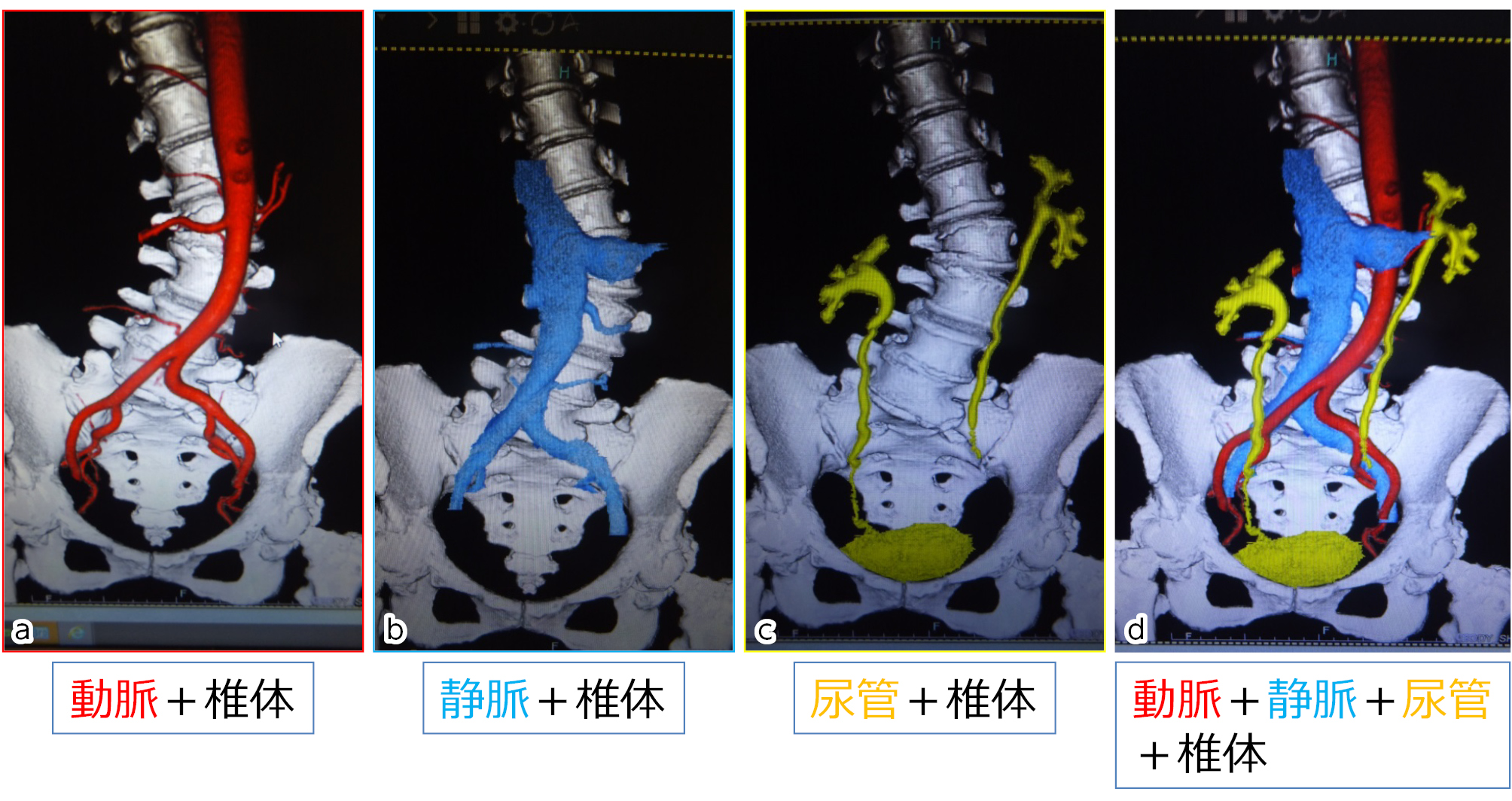
Introduction: In the narrow and deep surgical field for lateral lumbar interbody fusion (LLIF), even bleeding from the venous vessels can interfere with the operation. In this study, we investigated the running of inferior vena cava (IVC) and the frequency and blanching level of iliolumbar vein (ILV) by the three-phase contrast-enhanced CT.
Material and Methods: Subjects were 71 patients who underwent contrast-enhanced CT before LLIF surgery for adult spine deformity. After rapid administration of the contrast medium, three-phase CT angiography consisting of arterial, venous, and ureteral phases was performed to visualize the relationship between vertebral bodies and arteries, veins, and ureters. The horizontal distance (RLD) between the left lateral border of IVC and central sacral vertical line (CSVL), the number of ILVs, its inflow veins, and the running level of the vertebral bodies were studied.
Results: RLD between the left lateral border of IVC and CSVL significantly correlated with Cobb angle in the lumbar spine. ILV was identified in approximately 70% of cases. ILV was most frequently inflowing to common iliac vein (CIV), accounting for 80%. ILV running along L4/5 disc level occurred in nearly 5% cases.
Conclusion: It is difficult to visualize lumbar venous vessels by two-phase CT angiography in detail. The three-phase angiography we have devised provides clear delineation of the lumbar segmental vessels and ILVs and is useful for preoperative planning for LLIF to avoid the venous vessel injury.