Journal of Spine Research
Online ISSN : 2435-1563
Print ISSN : 1884-7137
Volume 14, Issue 10
Displaying 1-11 of 11 articles from this issue
Editorial
Review Article
  • Katsuji Shimizu
    2023 Volume 14 Issue 10 Pages 1268-1275
    Published: October 20, 2023
    Released on J-STAGE: October 20, 2023
    JOURNAL FREE ACCESS

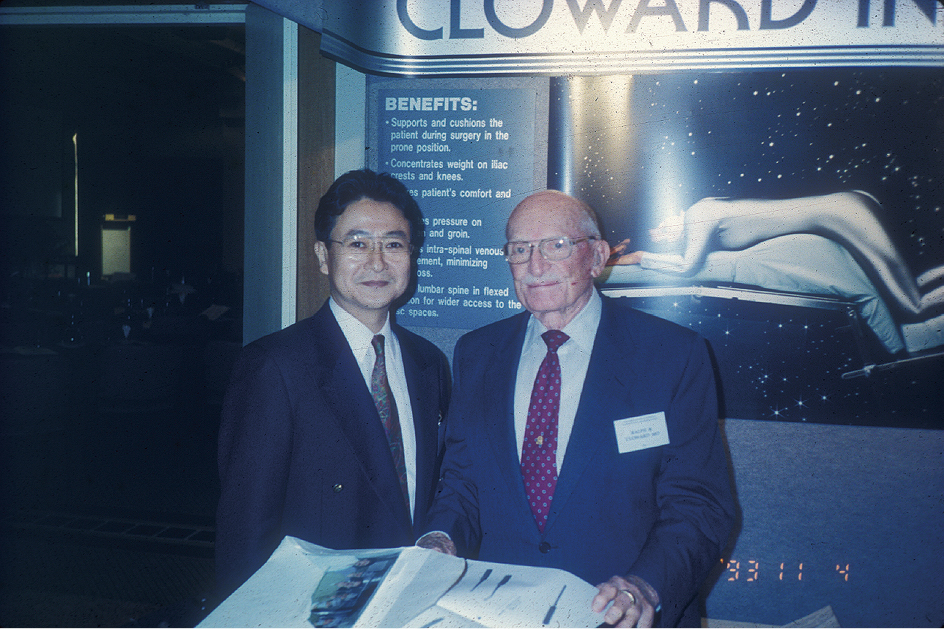
    To commemorate twentieth anniversary of Kumu Cloward Lecture, the birth history of the lecture was introduced and the lecture was delivered to review instrumentation for the treatment of spinal infection, specifically to show our experience on two-staged (PI-A) surgery. Anterior debridement and reconstruction with bone graft is a gold standard for surgical treatment of vertebral osteomyelitis. However, it was described "Open the gate of death" to perform anterior debridement and fusion before the appearance of antibiotics. It was after antibiotics that anterior surgery become popular. In modern era of antibiotic resistant bacteria, it seems to be as dangerous as pre-antibiotic era to perform anterior surgery. By analogy with the pre-antibiotic era, we performed posterior spinal instrumentation as the first stage operation, and then performed anterior surgery as the second operation. In 1997, we began to use a two-stage surgical treatment (first: posterior instrumentation; second: anterior debridement and bone graft). The advantages of this method is dual: surgical morbidity is divided and surgical effect is immediate. After the first procedure (posterior instrumentation), the patient is entertained with decrease of pain and is able to sit up from bed rest. Moreover, erythrocyte sedimentation rate (ESR) decreases after posterior stabilization and without drainage of anterior focus. This method is especially effective for thoracolumbar spondylitis of the patients with poor general conditions.

    Download PDF (2193K)
Original Article
  • Chikara Hayakawa, Ichiro Okano, Yoshifumi Kudo, Koki Tsuchiya, Ryo Yam ...
    2023 Volume 14 Issue 10 Pages 1276-1282
    Published: October 20, 2023
    Released on J-STAGE: October 20, 2023
    JOURNAL FREE ACCESS

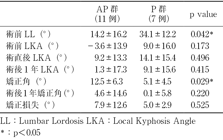
    Introduction: Surgical treatments for osteoporotic vertebral fractures (OVFs) at the thoracolumbar junctional level have been well documented, however there were few reports which described OVFs in the middle-low lumbar spine. In this study, we compared the surgical treatments of anterior combined surgery (AP group) and posterior surgery (P group) for OVFs in the middle-low lumbar spine.

    Methods: Eighteen patients (AP group 11, P group 7) were included in this study. Minimum follow up period was set at 1 year. Preoperative lumbar lordosis (LL), pre-and postoperative local kyphosis angle (LKA), correction angle, correction loss at the final follow-up, implant-related complications, operation time and total blood loss were compared between AP and P groups.

    Results: There was no significant difference in preoperative LKA between AP and P groups (the mean±standard deviations; AP group: −3.6±13.9°, P group: 9.0±16.0°; p=0.173). The mean postoperative correction angle was significantly higher in AP group (AP group: 12.5±6.3°, P group: 5.1±4.5°; p=0.029). There was no significant difference in the mean correction loss between two groups.

    Conclusions: Our results demonstrated that the mean correction angle was higher in AP group, and there was no significant difference in the mean correction loss in comparison between AP and P groups. Considering correction of the spinal alignment, anteroposterior combined surgery can be a better surgical option in the treatment of patients with OVFs in the middle-lower lumbar spine.

    Download PDF (1503K)
  • Taiki Yasukawa, Toshiyuki Shirahata, Tomoya Asakura, Yoshifumi Kudo, H ...
    2023 Volume 14 Issue 10 Pages 1283-1291
    Published: October 20, 2023
    Released on J-STAGE: October 20, 2023
    JOURNAL FREE ACCESS

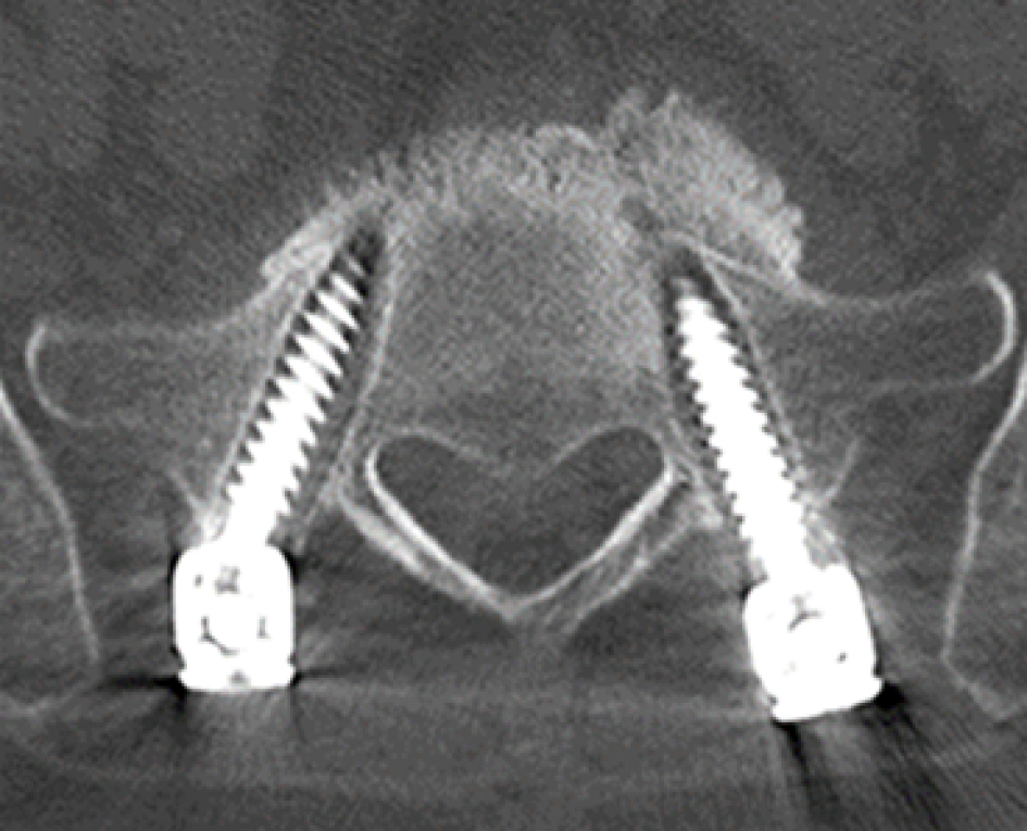
    Introduction: We have employed the sacral pedicle screw (S1PS) as the distal anchor in multi-level fixation of the lumbosacral spine combined with lateral lumbar interbody fusion (LLIF). However, S1PS loosening has been frequent. The present study aims to investigate the loosening of S1PS, cage subsidence, and fusion rate in multi-level lumbosacral fixation combined with LLIF.

    Methods: We evaluated fourteen patients diagnosed with lumbar canal stenosis and degenerative scoliosis who underwent multi-level lumbosacral fixation with LLIF. CT images were used to evaluate radiological findings, including radiolucent zones around sacral screws, cage subsidence, loss of correction in lumbosacral lordosis, and bone fusion at L5/S postoperative 1 year.

    Results: S1PS loosening was confirmed in 9 cases (9/14, 64%) at 3 months postoperatively, and in 13 cases (13/14, 92%) at 6 months postoperatively. The mean correction loss of lordosis at L5/S was 4.3° at 1 year postoperatively, and cage subsidence was observed in 10 cases (10/14, 71%). Bone fusion at L5/S was achieved in only 7 cases (7/14, 50%) at 1 year postoperatively. In the cases with no loosening of S1PS, the mean sacral Hounsfield unit was relatively high (215.3, Mean: 149.4), and both S1PS were inserted using a bicortical trajectory. In cases where both S1PS were inserted using a monocortical trajectory, a successful fusion of L5/S was only achieved in one out of six cases (16.6%).

    Conclusions: S1PS loosening occurred in 91% of the patients. Patients with multi-level lumbosacral fixation combined with LLIF have a high risk of S1PS loosening when used as the most distal anchor.

    Download PDF (3123K)
  • Masakazu Toi, Keishi Maruo, Fumihiro Arizumi, Kazuya Kishima, Tomoyuki ...
    2023 Volume 14 Issue 10 Pages 1292-1297
    Published: October 20, 2023
    Released on J-STAGE: October 20, 2023
    JOURNAL FREE ACCESS

    Introduction: Segmental lordosis is an important radiographic parameter as sagittal alignment has been correlated to clinical outcomes. However, there are few reports on the relationship between segmental lordosis angle (SLA) and global spinal alignment (GSA) after short segment lumbar fusion. This study retrospectively examined the relationship between SLA and GSA after L4/5 transforaminal lumbar interbody fusion (TLIF).

    Methods: The subjects were 91 patients who underwent consecutive mini-open L4/5 TLIF at our hospital between 2014 and 2020 with a minimum one-year follow-up. Subjects consisted of 48 men and 43 women with a mean age of 69.4±9.8 years. Patient factors (age, sex, BMI), cage factors (position, cage subsidence), radiographic parameters (LL, PI-LL, PT, SVA, and TPA), and SLA of L4/5 were investigated. In addition, the SLA change was compared between two groups. The group with increased SLA at 1 year postoperatively was defined as the SLA (+) group and the group with decreased SLA postoperatively as the SLA (−) group., The independent factor of SLA (+) was identified by logistic regression analysis.

    Results: The SLA increased significantly from 14.6 degrees before surgery to 16.9 degrees after surgery (P<0.001). SLA, LL, PI-LL, PT, SVA, and TPA also improved significantly after TLIF. There were 73 patients in the SLA (+) group and 18 patients in the SLA (−) group. There were no significant differences in SLA between the two groups with regard to patient background factors. However, there were significant differences in preoperative LL and SLA. Patients in the SLA (+) group had significantly lower preoperative SLA (13.7° vs. 18.4°, P=0.001) than the SLA (−) group, and there was a negative correlation between SLA change and preoperative SLA (r=−0.439). The cage subsidence was significantly lower (72% vs. 38%, P=0.010), and the cage was placed significantly more anteriorly in the SLA (+) group. However, there was no significant difference in the cage lordosis angle. There was no significant difference in GSA at 1 year postoperatively between the two groups, but there were significantly more cases of PI-LL mismatch in the SLA (−) group (50% vs. 24%, P=0.030). Logistic regression analysis showed that preoperative SLA (odds ratio 1.15, 95% confidence interval 0.02-0.19, P=0.024) and cage subsidence (odds ratio 6.69, 95% confidence interval −1.72-−0.18, P=0.009) were independent factors for SLA (+). The cutoff value via ROC analysis for preoperative SLA was 15.3°.

    Conclusions: Postoperative SLA increased in patients with low preoperative LL and SLA. Preoperative SLA and cage subsidence were independent factors for increased SLA. SLA change had no effect on GSA at 1 year postoperatively, however, there was less PI-LL mismatch in the SLA (+) group. Preoperative SLA and cage subsidence of L4/5 should be kept in mind for long-term outcomes.

    Download PDF (984K)
  • Masaru Hatano, Keishi Maruo, Shoji Nishio, Yoshiteru Nakamura, Nobuyos ...
    2023 Volume 14 Issue 10 Pages 1298-1307
    Published: October 20, 2023
    Released on J-STAGE: October 20, 2023
    JOURNAL FREE ACCESS

    Introduction: The S1 pedicle screw (PS) is anatomically and mechanically weak and prone to looseness, which has led to the proposal of bicortical or tricortical screws that penetrate the cortical bone of the anterior aspect of the sacrum. However, percutaneous pedicle screw (PPS) placement using a guide wire carries a risk of neurovascular injury. This study retrospectively investigated the trajectory of S1 PPS and the risk of vascular injury in L5/S interbody fusion surgery.

    Methods: 76 patients (49 males and 27 females, mean age 68±13 years) who underwent L5/S interbody fusion surgery including one or two vertebrae from January 2017 to December 2021 were included. A total of 152 S1 PS were placed, which were divided into three groups based on the trajectory: unicortical fixation (Uni) without anterior cortical breach, bicortical fixation (Bi) with anterior cortical breach, and modified penetrating endplate screw (mPES) group that inserted into the endplate of S1. The risk of vascular injury was evaluated based on the presence of vessels in the guide wire trajectory and the distance between the PS tip and vessels being ≤5 mm.

    Results: There were 10 cases in the mPES group, 15 cases with bilateral Bi, 21 cases with unilateral Bi, and 30 cases in the Uni group, with 19 PS in the mPES group, 51 PS in the Bi group, and 82 PS in the Uni group. The risk of guide wire trajectory was significantly different among the groups, with 5.26% in the mPES group, 58.8% in the Bi group, and 30.5% in the Uni group (p<0.01). The risk of PS tip was also significantly different among the groups, with 5.26% in the mPES group, 52.9% in the Bi group, and 7.32% in the Uni group (p<0.01). The presence of vascular injury risk and the PS medial angle showed significant differences for both guide wire trajectory and PS tip risk (guide wire trajectory: 12.1±6.3° vs 18.2±6.5°, p<0.01; PS tip: 7.2±7.2° vs 17.3±0.6°, p<0.01).

    Conclusions: Bicortical purchase of S1 PPS results in a smaller medial angle of the screw, which increases the risk of vascular injury. mPES is useful in S1 PPS because it keeps the PS tip within the disc space and reduces the risk of vascular injury.

    Download PDF (2495K)
  • Hiroaki Ishida, Masayuki Ishihara, Shinichiro Taniguchi, Takashi Adach ...
    2023 Volume 14 Issue 10 Pages 1308-1317
    Published: October 20, 2023
    Released on J-STAGE: October 20, 2023
    JOURNAL FREE ACCESS

    Purpose: To investigate the clinical results of lateral access surgery (LAS) such as lateral route lumbar interbody fusion (LLIF), lateral access corpectomy (LAC), and anterior column realignment (ACR) for osteoporotic vertebral fracture (OVF).

    Subjects and methods: Twenty-three patients who underwent LAS for KDOVF at our hospital after 2019 and were able to follow up for at least 18 months were included in the study. Surgical method, fractured vertebral body level, local kyphotic angle, number of fixed vertebral bodies, bone fusion rate, using of cement augmentation (CA), and complications were evaluated.

    Results: LLIF was performed in 6 cases (Group L), LAC in 14 cases (Group C), and ACR in 3 cases (Group A). Fractured vertebrae were L1 to L4 in Group L, T12 and L1 in Group C, and only L2 and 3 in Group A. The blood loss and operative time were significantly higher in Group C, and there was no significant difference in the number of fixed vertebra in the three groups. Changes in local kyphotic angle were significantly improved after surgery in all three groups. Correction loss was about 1 degree in Group L, 2.4 degrees in Group C, and 3 degrees in Group A, and was significantly lower in Group L. The average correction angle was 11 degrees for Group L, 25 degrees for Group C, and 18 degrees for Group A, and Group C was significantly larger than Group L. The bone fusion rate 12 months after surgery in Group A was the lowest, but the bone fusion rate 24 months after surgery was more than 90% in all three groups. Complications were endplate injury in one case each of Group C, and cage subsidence in one case of Group A. In the Group C, 4 cases of pleural injury and 1 case of proximal junctional failure (PJF) were observed, but no cases required reoperation.

    Conclusion: The clinical results of LAS for OVF was investigated. Good results were obtained in all three groups. In this study, it was suggested that LAS is a useful surgical technique for OVF kyphotic deformity with severe bone fragility.

    Download PDF (2735K)
  • Takumi Hashimura, Eijiro Onishi, Kouta Wada, Itsurou Yamane, Atsushi T ...
    2023 Volume 14 Issue 10 Pages 1318-1324
    Published: October 20, 2023
    Released on J-STAGE: October 20, 2023
    JOURNAL FREE ACCESS

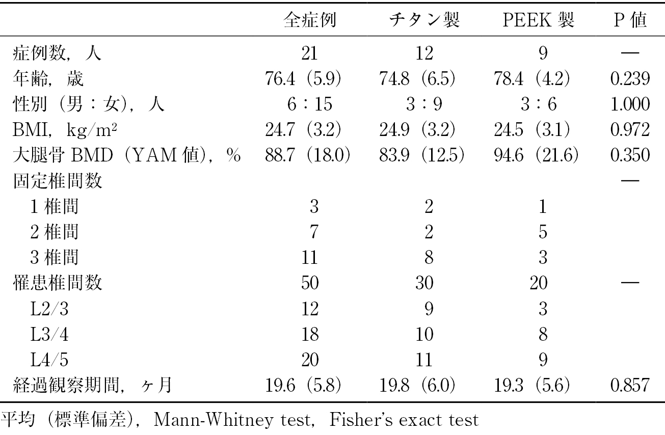
    Purpose: The purpose of this study was to compare the bony union of human demineralized bone matrix (DBM) and iliac crest bone graft (ICBG) in lateral lumbar interbody fusion (LLIF) cages.

    Subjects: Twenty-one patients (mean age 76.4 year) who underwent LLIF for lumbar degenerative disease from March 2019 to August 2020. The cage material was titanium for 30 intervertebral spaces in 12 patients and polyetheretherketone (PEEK) for 20 intervertebral spaces in 9 patients. The cage was filled with DBM alone and ICBG, divided into left and right sides with the midline as a border. Bony union was evaluated by CT images more than one year after operation. This time, we measured CT values of each grafted bone in the cage.

    Results: Intra-cage bone fusion was achieved in 62% for DBM and 90% for ICBG (P=0.001). There was no significant difference in DBM (87%) and ICBG (97%) for titanium cages (P=0.353), while there was a significant difference in DBM (25%) and ICBG (80%) for PEEK cages (P=0.001). The CT values of the grafted bone showed a similar trend to the degree of the examiner's visual assessment of the bone union.

    Conclusions: We observed a comparable ossification between DBM and ICBG in the titanium cages.

    Download PDF (1461K)
  • Masaaki Chazono, Sota Urimoto
    2023 Volume 14 Issue 10 Pages 1325-1331
    Published: October 20, 2023
    Released on J-STAGE: October 20, 2023
    JOURNAL FREE ACCESS

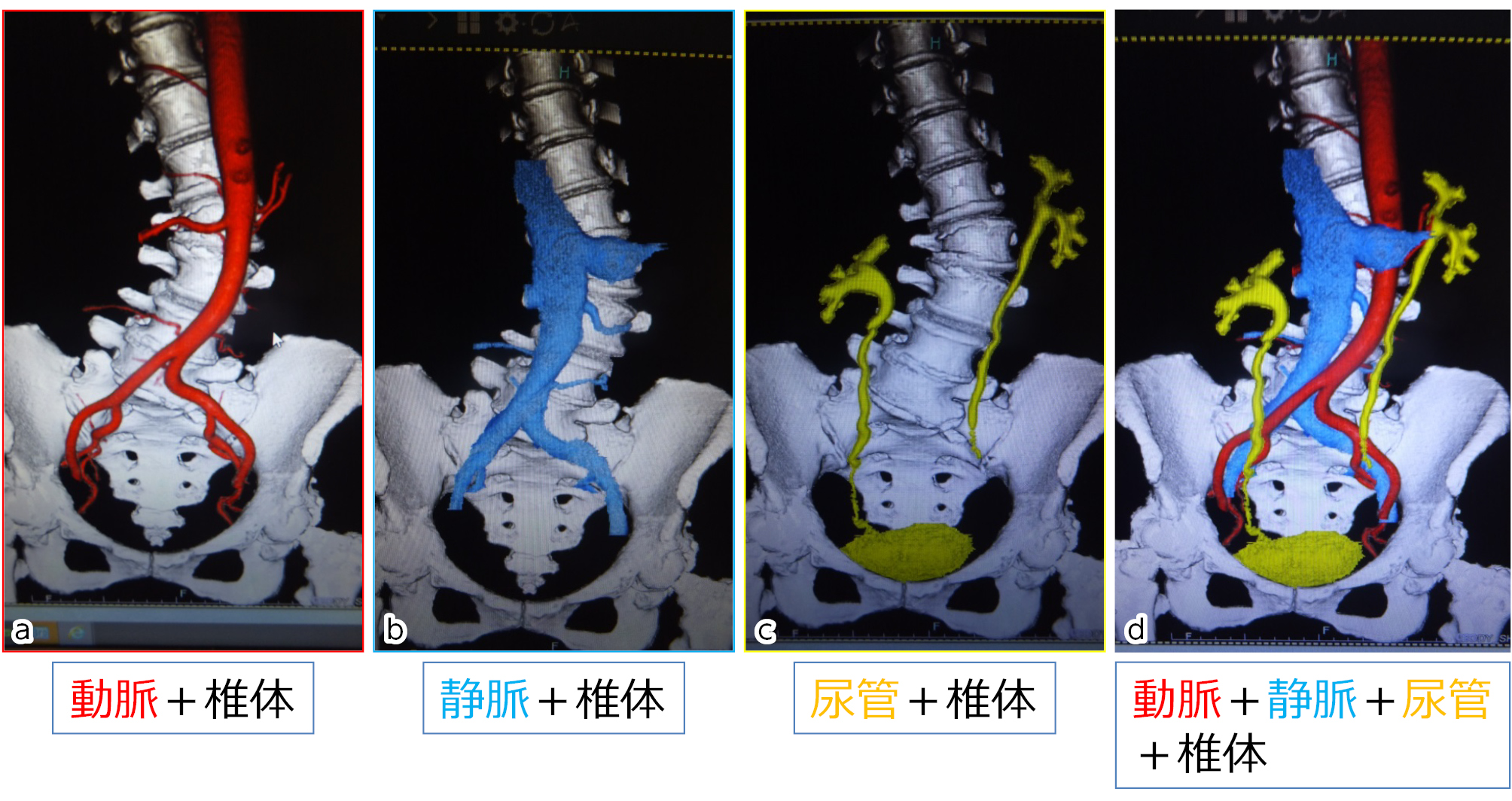
    Introduction: In the narrow and deep surgical field for lateral lumbar interbody fusion (LLIF), even bleeding from the venous vessels can interfere with the operation. In this study, we investigated the running of inferior vena cava (IVC) and the frequency and blanching level of iliolumbar vein (ILV) by the three-phase contrast-enhanced CT.

    Material and Methods: Subjects were 71 patients who underwent contrast-enhanced CT before LLIF surgery for adult spine deformity. After rapid administration of the contrast medium, three-phase CT angiography consisting of arterial, venous, and ureteral phases was performed to visualize the relationship between vertebral bodies and arteries, veins, and ureters. The horizontal distance (RLD) between the left lateral border of IVC and central sacral vertical line (CSVL), the number of ILVs, its inflow veins, and the running level of the vertebral bodies were studied.

    Results: RLD between the left lateral border of IVC and CSVL significantly correlated with Cobb angle in the lumbar spine. ILV was identified in approximately 70% of cases. ILV was most frequently inflowing to common iliac vein (CIV), accounting for 80%. ILV running along L4/5 disc level occurred in nearly 5% cases.

    Conclusion: It is difficult to visualize lumbar venous vessels by two-phase CT angiography in detail. The three-phase angiography we have devised provides clear delineation of the lumbar segmental vessels and ILVs and is useful for preoperative planning for LLIF to avoid the venous vessel injury.

    Download PDF (1462K)
Case Report
  • Takehiko Moroi, Atsuko Tachibana, Hironobu Watanabe, Takahito Iga, Kiy ...
    2023 Volume 14 Issue 10 Pages 1332-1339
    Published: October 20, 2023
    Released on J-STAGE: October 20, 2023
    JOURNAL FREE ACCESS

    Introduction: Posterior lumbar interbody fusion (PLIF) and posterolateral fusion (PLF) are standard surgical procedures for isthmic L5 spondylolisthesis. The risk of postoperative neurological deficit associated with surgical reduction may increase in the case of severe slippage. In patients with bony bridges anteriorly or laterally at the vertebral bodies, it is difficult to reduce the slippage. We performed posterior lumbosacral fusion using a S1 transdiscal screw to avoid postoperative neurological deficit and to obtain stronger fixation and bone union. We report three cases using this technique.

    Case 1: A 69-year-old woman had low back pain and right buttock pain. A lumbar radiograph and CT showed Meyerding classification grade 3, 56% slip, −3°slip angle. Disc degeneration at L5/S1 was severe and disc height was almost lost.

    Case 2: A 33-year-old man had low back pain and bilateral buttock pain. A lumbar radiograph and CT showed Meyerding classification grade 3, 55% slip, 8°slip angle. Disc degeneration at L5/S1 was severe and disc height was almost lost.

    Case 3: A 56-year-old man had low back pain and right buttock pain. A lumbar radiograph and CT showed Meyerding classification grade 2, 26% slip, −2°slip angle. An incomplete bony bridge at the vertebral body was formed anteriorly and laterally, and it seemed difficult to reduce the slippage and inserting the cage into the disc space.

    Conclusions: Low back pain and leg pain improved in all patients after surgery, and the bone union was obtained in 1 year, suggesting that this surgical procedure is useful.

    Download PDF (2379K)
  • Takeshi Tezuka, Takeo Furuya, Satoshi Maki, Takaki Inoue, Atsushi Yund ...
    2023 Volume 14 Issue 10 Pages 1340-1344
    Published: October 20, 2023
    Released on J-STAGE: October 20, 2023
    JOURNAL FREE ACCESS

    We report a case of posterior decompression with instrumented fusion in patient with cervical radiculopathy refractory to conservative treatment caused by foraminal stenosis due to cervical posterior spondylolisthesis.

    A 47-year-old woman presented to with a chief complaint of right upper extremity pain and numbness.

    She was diagnosed cervical radiculopathy based on neurological examination and imaging tests.

    The neurological symptoms were numbness and pain from the right neck to the right upper extremity.

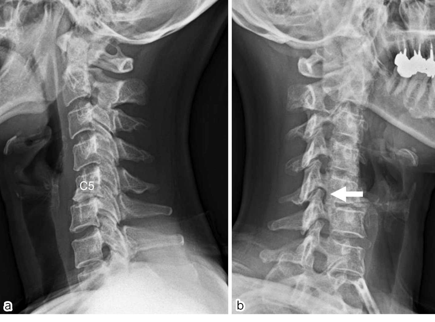
    X-ray images showed a C5 posterior spondylolisthesis and MRI showed right foraminal stenosis between C5 and C6.

    CT showed degeneration of the C5/C6 disc and bone spurs.

    The patient was diagnosed with right C6 radiculopathy due to right C5/C6 intervertebral foramen narrowing caused by posterior slip of the cervical spine, which was thought to have resulted in radiculopathy.

    Surgical treatment was planned, and a C5 total laminectomy, C6 partial laminectomy, right C5/C6 foraminotomy, and C5-6 posterior decompression and corrective fixation were performed to correct the slipped right C5.

    After the surgery, symptoms completely disappeared immidiately, and No recurrence of the symptom was observed as of two years after surgery.

    Posterior decompression with instrumented fusion was effective in treating cervical radiculopathy due to intervertebral foraminal stenosis with cervical posterior spondylolisthesis.

    Download PDF (1347K)
feedback
Top